- Anatomical terminology
- Skeletal system
- Skeleton of trunk
-
Skull
- Neurocranium
- Viscerocranium
- Auditory ossicles
- Sutures of skull
- Topography of skull
- Skeleton of upper limb
- Skeleton of lower limb
- Joints
- Muscles
- Heart
- Blood vessels
- Lymphatic system
- Nervous system
- Respiratory system
- Digestive system
- Urinary system
- Female reproductive system
- Male reproductive system
- Endocrine glands
- Eye
- Ear
Skull
The human skull (Latin: cranium) is the skeleton of the head composed of 22 bones. Bones of the skull are joined together primarily by sutures. The primary function of the skull is to provide protection for the brain and sensory organs connected with it.
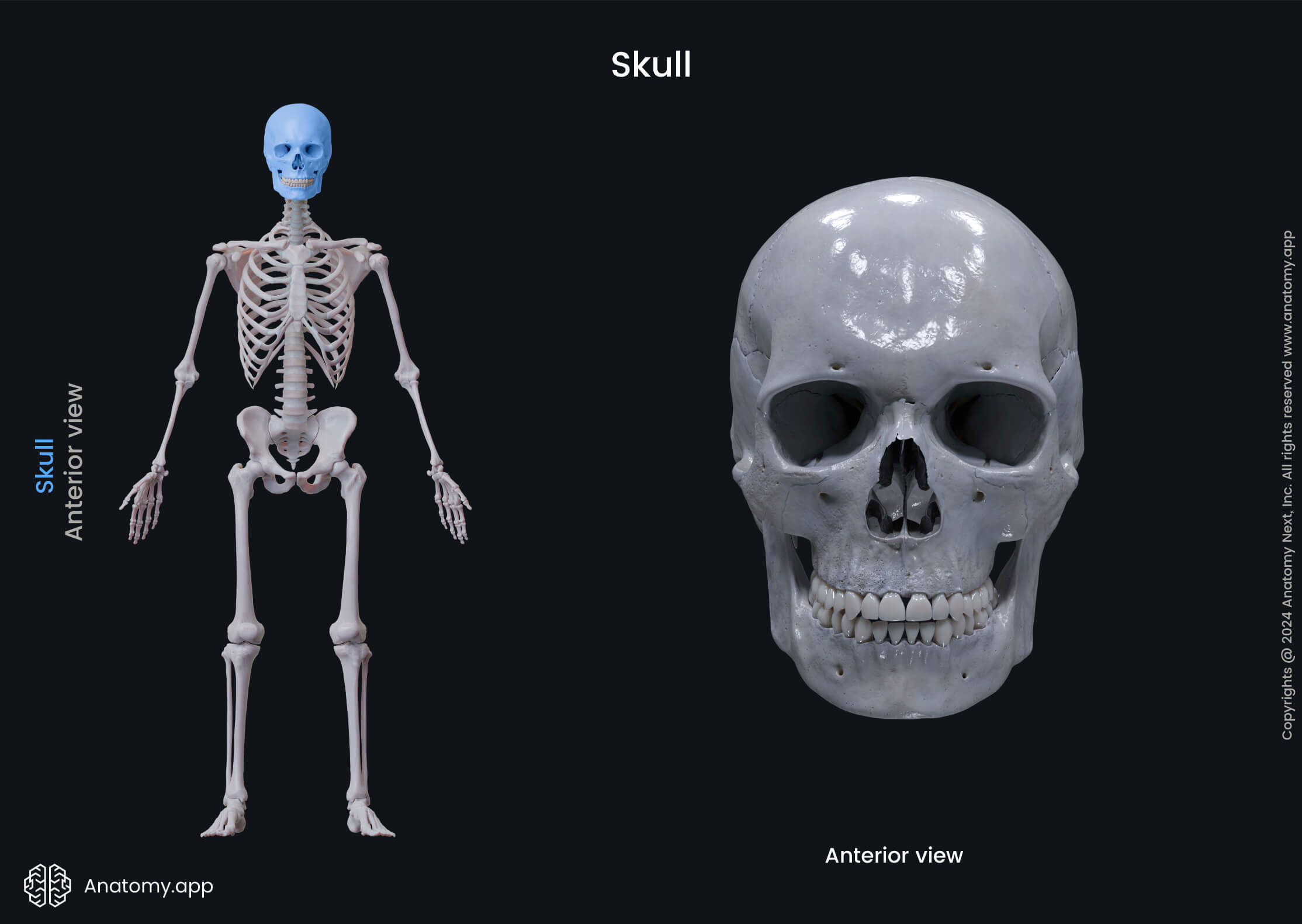
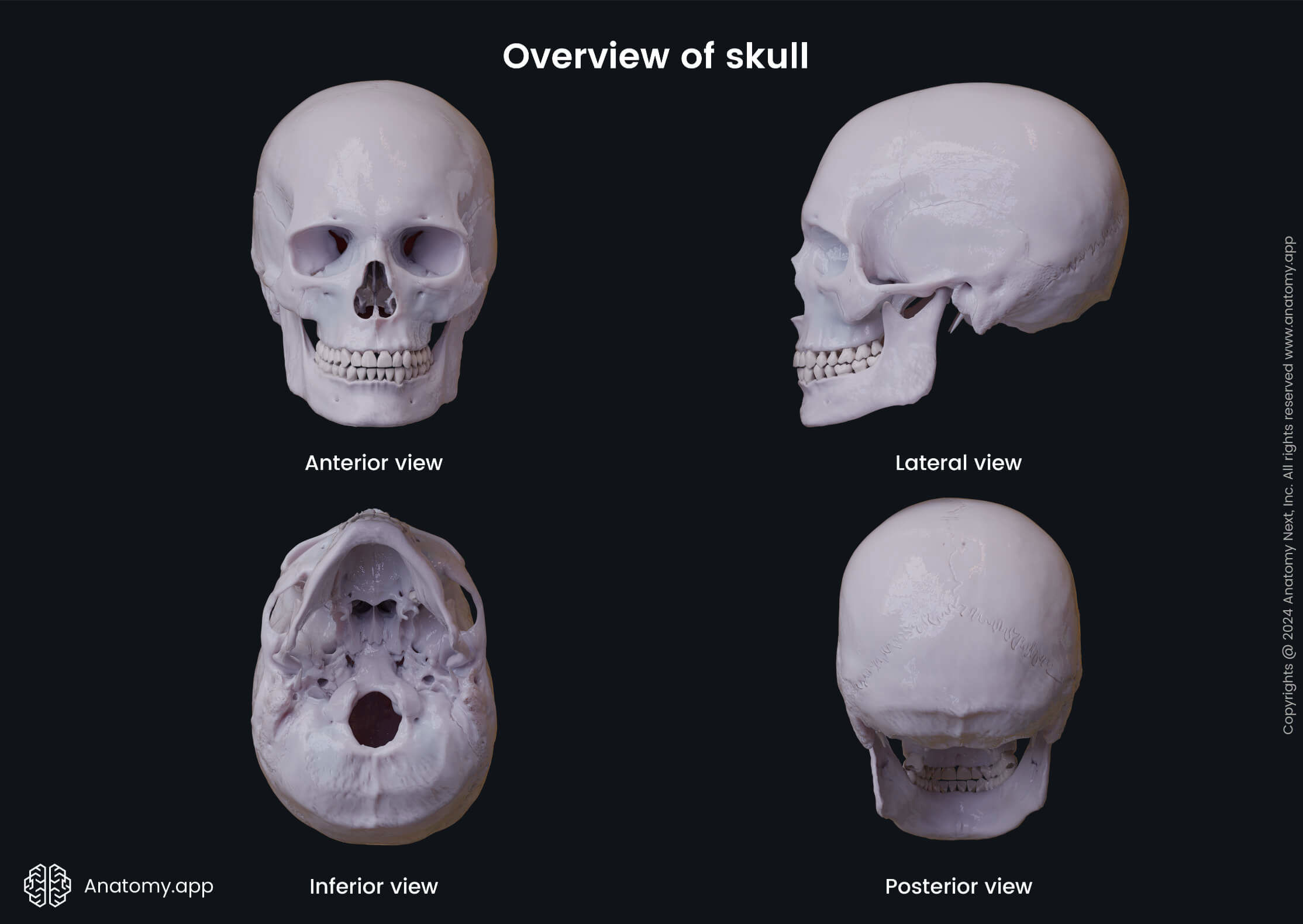
The skull incorporates the upper part of the digestive tract including the oral cavity, as well as the nasal cavity, which is the initial part of the respiratory tract. The skull also supports soft tissue of the head and facial anatomical structures.
Most of the bones that form the skull are immobile and are attached to each other by sutures to form the cranium. An exception is the mandible, which forms the lower jaw. It is a mobile bone that is connected to the skull at the temporomandibular joints.
Several bones of the skull contain air-filled cavities called the paranasal sinuses. These sinuses help to create voice resonance and aid in warming and moistening the inhailed air.
Parts of the skull
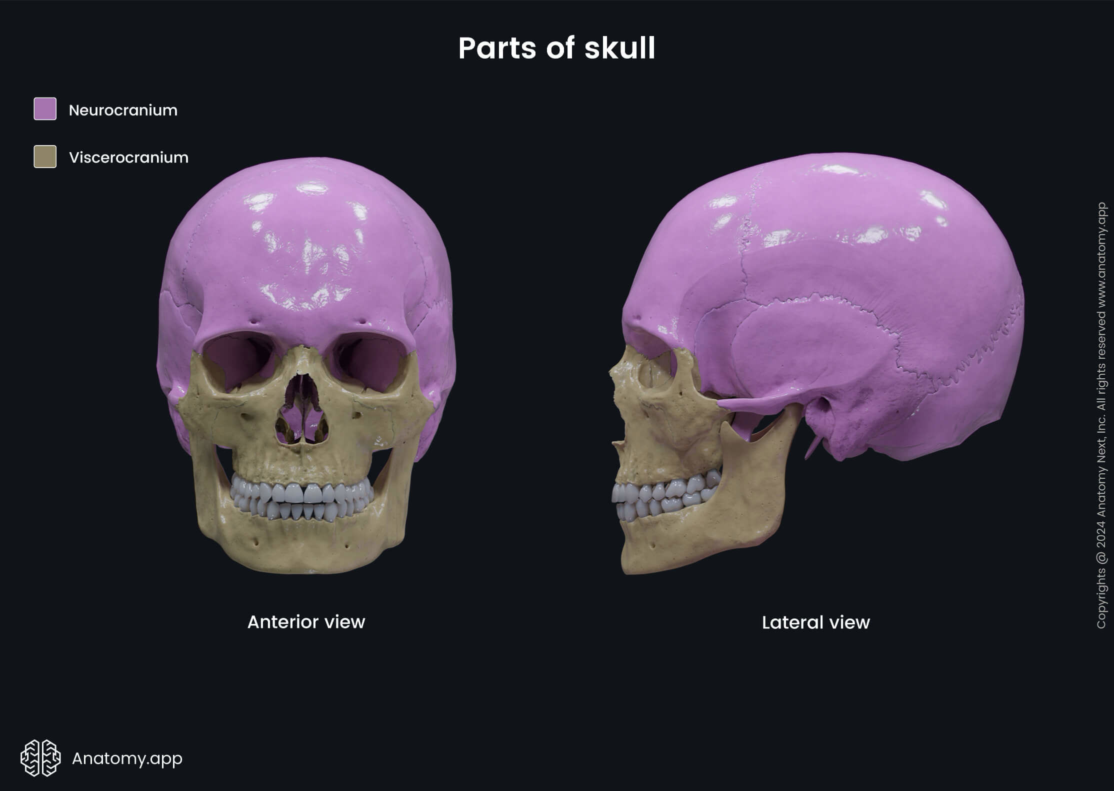
The human skull consists of two main parts. The neurocranium or braincase is the part which encloses the brain, while the viscerocranium is the portion that forms the facial skeleton. The skull may also be divided into calvaria (skullcap or skull vault) and cranial base (or skull base). The calvaria is the upper part of the neurocranium covering the cranial cavity. The cranial base includes the floor of the cranial cavity and the inferior part of the viscerocranium.
Neurocranium
The neurocranium encloses the cranial cavity. It protects all parts of the brain, including the cerebrum, cerebellum, brainstem, and sensory organs connected with the brain. These organs include the eyes, ears, tongue (with taste buds) and olfactory receptors in the nasal cavity.
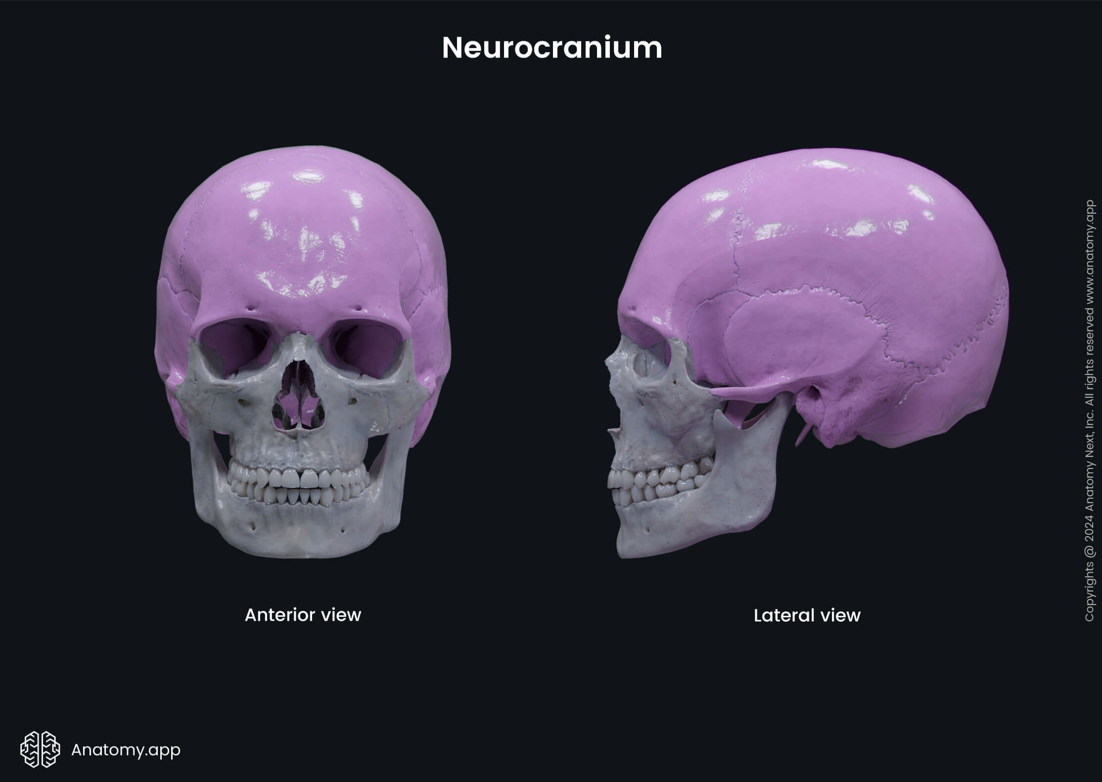
The inner surfaces of neurocranial bones contain grooves for arteries or arterial grooves, as well as depressions corresponding to the convolutions of the brain (cerebral gyri). These depressions are called the digitate impressions or impressions of cerebral gyri.
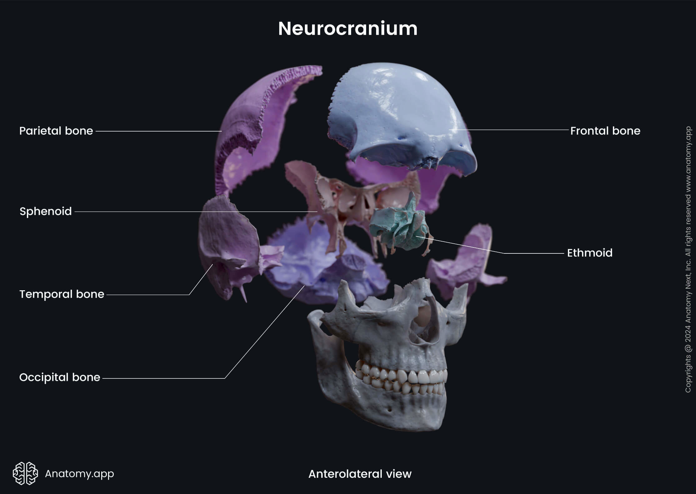
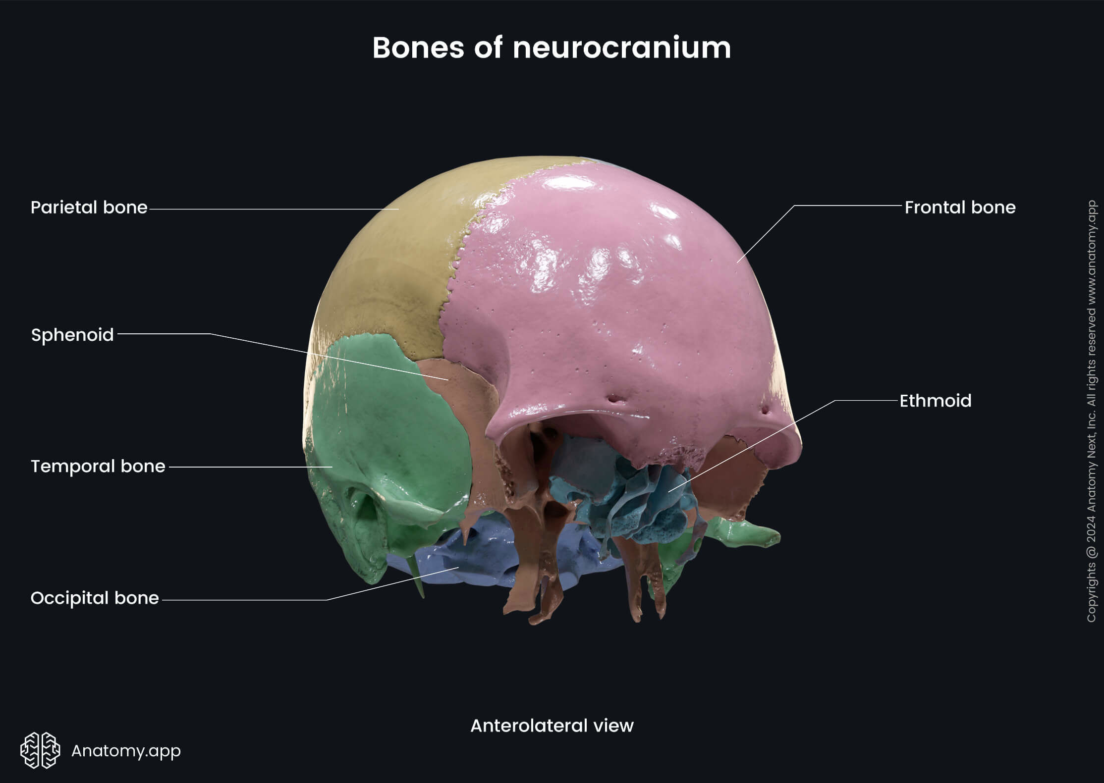
Bones of neurocranium
The neurocranium is formed by four unpaired (single) bones and two paired bones. The unpaired bones are the following:
- Sphenoid bone - situated in the middle of the skull towards the front, it forms a rear of the orbit;
- Frontal bone - forms the skeleton of the forehead;
- Occipital bone - forms the back part and base of the skull;
- Ethmoid bone - located in front of the skull and separating the nasal cavity from the cranial cavity and brain.
The paired bones of the neurocranium are:
- Temporal bones - located to the base and sides of the skull;
- Parietal bones - superior to temporal bones, make up the roof part and sides of the skull.
Only the sphenoid, frontal, and ethmoid bones contain air-filled spaces called the paranasal sinuses.
Viscerocranium
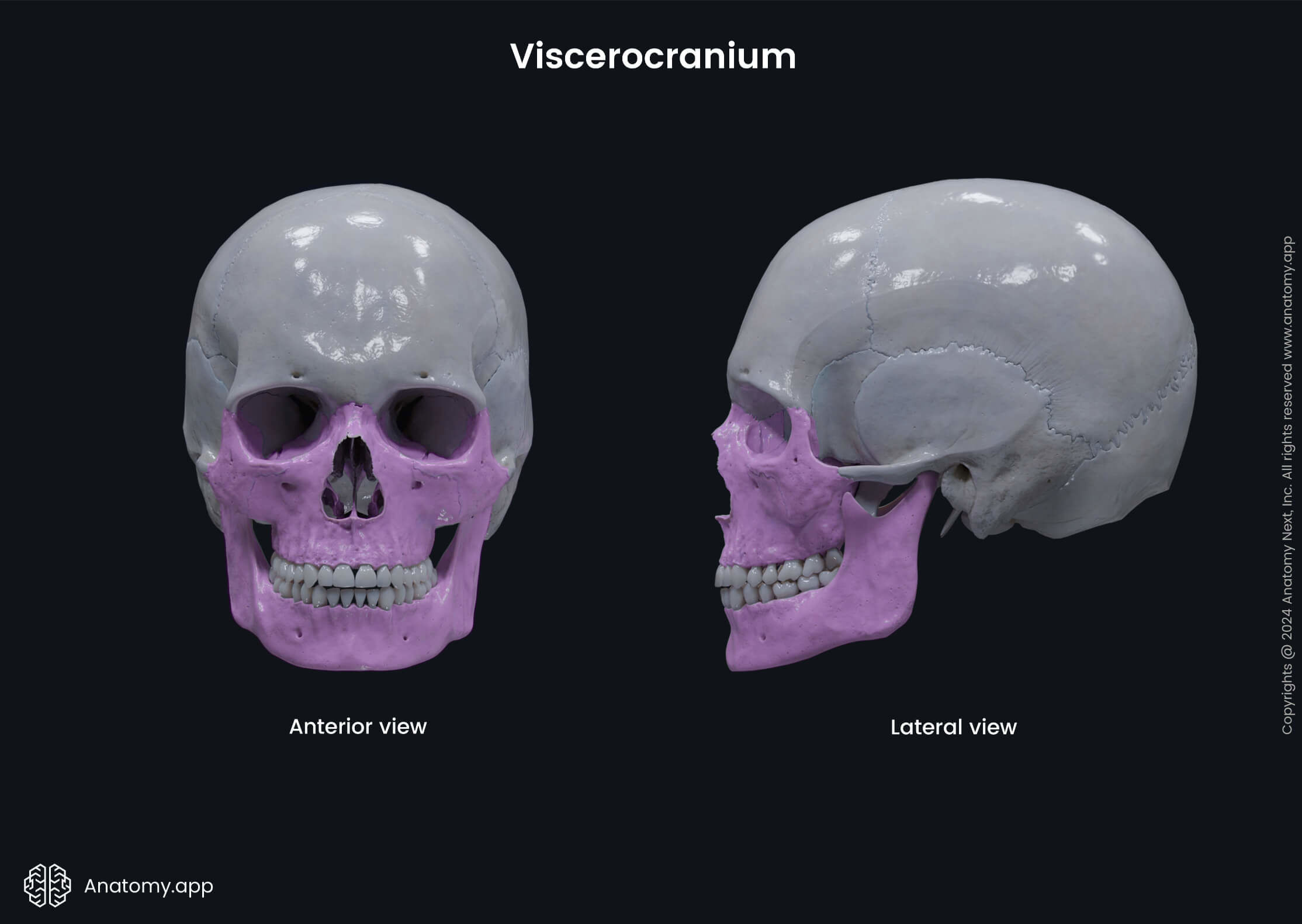
The viscerocranium is the part situated anterior to the neurocranium. It forms the facial skeleton and skeleton of the jaw. The viscerocranium also supports the soft tissue of the face.
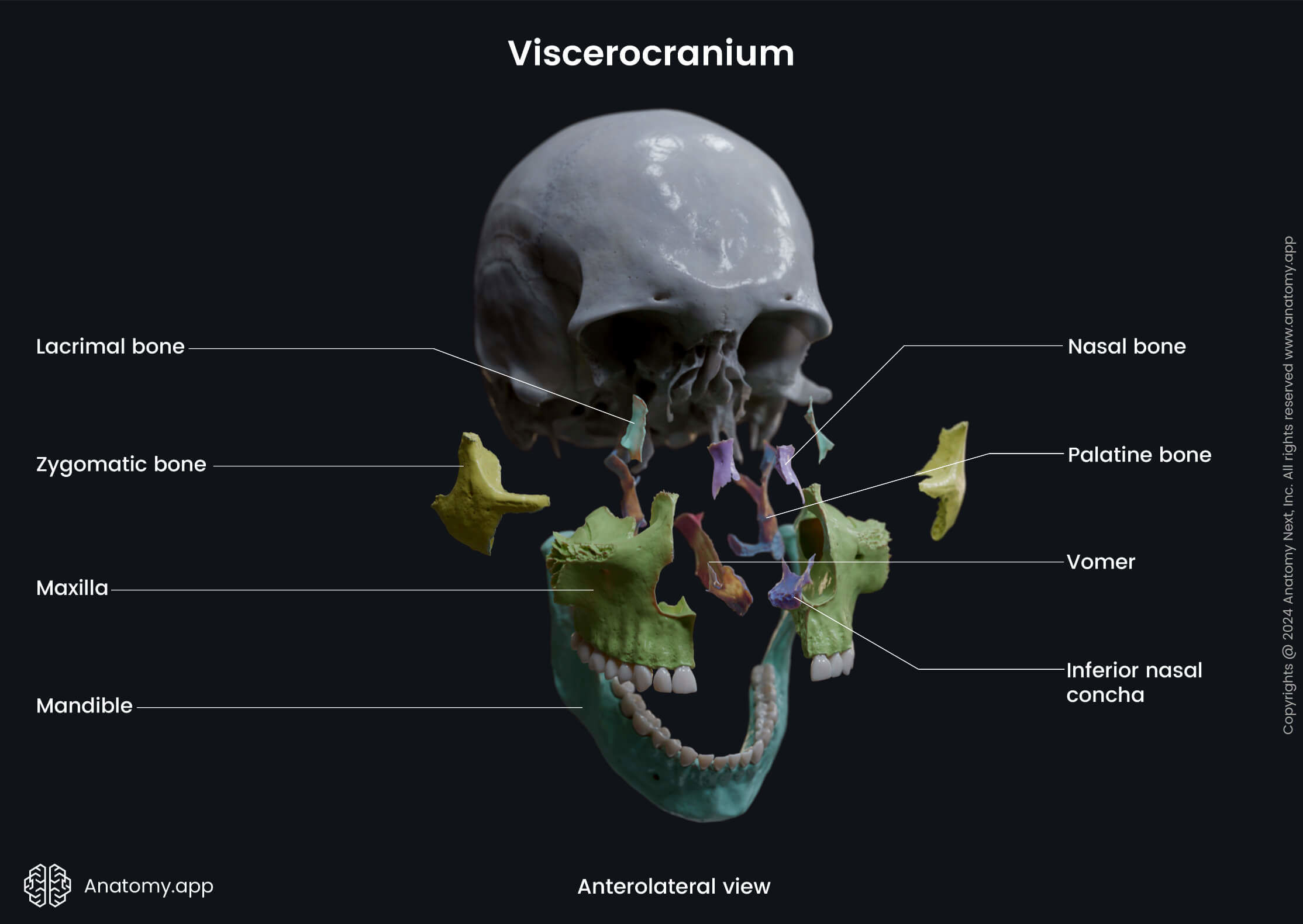
Bones of viscerocranium
The viscerocranium is formed by six paired and two unpaired bones. The paired bones are the following:
- Lacrimal bones - these are the smallest bones of the face, and they form the medial wall of the orbit;
- Nasal bones - located in the midline of the face, these bones form the nose bridge;
- Inferior nasal conchae - located within the lateral wall of the nasal cavity;
- Zygomatic bones - form the cheeks and also contribute to the orbits;
- Maxillae - located in the midline of the face, both bones are fused in a single bone called the maxilla; the maxilla forms the upper jaw and contributes to the hard palate;
- Palatine bones - both bones fuse in the midline and participate in creating the hard palate, which forms floor of the nasal cavity and the roof of the oral cavity.
And the unpaired bones of the viscerocranium are:
- Vomer - forms the posterior part of the nasal septum;
- Mandible - forms the lower jaw and is connected to the skull with the temporomandibular joint.
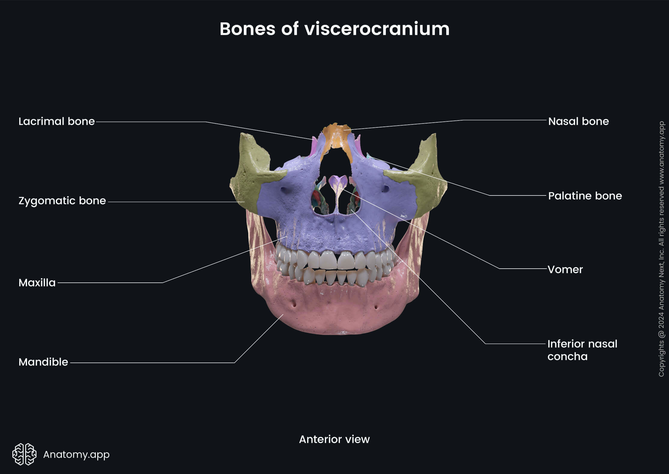
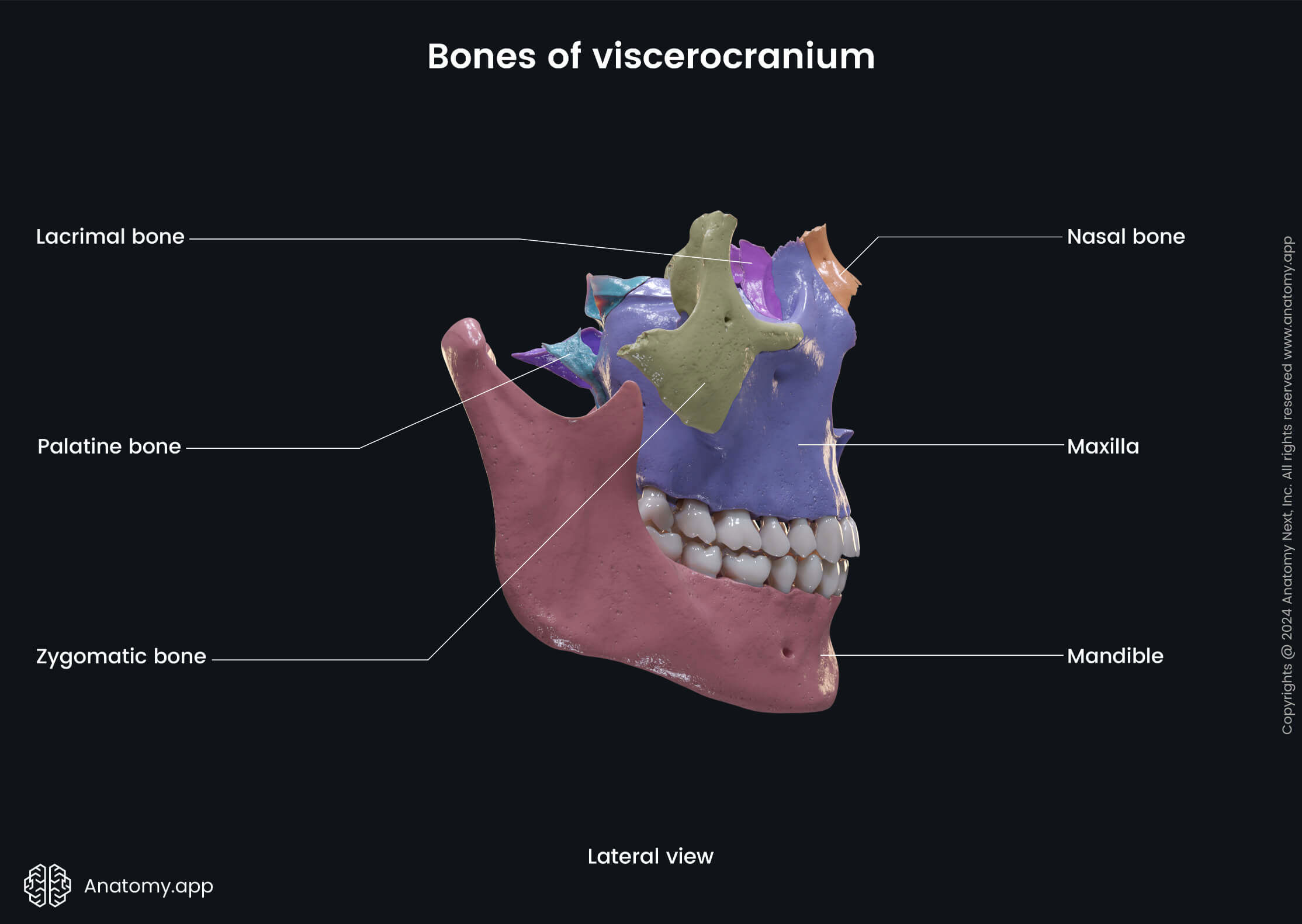
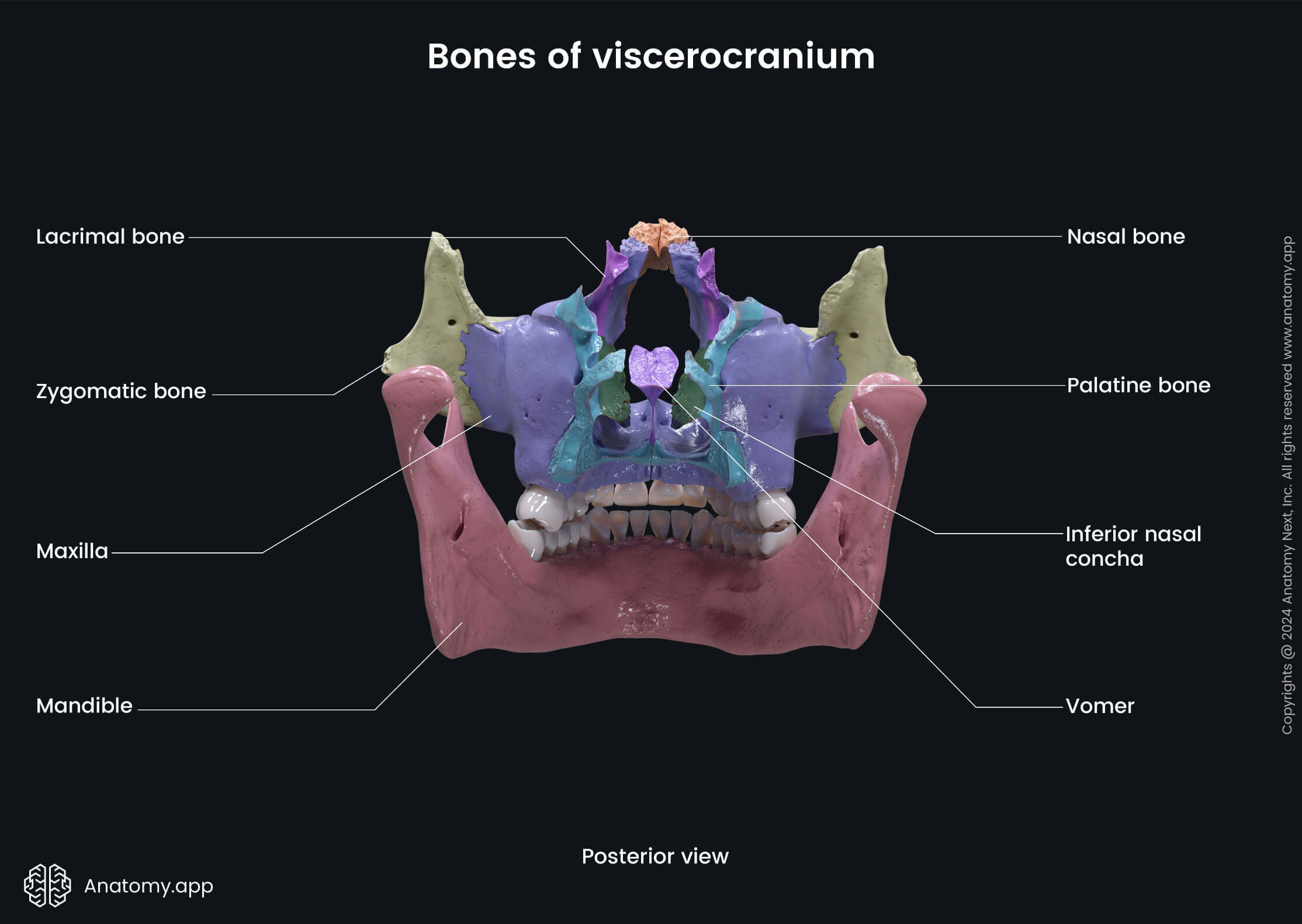
Only one bone of the viscerocranium, the maxilla, contains a paranasal sinus (maxillary sinus). The maxilla is also the most prominent immovable bone of the facial skeleton.
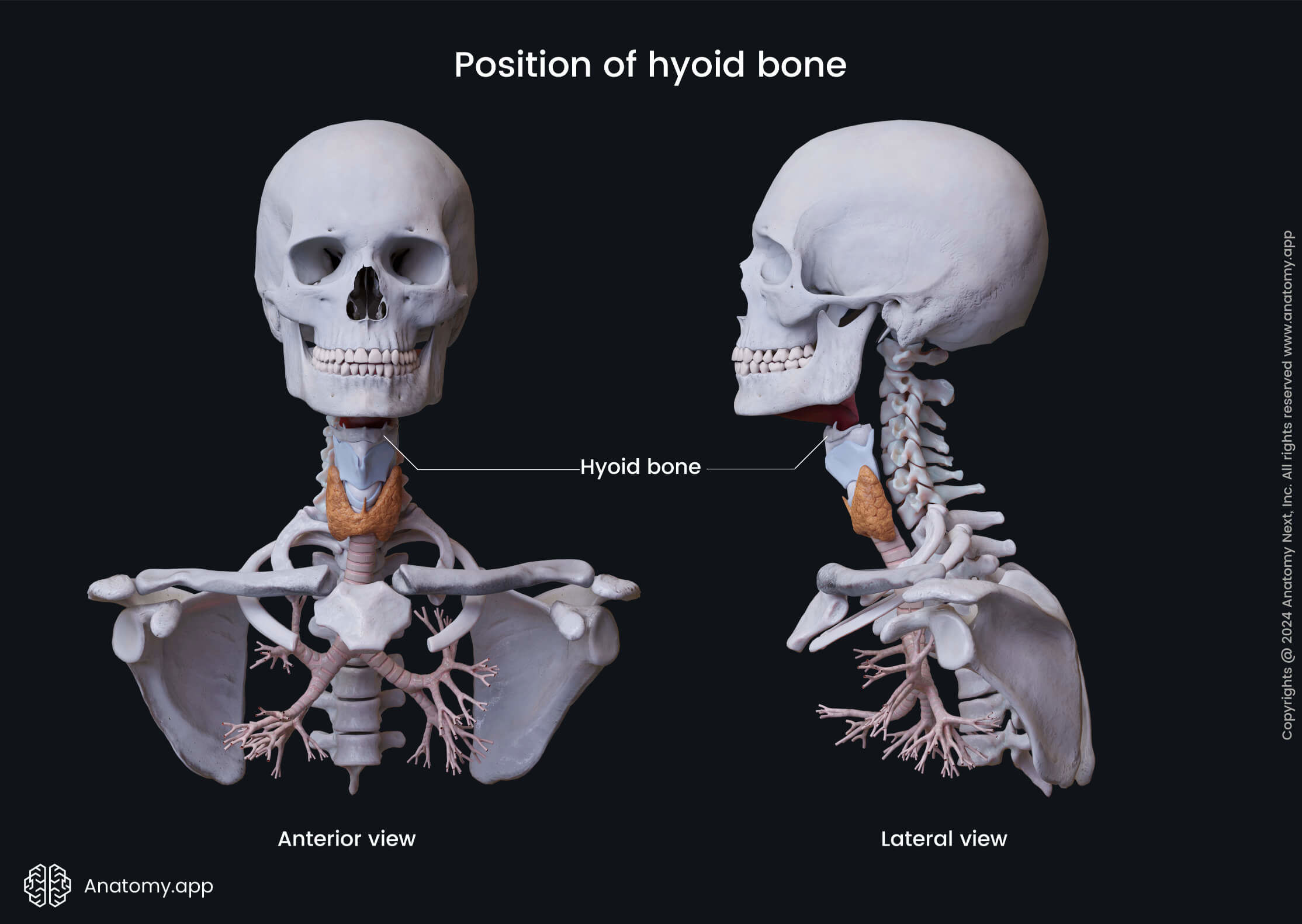
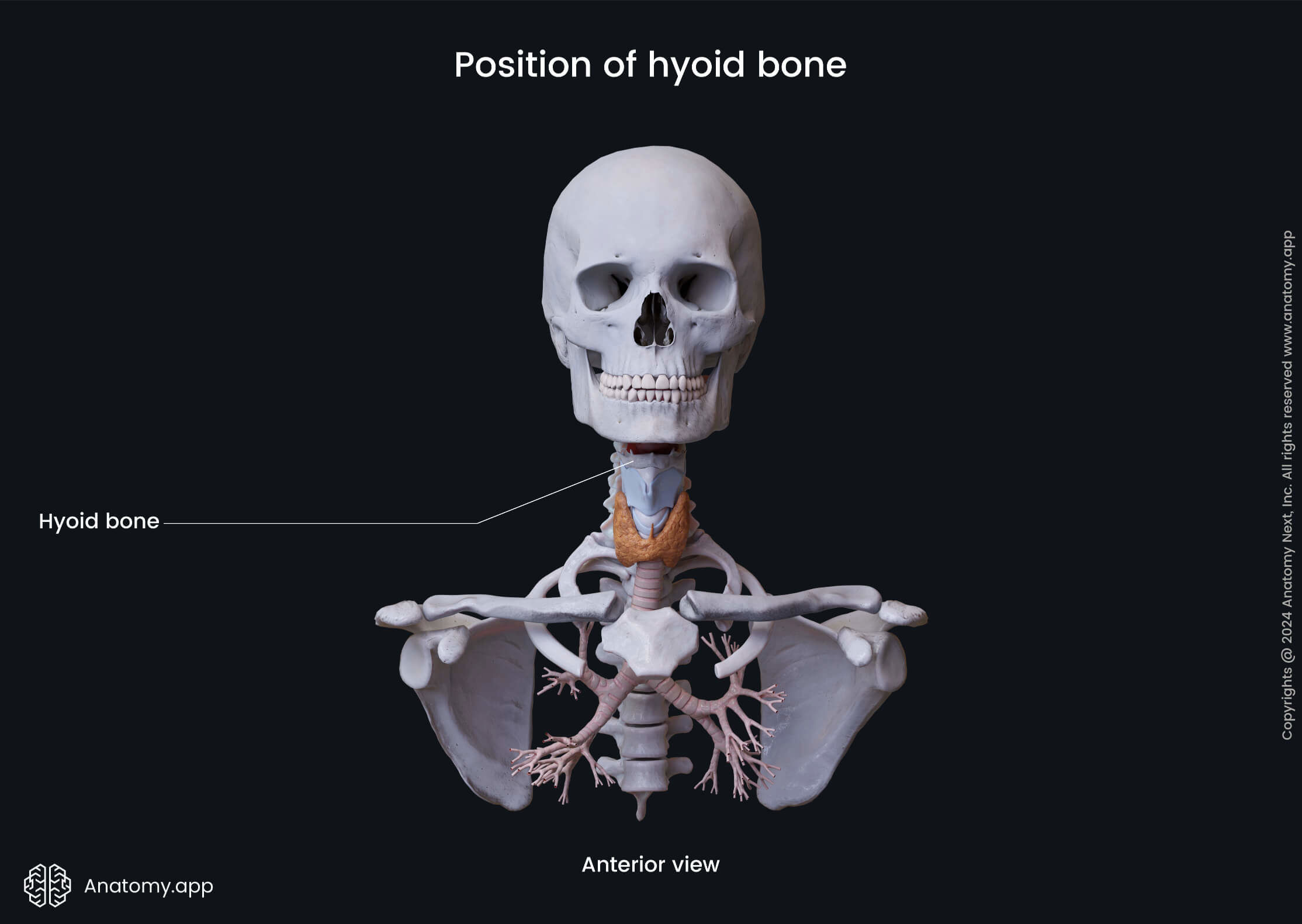
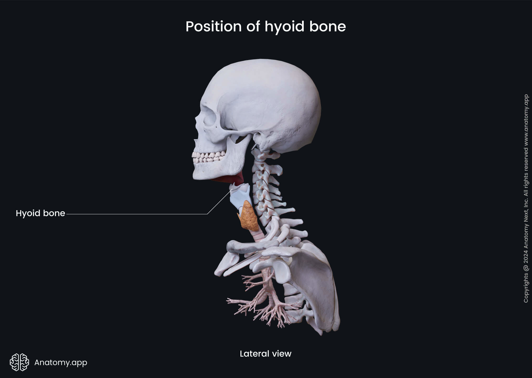
Sometimes the unpaired hyoid bone is also classified as a bone of the viscerocranium, although located in the upper neck region. The hyoid bone is connected with the skull with the help of ligaments.
Calvaria
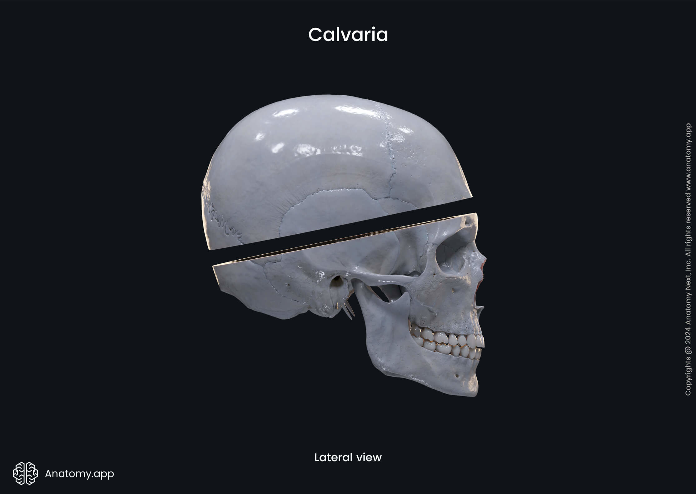
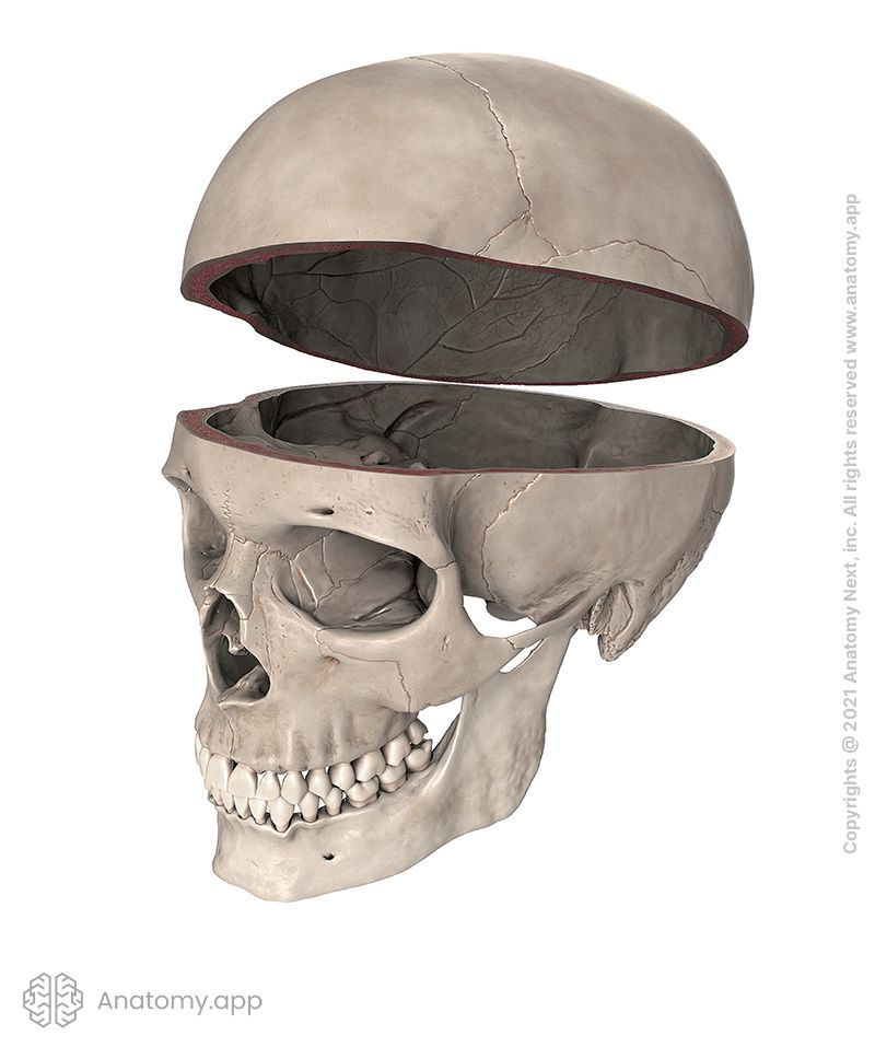
As mentioned above, the skull may also be divided into the calvaria and the cranial base. The calvaria is the top part of the neurocranium. It covers the cranial cavity, which contains the brain. It is also known as the skullcap or cranial roof. The calvaria is robust, it surrounds and protects the brain. But the cranial base is more delicate, composed mainly of thin-walled bones.
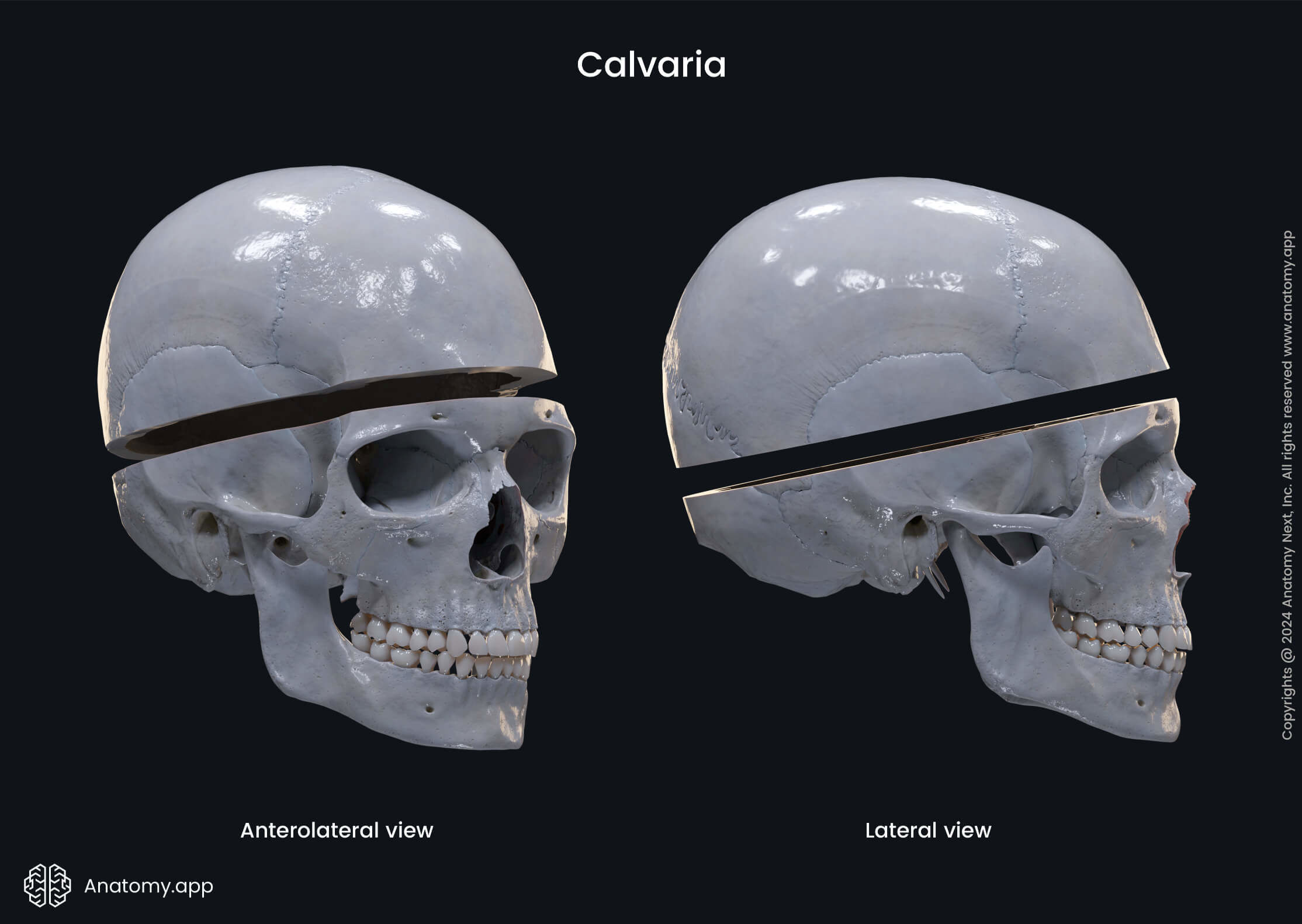
The calvaria is formed by the parietal, frontal, and occipital bones joined together with sutures. More precisely, it is composed of the squamous part of the frontal bone, squamous part of the occipital bone (above the superior nuchal line), parietal bones, squamous part of the temporal bone, and the greater wing of the sphenoid bone.
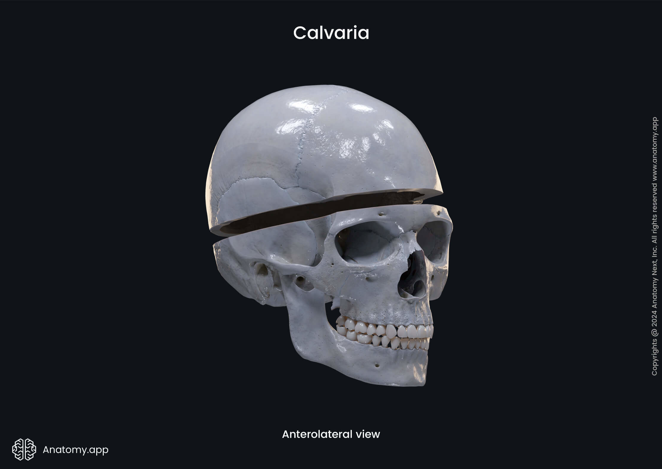
The border between the calvaria and cranial base is marked by the following structures:
- Supraorbital margin of the frontal bone
- Zygomatic process of the frontal bone
- Infratemporal crest on the greater wing of the sphenoid
- Zygomatic process of the temporal bone
- Mastoid process of the temporal bone
- Superior nuchal line of the occipital bone
- External occipital protuberance of the occipital bone
Cranial cavity
The space within the skull is known as the cranial cavity or the intracranial space. The cranial cavity houses the brain and meninges, as well as the intracranial portions of the cranial nerves. It also contains vessels supplying the brain with arterial blood (arteries) and vessels collecting venous blood (veins and venous sinuses).
Auditory ossicles
There are also three tiny bones called the auditory ossicles. They are known as the malleus, incus, and stapes. They are located within each middle ear housed in the skull. Specifically, these tiny bones are found within the right and left temporal bones. The stapes is the smallest bone in the human body.
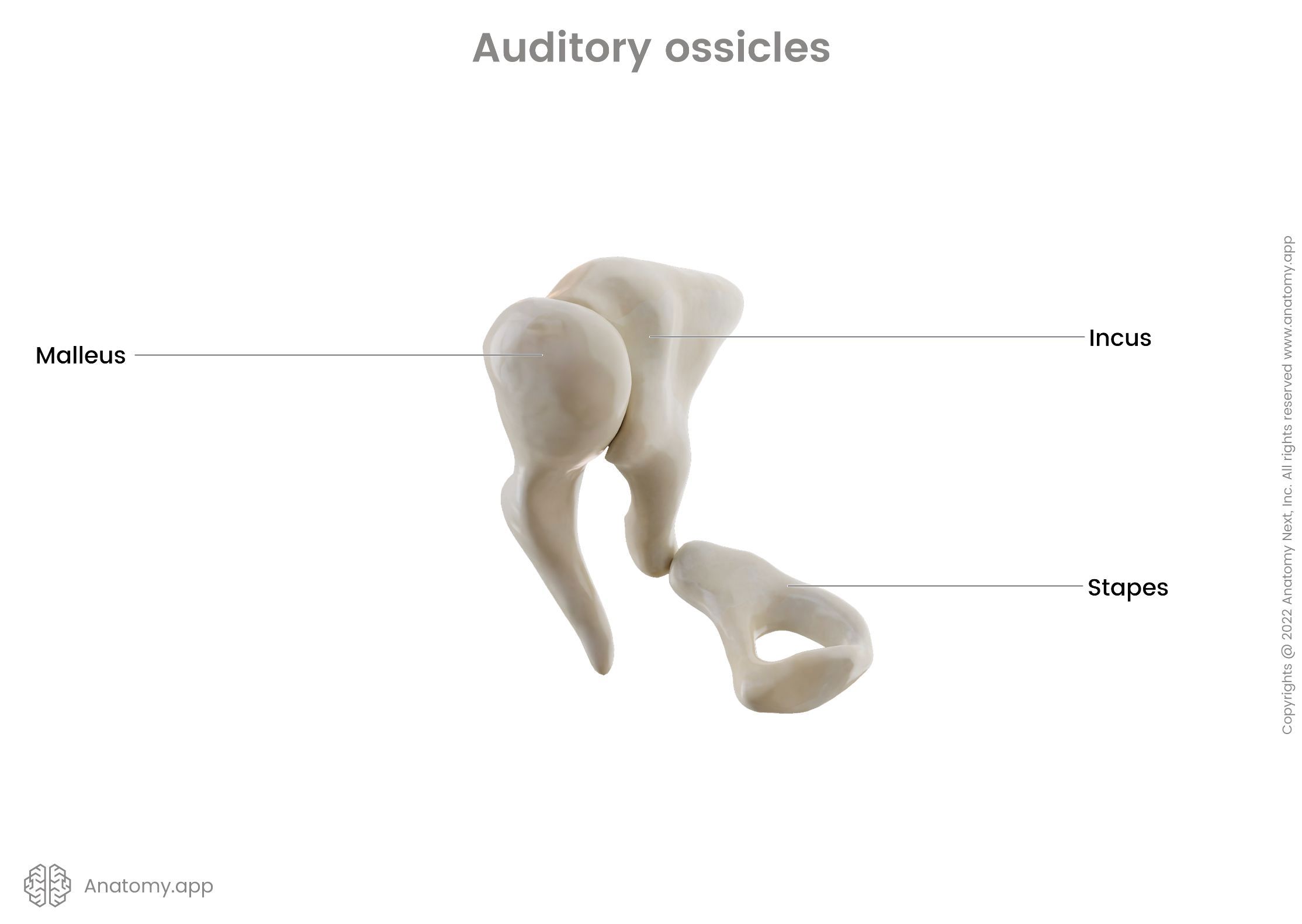
As mentioned above, the skull is formed by 22 bones. Including the hyoid bone, it is 23 bones. And if auditory ossicles are also included, then actually, in total, there are 29 bones that count as bones of the skull. This is because there are three pairs of auditory ossicles: 22 + 1 (hyoid bone) + 6 (3 pairs of auditory ossicles).
Junctions of the skull
The human skull contains two main junction types:
- Synarthroses or synarthrodial joints - junctions that allow no movement;
- Diarthroses or diarthrodial joints - joints that allow free activities.
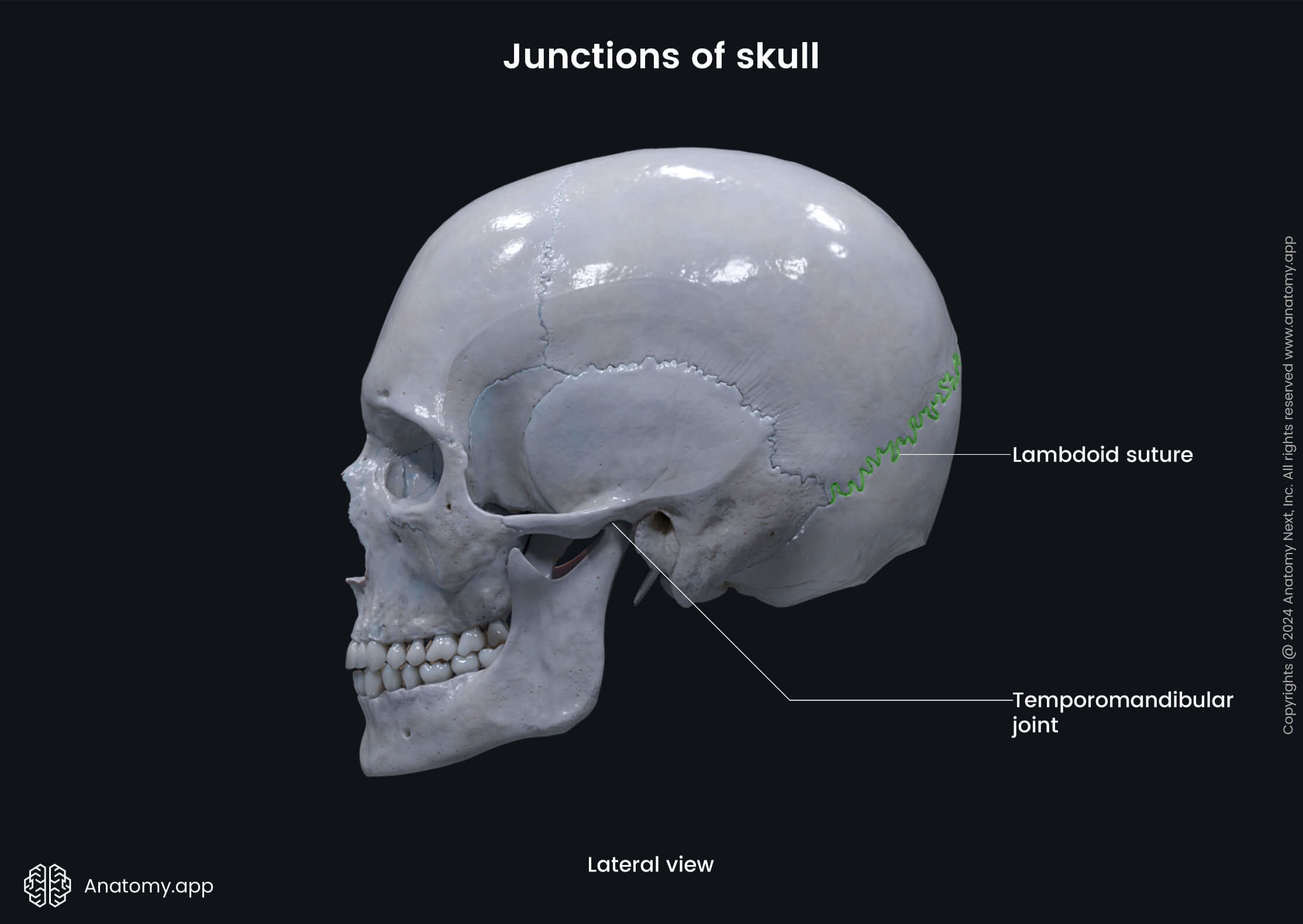
Sutures and synchondroses (cartilaginous joints) are classified as synarthrodial joints. The only joint in the skull allowing free movements is called the temporomandibular joint. And it is classified as a diarthrodial joint type.
Sutures
Almost all bones of the skull are connected with the help of fibrous junctions called sutures. Sutures are rigid joints between two or more bones. Within the human body, only the skull contains sutures. The cranial bones grow and fuse together during fetal and childhood development, forming a single skull. However, the mandible remains separate from the rest of the skull. In newborns and infants, the bones of the skull are fused incompletely. In these sites, membranous gaps called fontanelles are located.
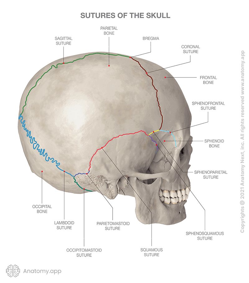
There are around 33 sutures in the human skull. The most important sutures in the skull are the following:
- Coronal suture - between the frontal and the two parietal bones;
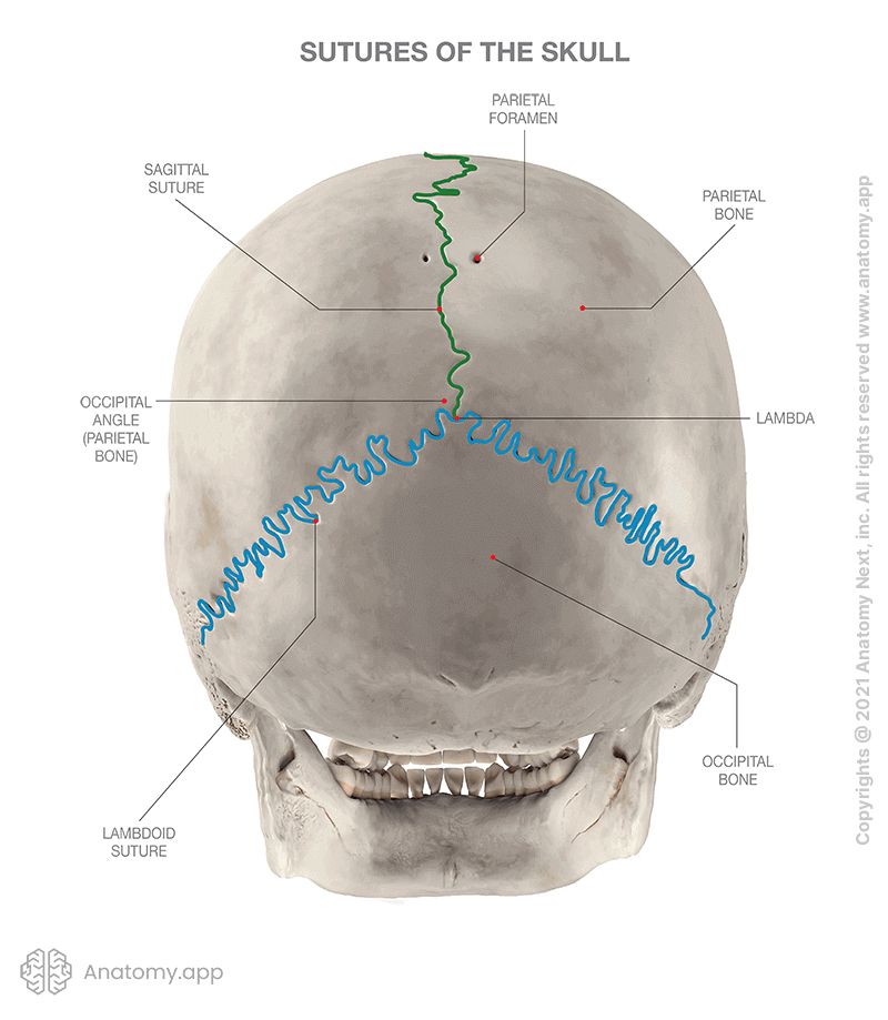
- Sagittal suture - a median suture situated between the right and left parietal bones;
- Lambdoid suture - between the occipital and the two parietal bones;
- Squamous suture - between the parietal bone and temporal bone.
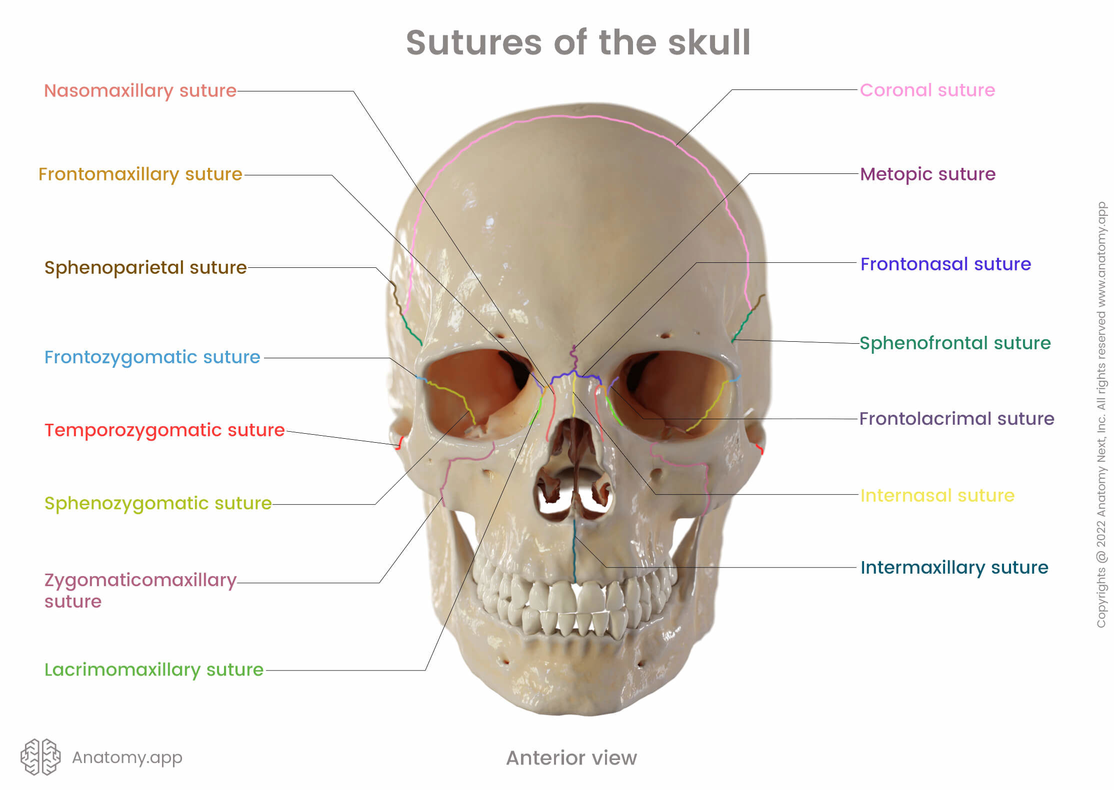
Other sutures of the skull include the following:
- Nasomaxillary suture
- Lacrimoomaxillary suture
- Zygomaticomaxillary suture
- Intermaxillary suture
- Sphenozygomatic suture
- Sphenofrontal suture
- Sphenosquamous suture
- Frontonasal suture
- Frontozygomatic suture
Most sutures between bones forming the viscerocranium are plane sutures, as the edges of the articulating bones are relatively smooth. For example, the bony base of the hard palate is formed by two horizontal plates of the palatine bone and two palatine processes of the maxilla. Both parts are connected by a sagittal oriented median palatine suture in the midline and frontal oriented transverse palatine suture located between the anterior two-thirds and posterior one-third of the hard palate.
Fontanelles
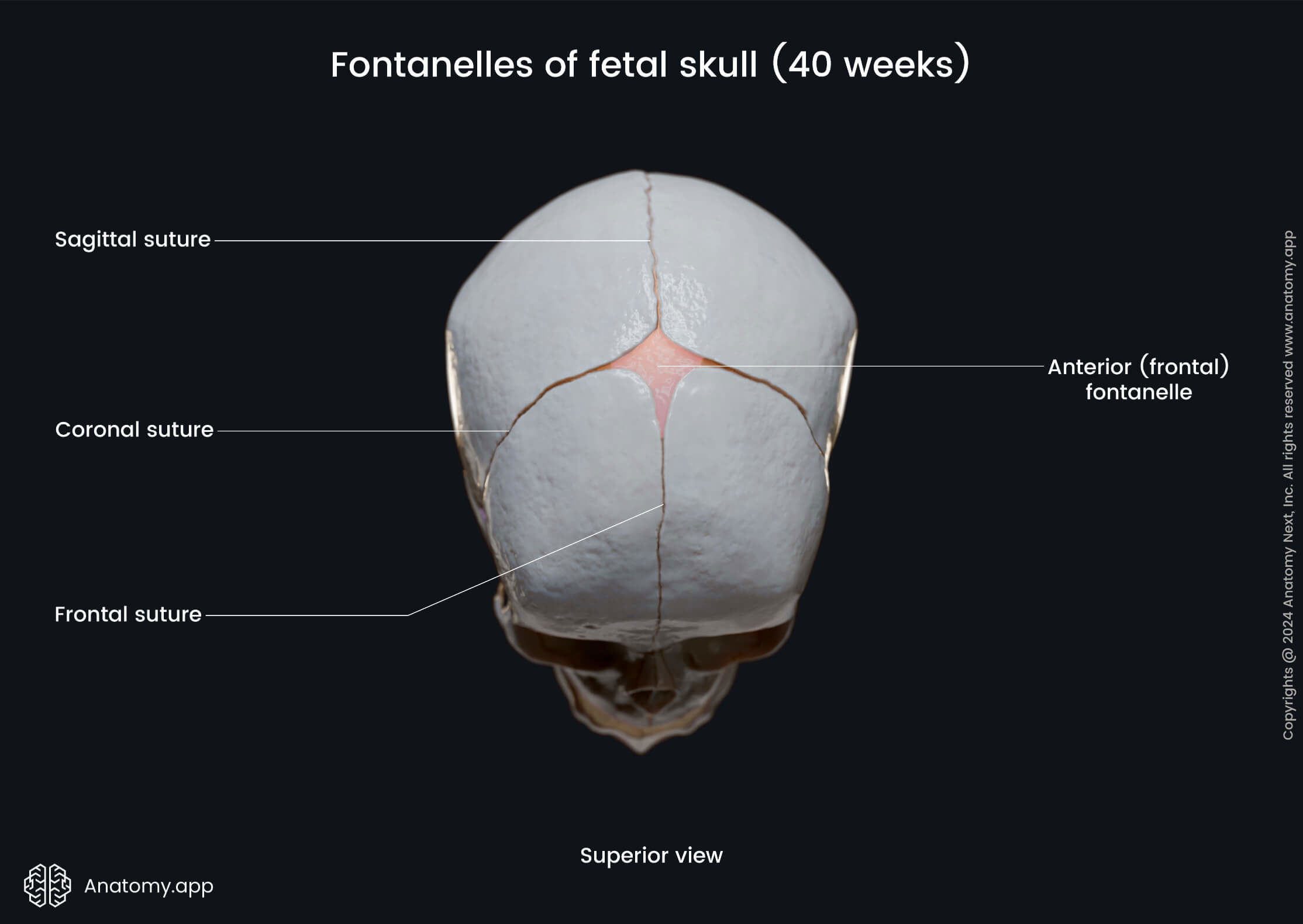
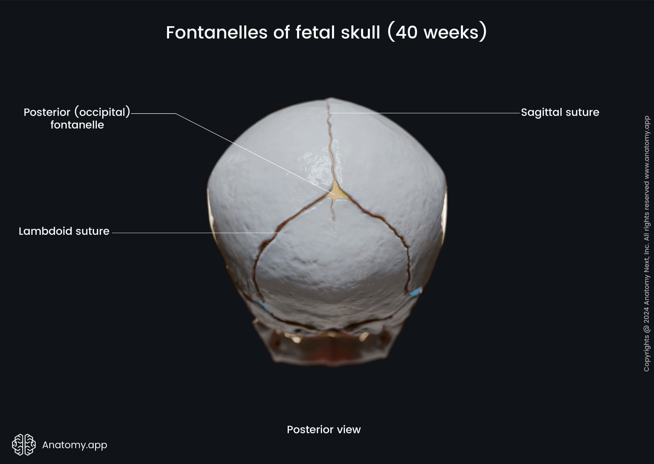
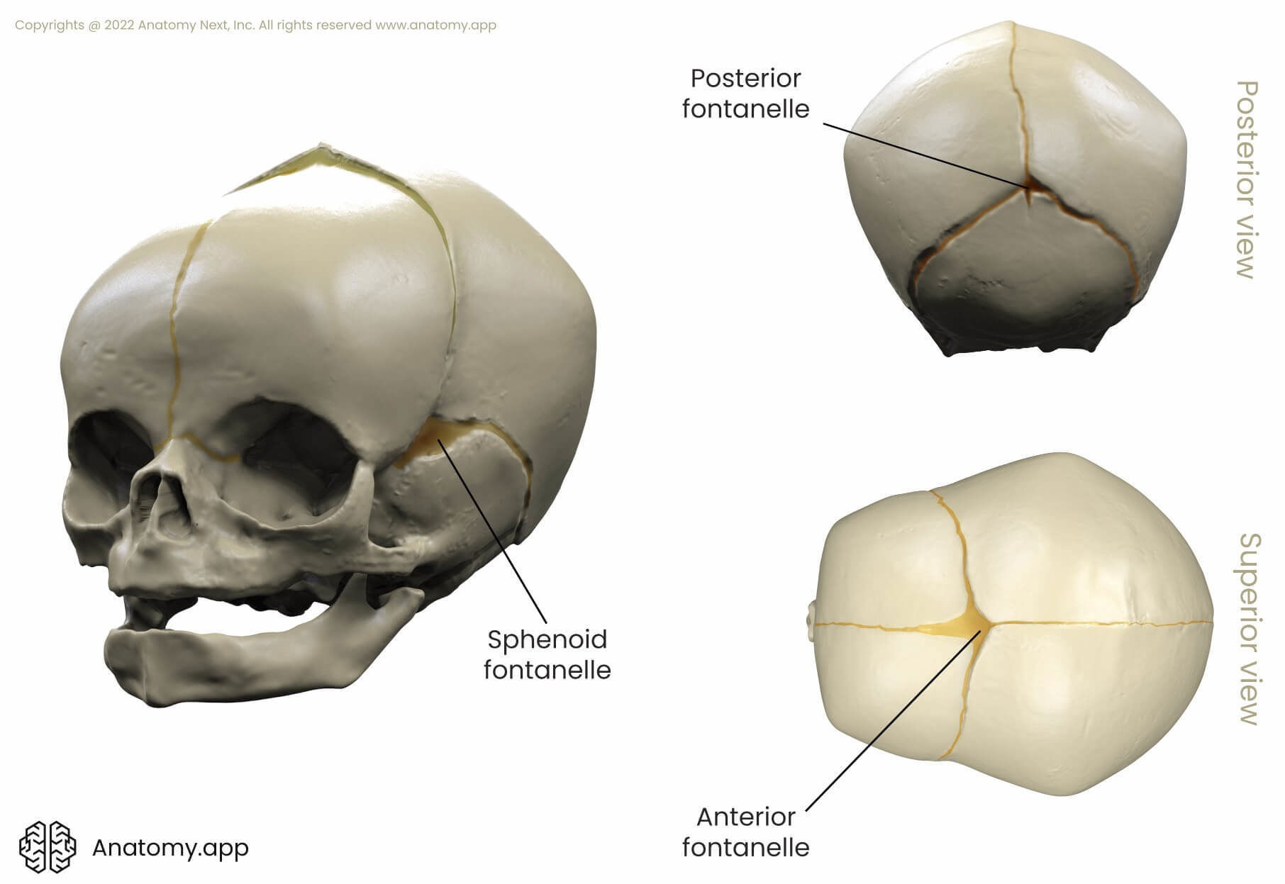
In newborn babies and infants, sutures are fused incompletely. Between bones of the skull are soft spots - membranous gaps called the fontanelles. They allow the child's head to mold, so that it can pass through the birth canal. The fontanelles slowly close by a process called intramembranous ossification. The fontanelles can be divided into two groups - major and minor fontanelles.
There are two major fontanelles. The frontal (anterior fontanelle) is the largest one, and its shape resembles a diamond. It is located at the junction of the sagittal and coronal sutures. It stays open until the age two. The occipital (posterior fontanelle) is triangular-shaped. It is found at the junction of the lambdoid and sagittal sutures. It closes during the first three months after birth.
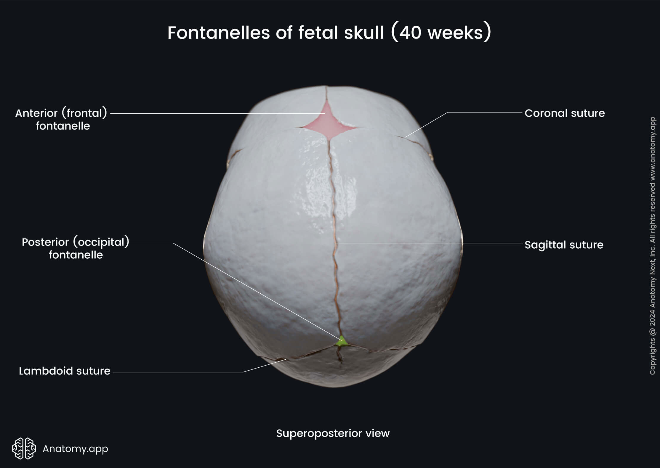
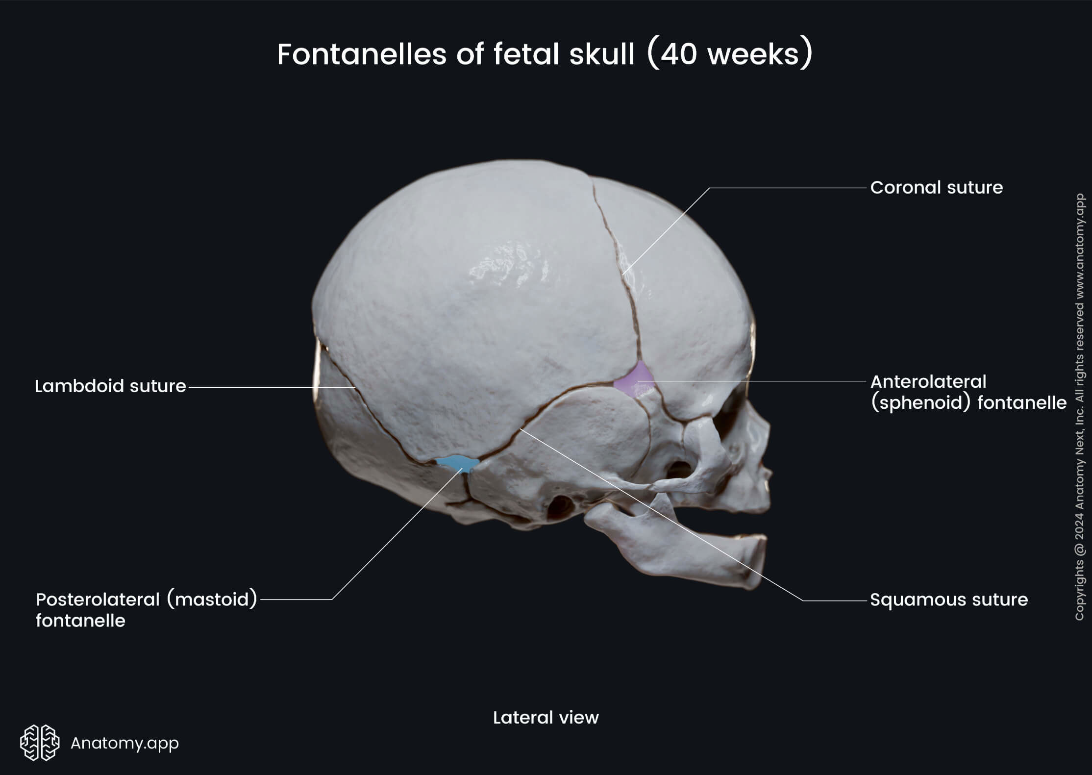
The minor fontanelles are smaller fontanelles situated on the sides of the skull. These are the sphenoidal and mastoid fontanelles. The sphenoid fontanelle is located between the sphenoid, temporal, frontal, and parietal bones. But the mastoid fontanelle is located between the temporal, occipital, and parietal bones. Both mentioned fontanelles have pairs.
Synchondroses
Besides sutures that are described above, the skull contains other synarthrodial joints as well. There are several synchondroses between bones that form the skull. Synchondroses are cartilaginous joints. For example, there is a synchondrosis between the sphenoid bone and occipital bone. It is called the sphene-occipital synchondrosis. Other synchondroses of the skull include:
- Spheno-petrosal synchondrosis
- Petro-occipital synchondrosis
- Spheno-ethmoidal synchondrosis
- Inter-sphenoid synchondrosis
- Anterior intra-occipital synchondrosis
- Posterior intra-occipital synchondrosis
Temporomandibular joint
There is only one synovial joint between the bones of the skull, and it is the temporomandibular joint (TMJ) formed between the head of the mandible and the mandibular fossa and the articular tubercle of the temporal bone. The TMJ is classified as a diarthrodial joint or diarthrosis. It is located anterior to the outer ear, to be more precise - anterior to the tragus of the ear on the lateral side of the face.
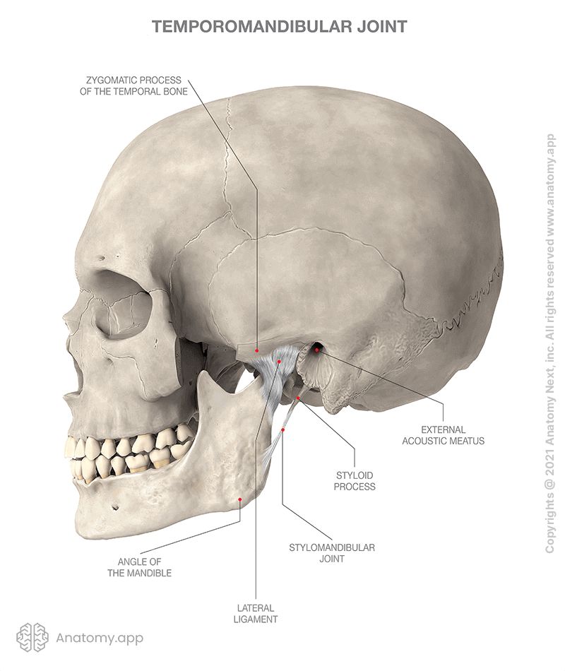
An articular disc separates the articular surfaces of the TMJ, so they do not have contact with each other. With the help of this disc, the joint is divided into two cavities or compartments. A fibrous articular capsule surrounds the joint. Several ligaments help in joint stabilization and fixation. The ligaments of the TMJ include the lateral and medial collateral ligaments, sphenomandibular ligament, as well as the stylomandibular and temporomandibular ligaments.
The temporomandibular joint is divided into two floors, and the movement takes place in both. Thus, it acts as a multi-axis joint - a joint that allows several directions of motion, more precisely, movement around three axes. Movements permitted by the TMJ include depression and elevation of the lower jaw, moving the lower jaw forward and backward, and side to side. It also allows rotating movements of the lower jaw, for example, during chewing.
Muscle attachments on the skull
Bones of the skull provide attachment for muscles of the head and neck. The facial muscles arise from the bones of the facial skeleton. But the base of the skull serves as attachment site for the following muscle groups:
- Masticatory muscles
- Extrinsic muscles of the tongue
- Superior constrictor of the pharynx
- Muscles of the soft palate
Paranasal sinuses
Several bones of the skull contain air-filled spaces surrounding the nasal cavity known as the paranasal sinuses. There are four sets of bilateral paranasal sinuses named according to the bone they are located. All of these sinuses open into the nasal cavity and they are considered to belong to the respiratory system. The four paranasal sinuses are the: maxillary sinus, frontal sinus, sphenoidal sinus, and ethmoid sinus (also known as the ethmoidal air cells).
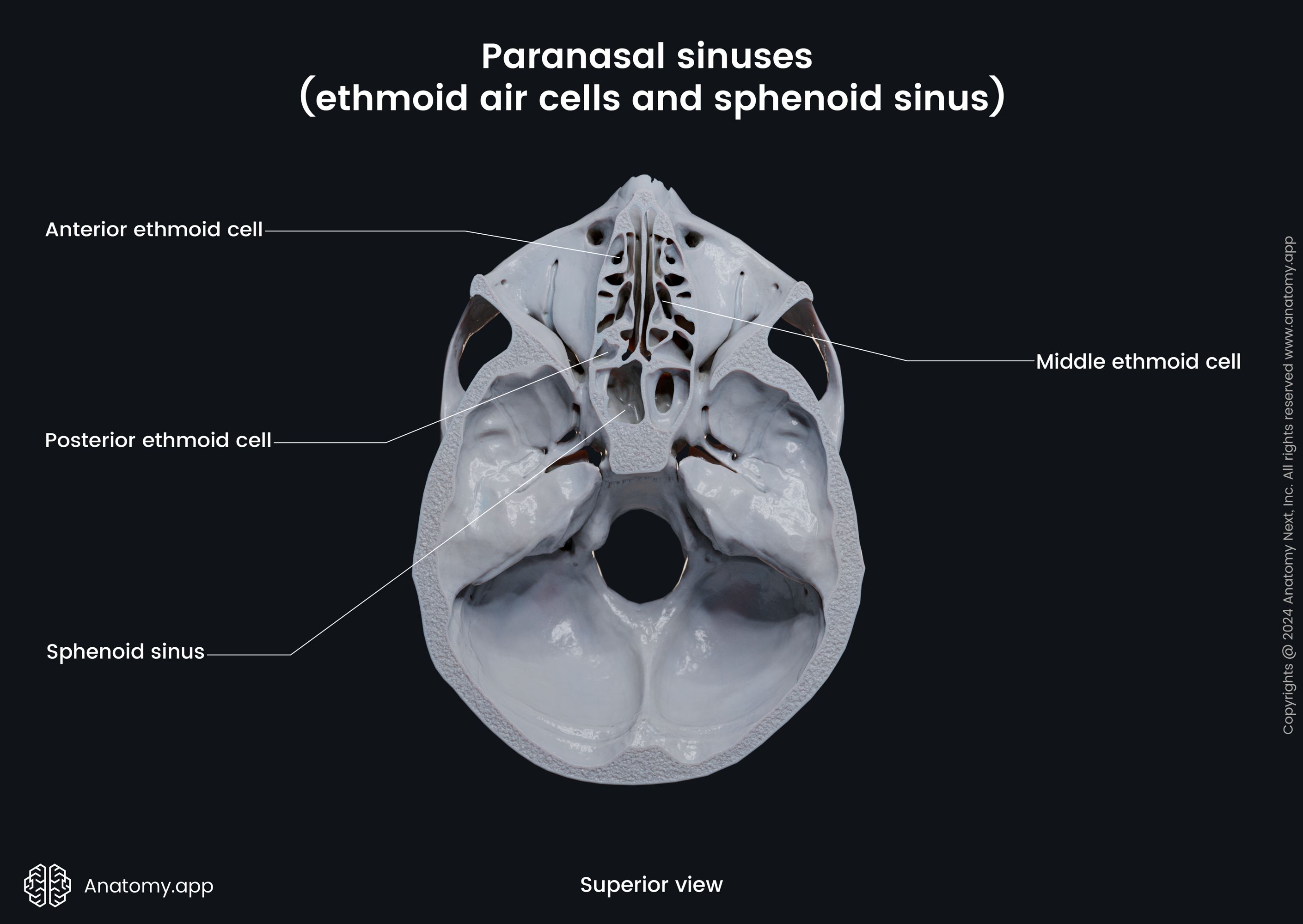
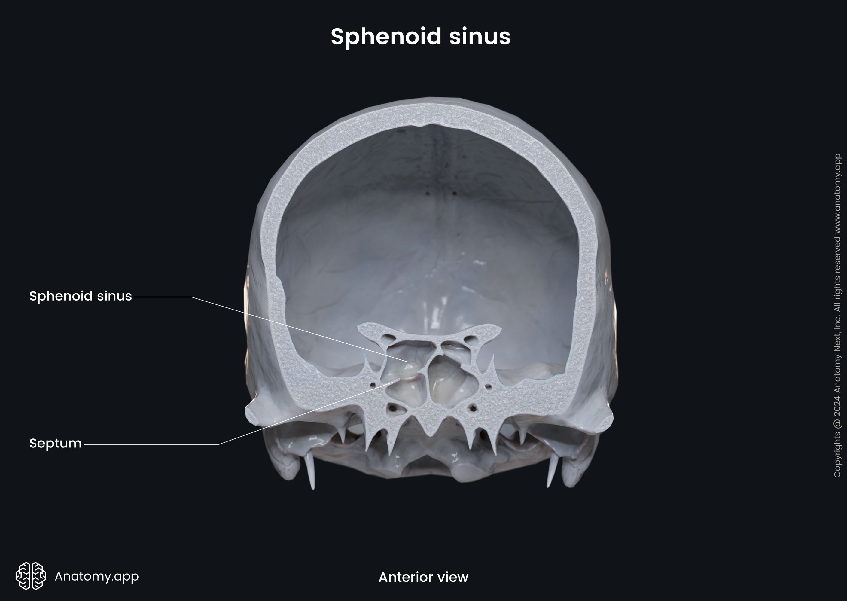
The maxillary sinus is the most significant one. It is located under the orbit laterally to the nasal cavity. The frontal sinus is triangular-shaped and located within the frontal bone in the forehead region. It is the most superior sinus. The sphenoidal sinus is located within the body of the sphenoid bone. Therefore, it is located behind the eyes and nasal cavity. It is the most posterior one. The ethmoidal air cells are situated within the ethmoid bones, between the orbits and in the superior aspect of the nose.
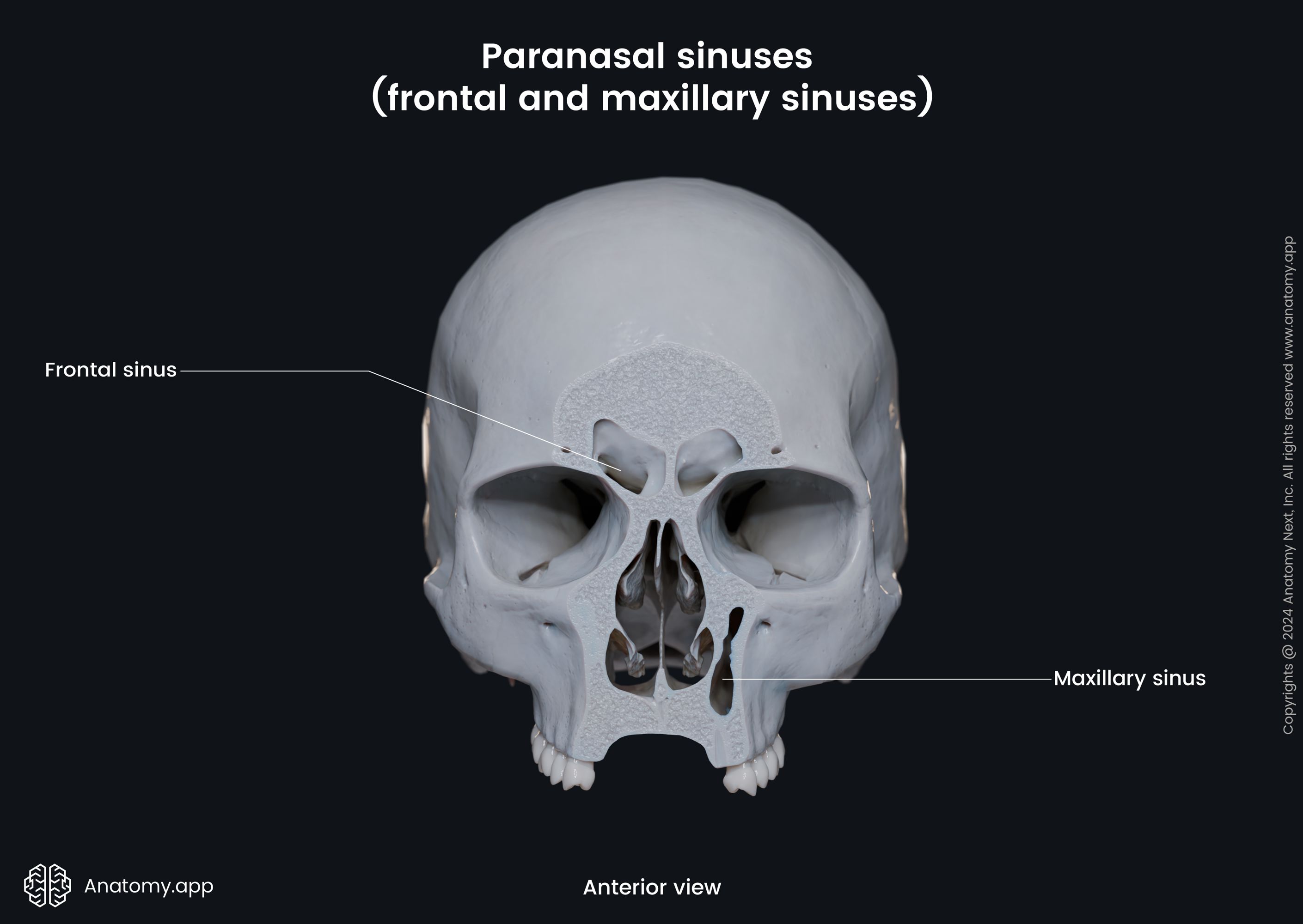
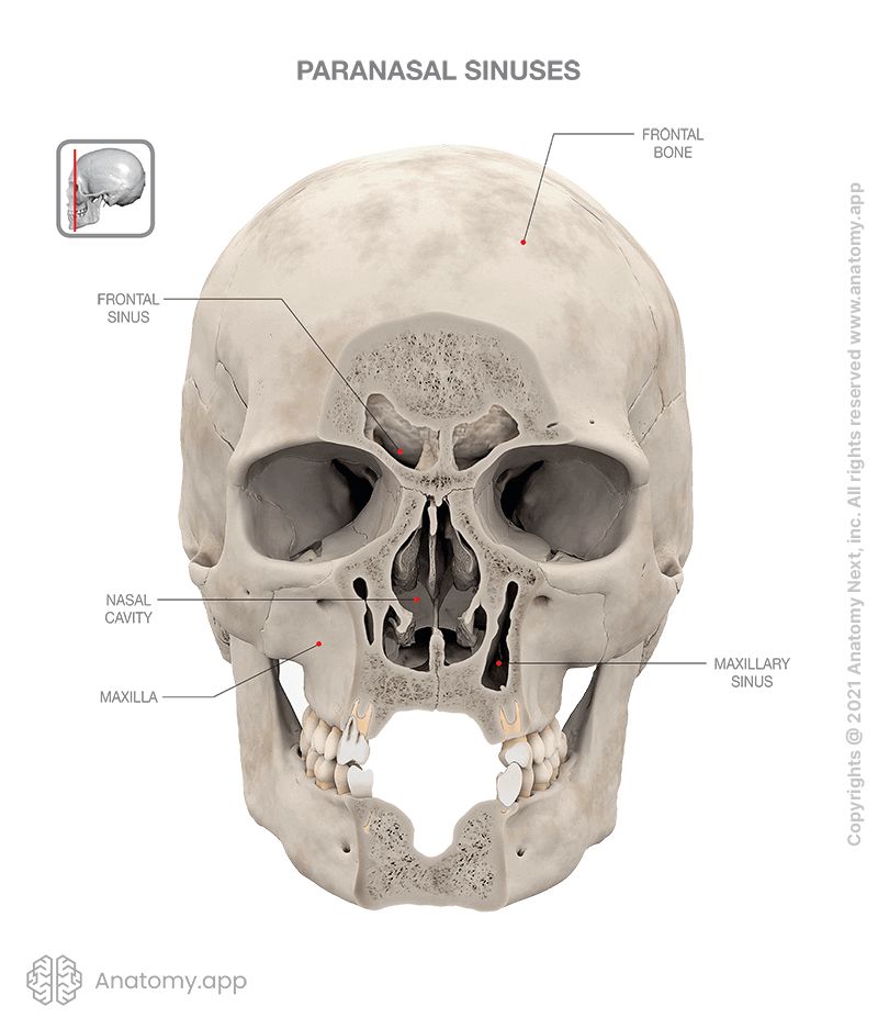
Functions of the paranasal sinuses include the following. The sinuses reduce weight of the front of the skull and facial bones. They are believed to humidify and heat inspired air. They serve to buffer against blows. Regulation of gas pressures, as well as defense agains various antigens are among their functions. The paranasal sinuses also aid in voice resonance.
Openings of the skull
The skull is rich with openings, including foramina (singular: foramen) and canals that connect one anatomical structure or region with another. These openings serve primarily as passages for cranial nerves, their branches, and accompanying blood vessels (arteries and veins). Nerves and vessels form neurovascular bundles. They may be harmed at the sites of skull openings by pathological processes or trauma.
Foramina are found in the frontal, sphenoid, ethmoid, temporal, occipital, palatine bones, and the maxilla. The largest opening of the skull is the foramen magnum in the cranial base. Through this opening, the spinal cord exits the cranial cavity as the continuation of the medulla oblongata. The foramen magnum is a single opening. But most of the skull openings are paired (bilateral). Some are multiple in number, for example, the foramina of the cribriform plate.
Listed below are essential openings in specific bones of the skull, with the parts of the skull they connect and the structures they carry.
| Frontal bone | |||
| Opening | Location | Connection | Content |
| Supraorbital foramen or notch | Central part of the supraorbital margin | Orbit with the forehead region on the external face | Supraorbital nerve, artery and vein |
| Frontal foramen or notch | Medially to the supraorbital foramen or notch | Orbit with the forehead region on the external face | Supratrochlear artery Medial branch of the supraorbital nerve |
| Foramen cecum | Anterior to the cribriform plate of the ethmoid bone in the anterior cranial fossa | Anterior cranial fossa with the nasal cavity | Emissary vein connecting the nasal cavity with the superior sagittal sinus |
| Ethmoid bone | |||
| Opening | Location | Connection | Content |
| Foramina of the cribriform plate | Cribriform plate | Nasal cavity with the anterior cranial fossa | Bundles of the olfactory nerves (CN I) |
| Anterior ethmoidal foramen | Between the anterior and posterior ethmoidal air cells | Anterior cranial fossa with the orbit | Anterior ethmoidal nerve, artery and vein |
| Posterior ethmoidal foramen | Posterior to the anterior ethmoidal foramen | Anterior cranial fossa with the orbit | Posterior ethmoidal nerve, artery and vein |
| Sphenoid bone | |||
| Opening | Location | Connection | Content |
| Optic canal | Lesser wing | Middle cranial fossa with the orbit | |
| Superior orbital fissure | Between the body, lesser and greater wings | Middle cranial fossa with the orbit | Lacrimal, frontal and nasociliary nerves (from the ophthalmic nerve (CN V1))
|
| Foramen rotundum | Anterior part of the greater wing | Middle cranial fossa with the pterygopalatine fossa | Maxillary nerve (CN V2) |
| Foramen ovale | Greater wing, posterior to the foramen rotundum | Middle cranial fossa with the infratemporal fossa | Emissary vein connecting the cavernous sinus with the pterygoid venous plexus |
| Foramen spinosum | Greater wing, posterior and on sides from the foramen ovale | Middle cranial fossa with the infratemporal fossa | Spinous branch of the mandibular nerve (CN V3) |
| Temporal bone | |||
| Opening | Location | Connection | Content |
| Internal acoustic meatus | Petrous part | Posterior cranial fossa with the inner ear | Vestibulocochlear nerve (CN VIII) Vestibular ganglion |
| Stylomastoid foramen | Petrous part between the styloid and mastoid processes | Posterior cranial fossa with the external cranial base | Facial nerve (CN VII) |
| Carotid foramen | Petrous part | Middle cranial fossa with the external cranial base | Carotid plexus of nerves |
| Occipital bone | |||
| Opening | Location | Connection | Content |
| Hypoglossal canal | Lateral part | Posterior cranial fossa with the external cranial base | Hypoglossal nerve (CN XII) |
| Foramen magnum | Most inferior part of the bone | Posterior cranial fossa with the external cranial base | Rostral part of the medulla oblongata Ascending fibers of the spinal root of the accessory nerve (CN XI) Anterior and posterior spinal arteries Tectorial membrane |
| Maxilla | |||
| Opening | Location | Connection | Content |
| Incisive foramen | Median line of the palatine process, posterior to the incisor teeth | Oral cavity with the nasal cavity (incisive canal) | Nasopalatine nerve (branch of the CN V2) Greater palatine artery and vein |
| Infraorbital foramen | Body under the infraorbital margin | Infraorbital canal with the external face | Infraorbital nerve, artery and vein |
| Alveolar foramina | Body part | Infratemporal fossa with the alveolar canals | Contain alveolar canals through which pass nerves and blood vessels to the teeth |
| Palatine bone | |||
| Opening | Location | Connection | Content |
| Greater palatine foramen | Horizontal plate | Oral cavity with the pterygopalatine fossa | Greater palatine nerve, artery and vein |
| Lesser palatine foramina | Horizontal plate, posterior to the greater palatine foramen | Oral cavity with the pterygopalatine fossa | Lesser palatine nerves, arteries and veins |
| Mixed bones | |||
| Opening | Location | Connection | Content |
| Sphenopalatine foramen | Between the sphenoid and palatine bones | Nasal cavity with the pterygopalatine fossa | Superior posterior nasal branches of the maxillary nerve (CN V2) Sphenopalatine artery and vein |
| Inferior orbital fissure | Inferior to the superior orbital fissure in the orbital floor between the sphenoid bone and maxilla | Orbit with the pterygopalatine and infratemporal fossae | Orbital branches of the pterygopalatine ganglion Emissary veins connecting the previous vein with the pterygoid venous plexus Infraorbital artery and vein |
| Foramen lacrum | Between the sphenoid and temporal bones and basilar part of the occipital bone | Middle cranial fossa with the external cranial base | Nerve and artery of the pterygoid canal Meningeal branch of the ascending pharyngeal artery Emissary veins from the cavernous sinus to the pterygoid venous plexus |
| Jugular foramen | Between the petrous part of the temporal bone and occipital bone | Posterior cranial fossa with the external cranial base | Glossopharyngeal nerve (CN IX) Inferior petrosal sinus Meningeal branches of the occipital and ascending pharyngeal arteries |
Skull topography
The skull is divided into several important topographical regions, including the following:
Most of these regions are formed by different cranial fossae.
Cranial base
The cranial base, also known as the base of the skull or skull base, is the most inferior part of the skull. It forms the floor of the cranial cavity. The cranial base is formed by six different bones. These are the ethmoid, sphenoid, occipital, frontal, paired parietal and temporal bones.
The cranial base can be inspected from two sides - from inside and outside. The inner aspect of the skull base is known as the internal cranial base and is topographically divided further into three regions - anterior, middle, and posterior cranial fossae. The outside of the cranial base is called the external cranial base.
Internal cranial base
The internal cranial base is bounded anteriorly by parts of the frontal, sphenoid and ethmoid bones. Laterally, it borders with the parietal and temporal bones and posteriorly with the squamous part of the occipital bone. The internal cranial base contains the brain, the intracranial parts of cranial and spinal nerves, meninges, blood vessels, and cerebrospinal fluid.
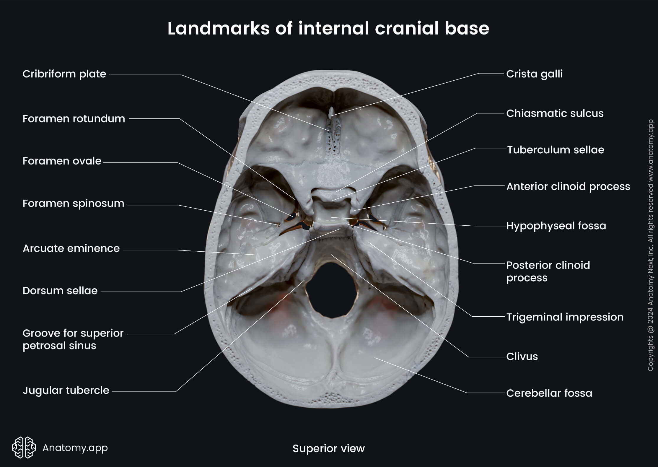
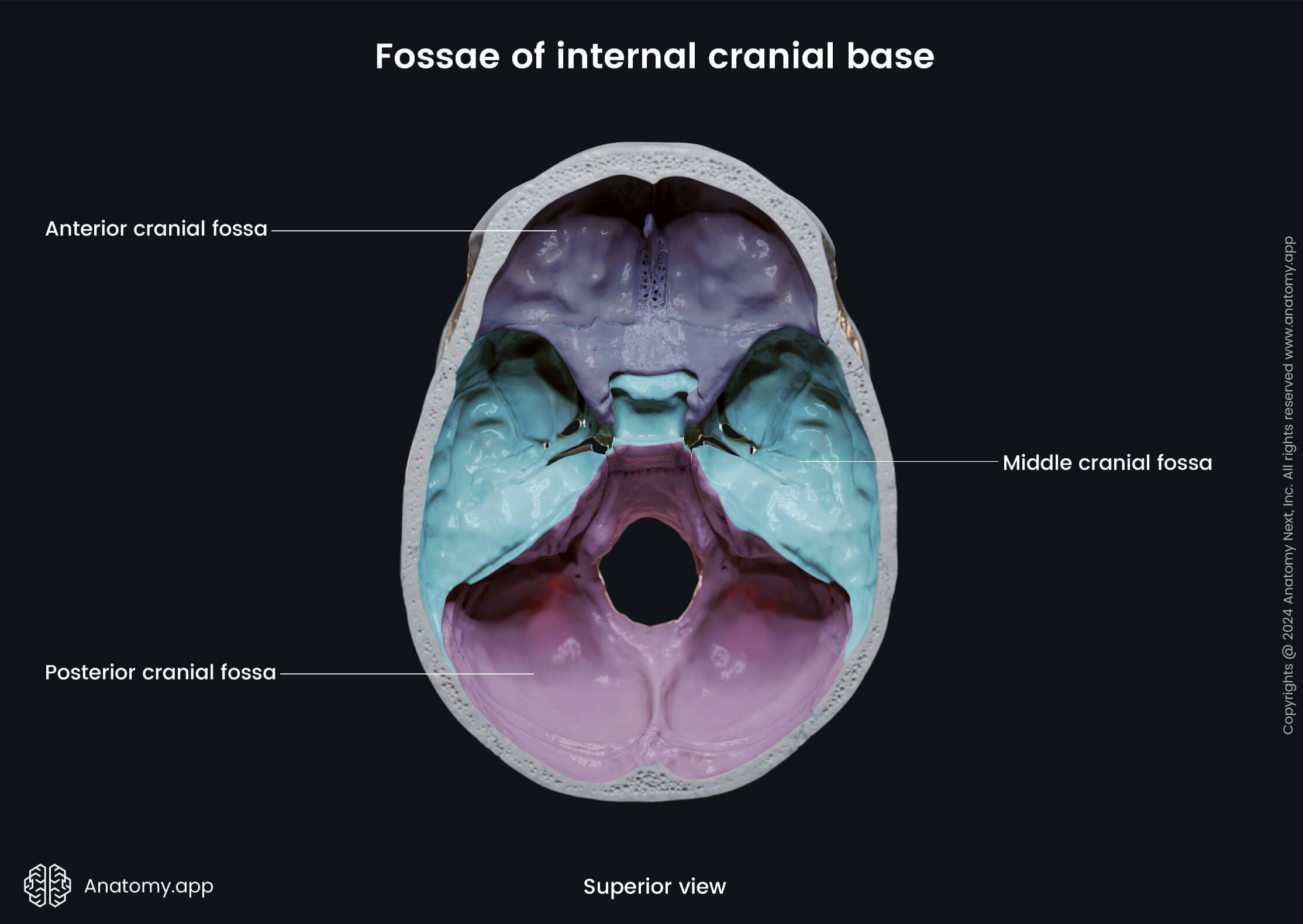
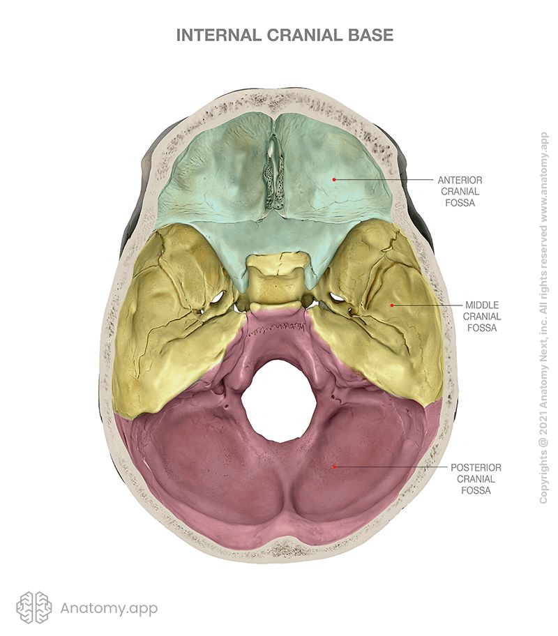
The internal cranial base is subdivided into three distinct regions called cranial fossae. Each cranial fossa houses a different part of the brain. The three fossae are:
Anterior cranial fossa
The anterior cranial fossa lies at the highest and most anterior level of the internal cranial base. It is made of three bones: ethmoid, frontal, sphenoid. It is formed by the cribriform plate of the ethmoid bone, the orbital part of the frontal bone, and the lesser wings of the sphenoid.
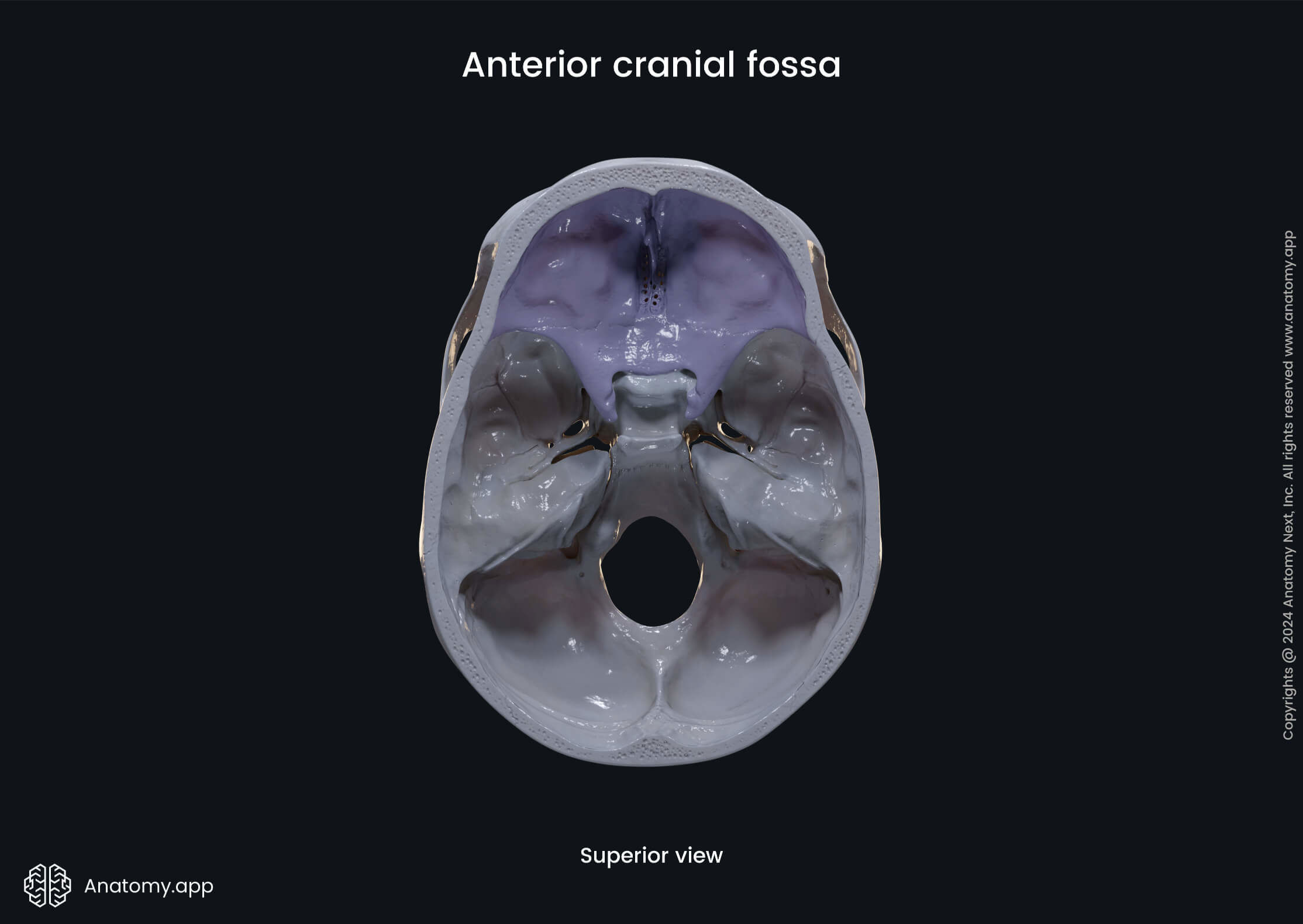
| Anterior cranial fossa | ||||||||||||||||||||||
| Anterior and lateral - inner surface of the frontal bone Posterior - sphenoid bone Posteromedial - anterior border of the chiasmatic sulcus Posterolateral - lesser wings of the sphenoid bone | |||||||||||||||||||||
| Frontal lobe of the cerebral cortex Orbital gyri | |||||||||||||||||||||
| Cribriform foramina (olfactory foramina) Foramen cecum | |||||||||||||||||||||
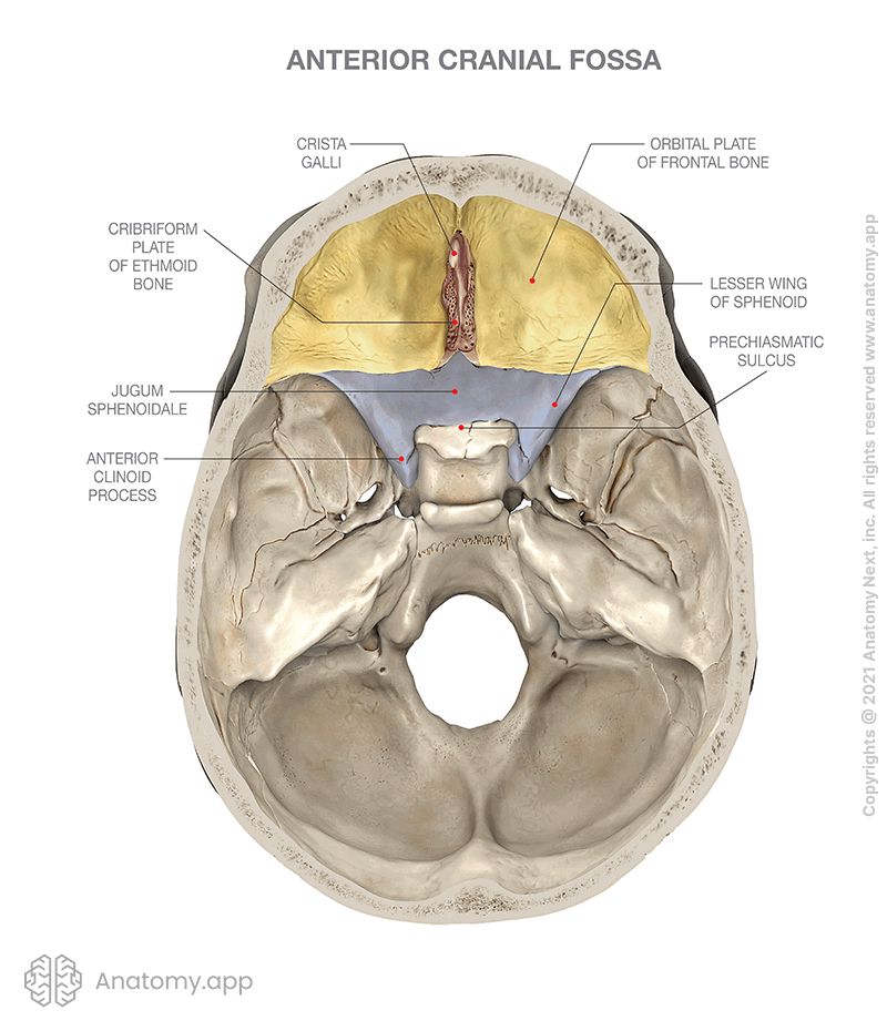
Middle cranial fossa
The middle cranial fossa lies deeper within the skull base. It is wider than the anterior cranial fossa. It is butterfly-shaped and located in the center of the cranial floor. The middle cranial fossa is made up of sphenoid and temporal bones. The middle cranial fossa is formed by the body and greater wings of the sphenoid, the squamous part of the temporal bone, and the anterior surface of the petrous portion of the temporal bone.
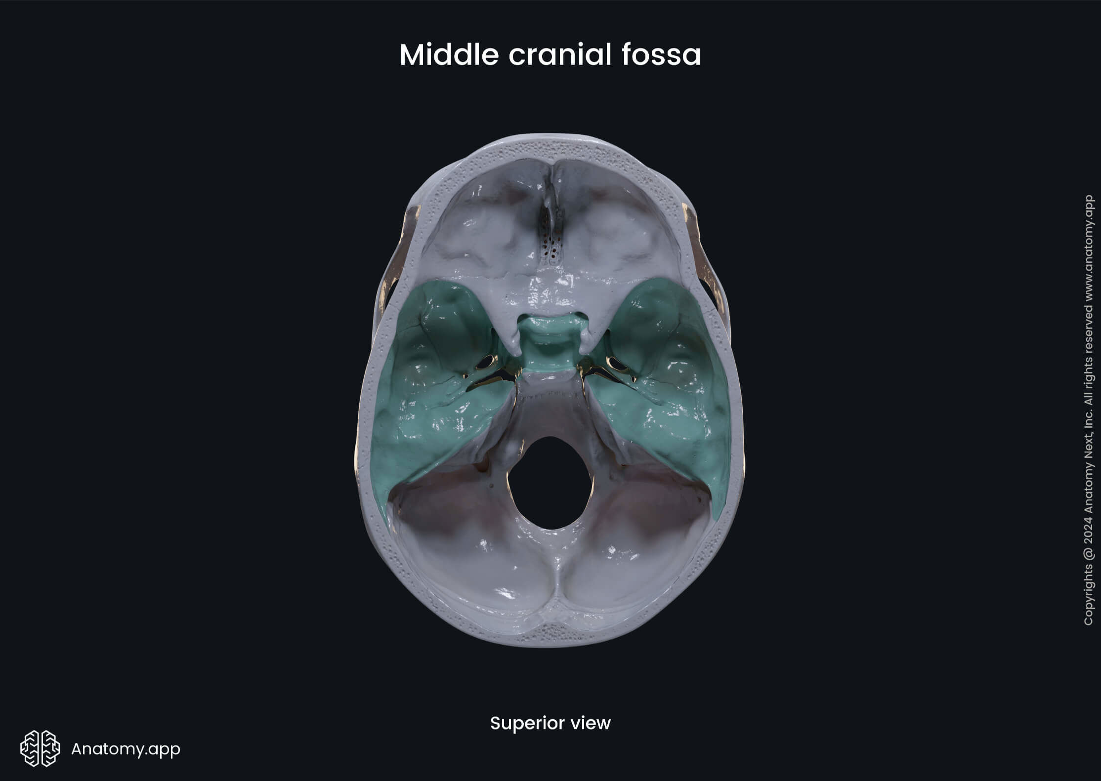
| Middle cranial fossa | ||||||||||||||||||||||
| Anteriorly - anterior margin of the chiasmatic groove, posterior margins of the lesser wings, and part of the body of sphenoid bone Posteriorly - superior borders of the petrous part of the temporal bones, dorsum sellae of the sphenoid Laterally - squamous parts of the temporal bones, greater wings of the sphenoid bone, and part of the parietal bones | |||||||||||||||||||||
| Pituitary gland Temporal lobes of the cerebral cortex | |||||||||||||||||||||
| ||||||||||||||||||||||
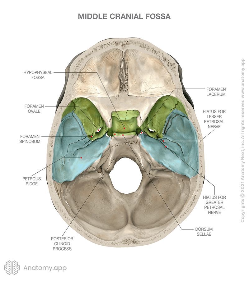
Posterior cranial fossa
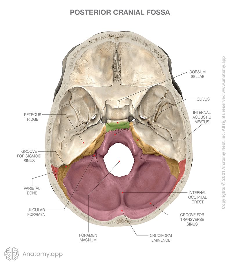
The posterior cranial fossa lies at the lowest level of the internal cranial base and is the largest of the three cranial fossae. It is made of the occipital, temporal, and parietal bones. It is formed by all parts of the occipital bone, the posterior surface of the petrous portion of the temporal bone, and the mastoid angle of the parietal bone.
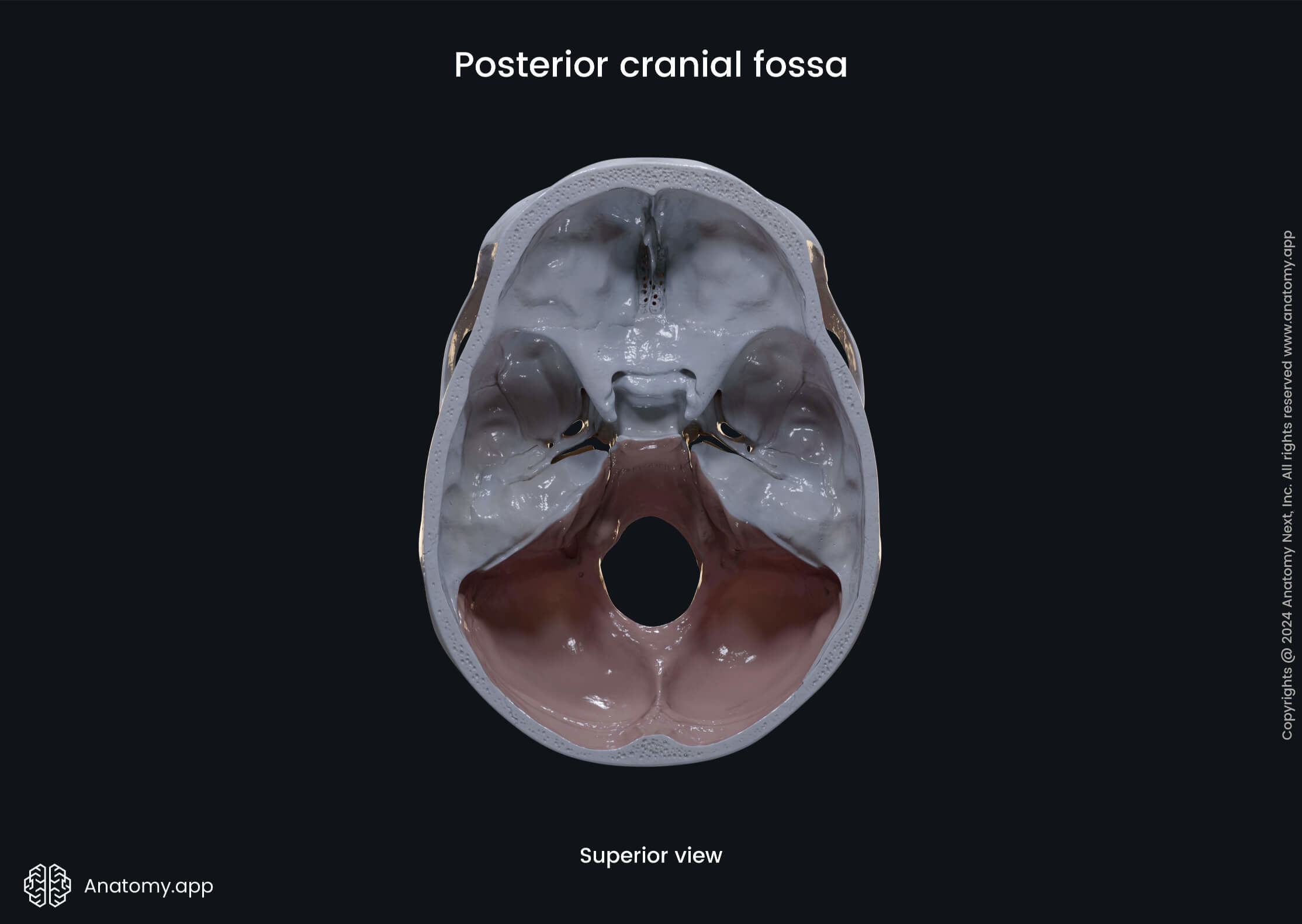
| Posterior cranial fossa | |||||||||
| Anteriorly - dorsum sellae, superior margins of the petrous parts of the temporal bones Posteriorly - squamous part and internal occipital protuberance of the occipital bone, groove for the transverse sinus Laterally - petrous and mastoid parts of the temporal bones, lateral parts of the occipital bone | ||||||||
| |||||||||
| |||||||||
External cranial base
The external cranial base is the outer aspect of the skull base. It extends from the superior incisor teeth to the superior nuchal line of the occipital bone. The external cranial base can be subdivided into anterior, middle, posterior, and two lateral parts.
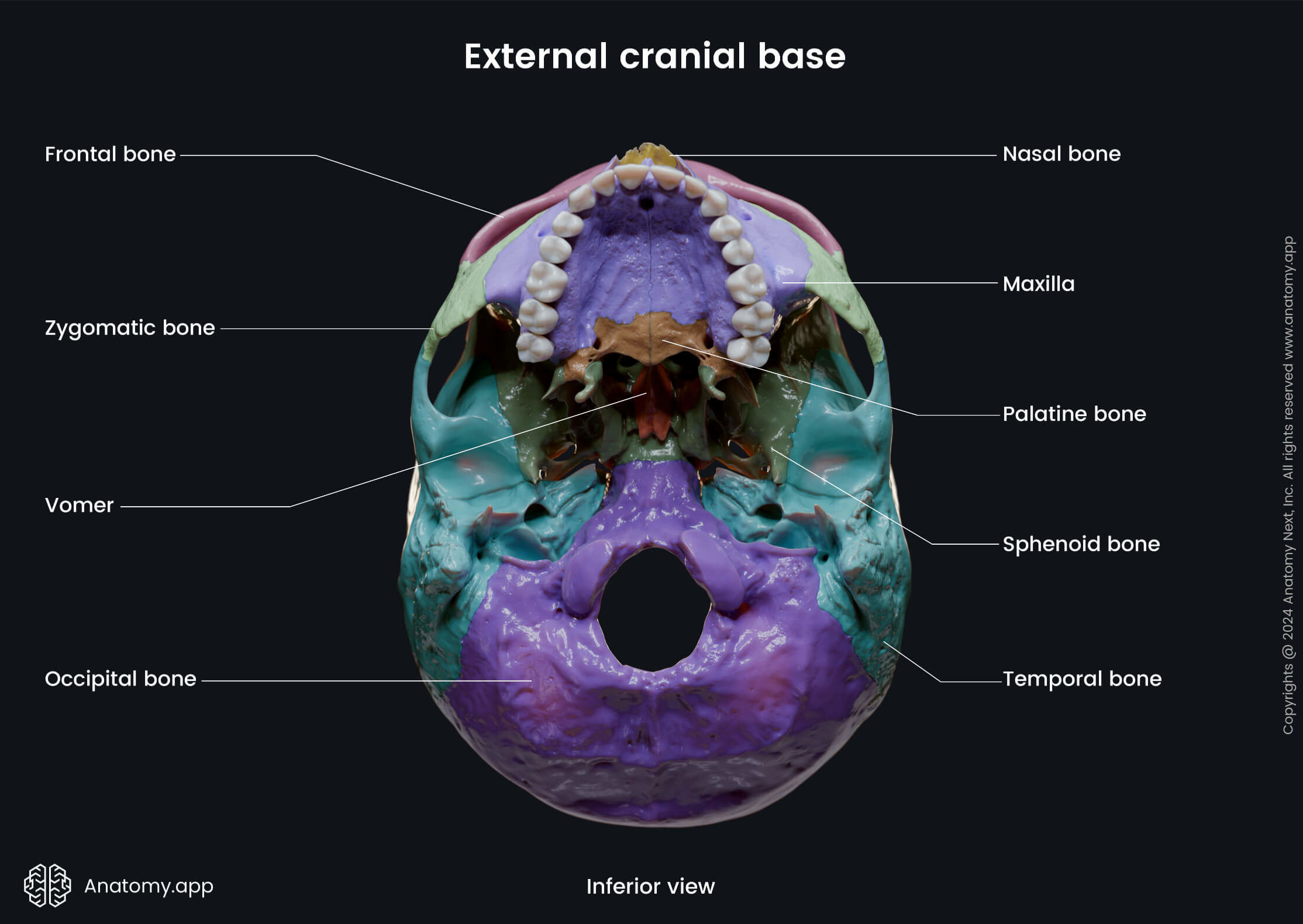
The anterior part of the external cranial base is comprised of the superior alveolar arch, the bony palate, and the choanae. The alveolar process of the maxilla forms the superior alveolar arch, and it contains the dental alveoli or sockets for the teeth. The bony palate is formed by the palatine process of the maxillae anteriorly and horizontal plates of the palatine bones posteriorly. median palatine suture in the midline, and the frontally oriented transverse palatine suture located between the anterior two-thirds and posterior one-third of the hard palate.
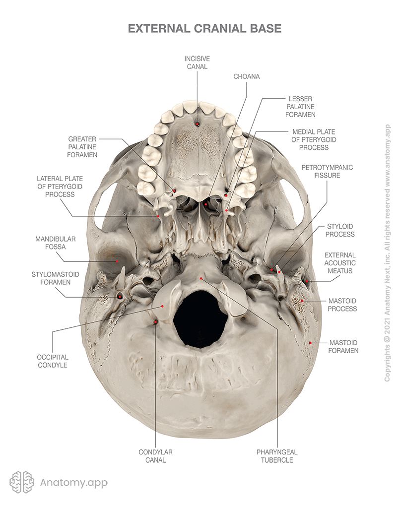
The hard palate forms the roof of the oral cavity and the inferior wall of the nasal cavity. Openings presented on the bony palate include the incisive foramen and greater and lesser palatine foramina. The choana (plural: choanae) is a paired posterior aperture of the nasal cavity that opens into the nasopharynx. It is delimited by the horizontal plate of the palatine bone, a medial plate of the pterygoid process, the body of the sphenoid, and the vomer.
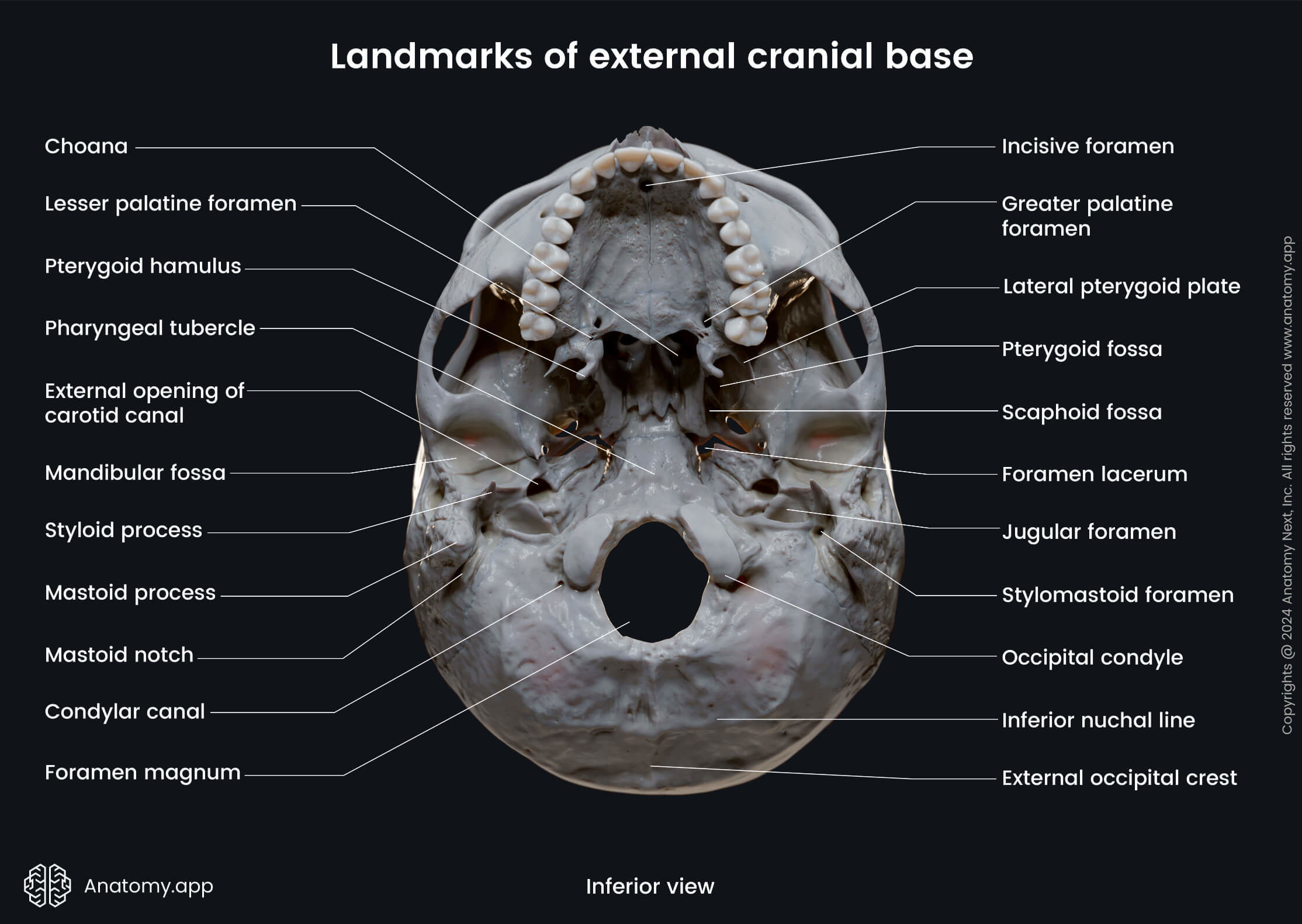
The middle part of the external cranial base is formed by the body of the sphenoid, the petrous portion of the temporal bones, and the basilar part of the occipital bone. It extends from the choanae anteriorly to the anterior margin of the foramen magnum posteriorly. The middle part of the external cranial base presents with several openings, including the foramen lacerum, pterygopalatine fissure, foramen ovale, foramen spinosum, and the carotid canal.
The occipital bone mainly forms the posterior part of the external cranial base. It features such openings as the single foramen magnum and the paired jugular foramen, hypoglossal canal, condylar canal, as well small openings - the mastoid canaliculus and the canaliculus for the tympanic nerve.
Each lateral part of the external cranial base consists of the zygomatic arch and the infratemporal fossa anterior, but the mandibular fossa, the tympanic part of the temporal bone, and the styloid and mastoid processes are located posteriorly. The lateral parts of the external cranial base present such openings as the external acoustic meatus, petro-tympanic fissure, stylomastoid foramen, and other smaller apertures.
Temporal fossa
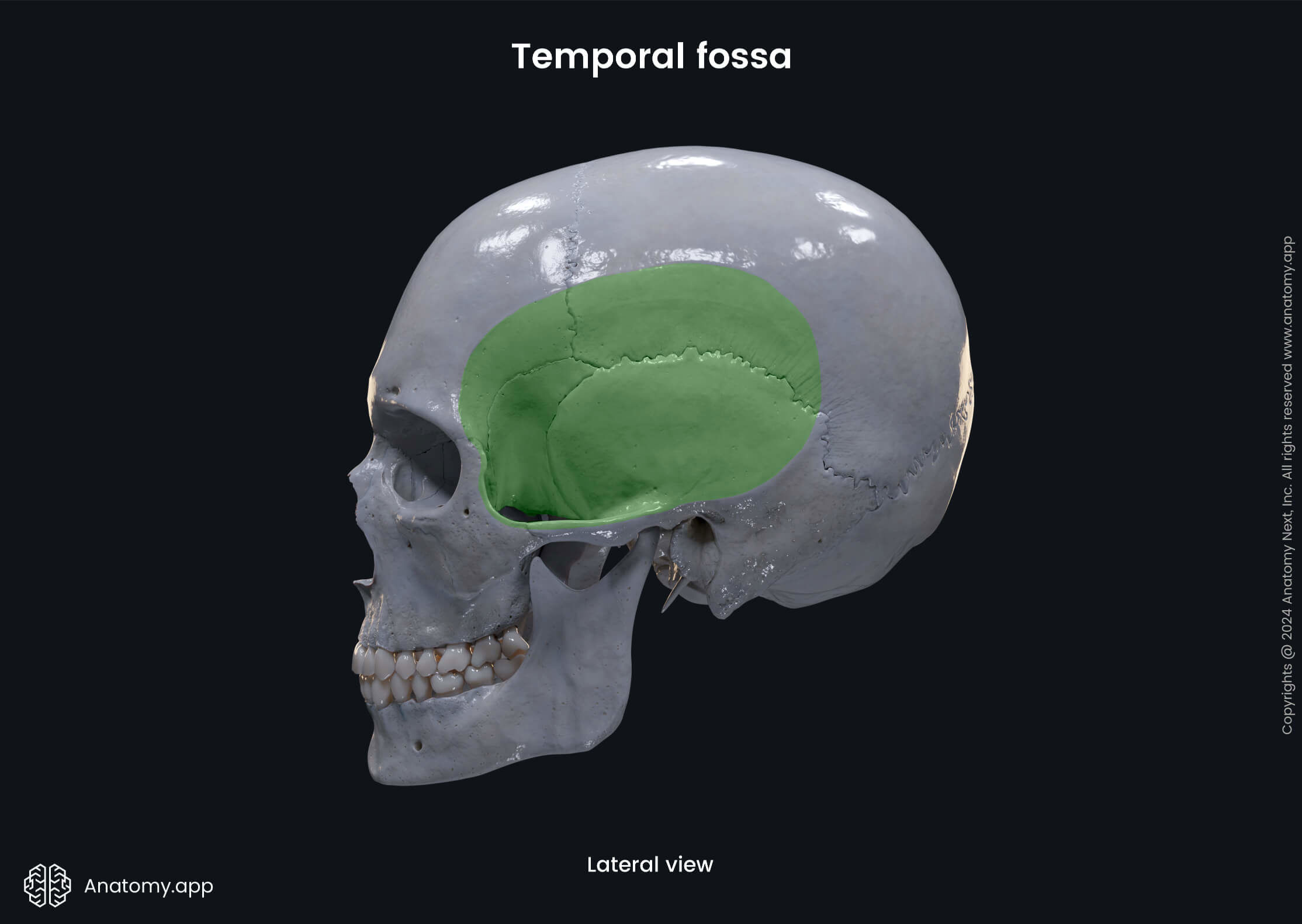
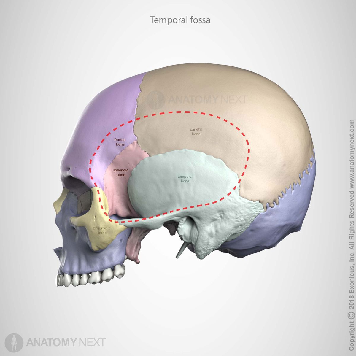
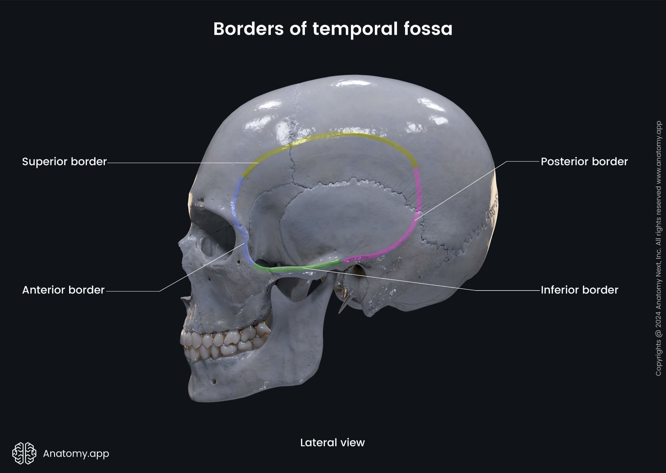
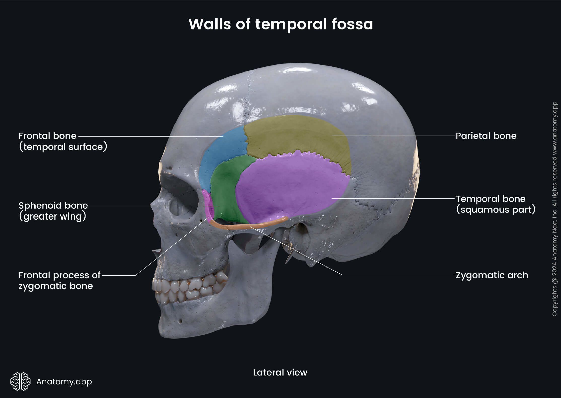
The temporal fossa is a depressed area on each side of the skull bounded by the temporal lines and the zygomatic arch. It is formed by the temporal, sphenoid, parietal, and frontal bones. The temporal fossa is located superiorly to the infratemporal fossa.
| Temporal fossa | |||||||||
| Anteriorly - frontal process of the zygomatic bone, zygomatic process of the frontal bone Superiorly and posteriorly - superior and inferior temporal lines Inferiorly - infratemporal crest on the greater wing of the sphenoid, zygomatic arch Laterally - temporal fascia | ||||||||
| Anterior - temporal surface of the frontal and zygomatic bones Medial - temporal surface of the frontal, parietal and temporal bones, and the greater wing of the sphenoid Lateral - zygomatic arch | ||||||||
| Temporal muscle and fascia Zygomaticotemporal nerve Temporal branch of the facial nerve (CN VII) Superior temporal artery Corresponding veins | ||||||||
| Foramina | Zygomaticotemporal foramen | ||||||||
Infratemporal fossa
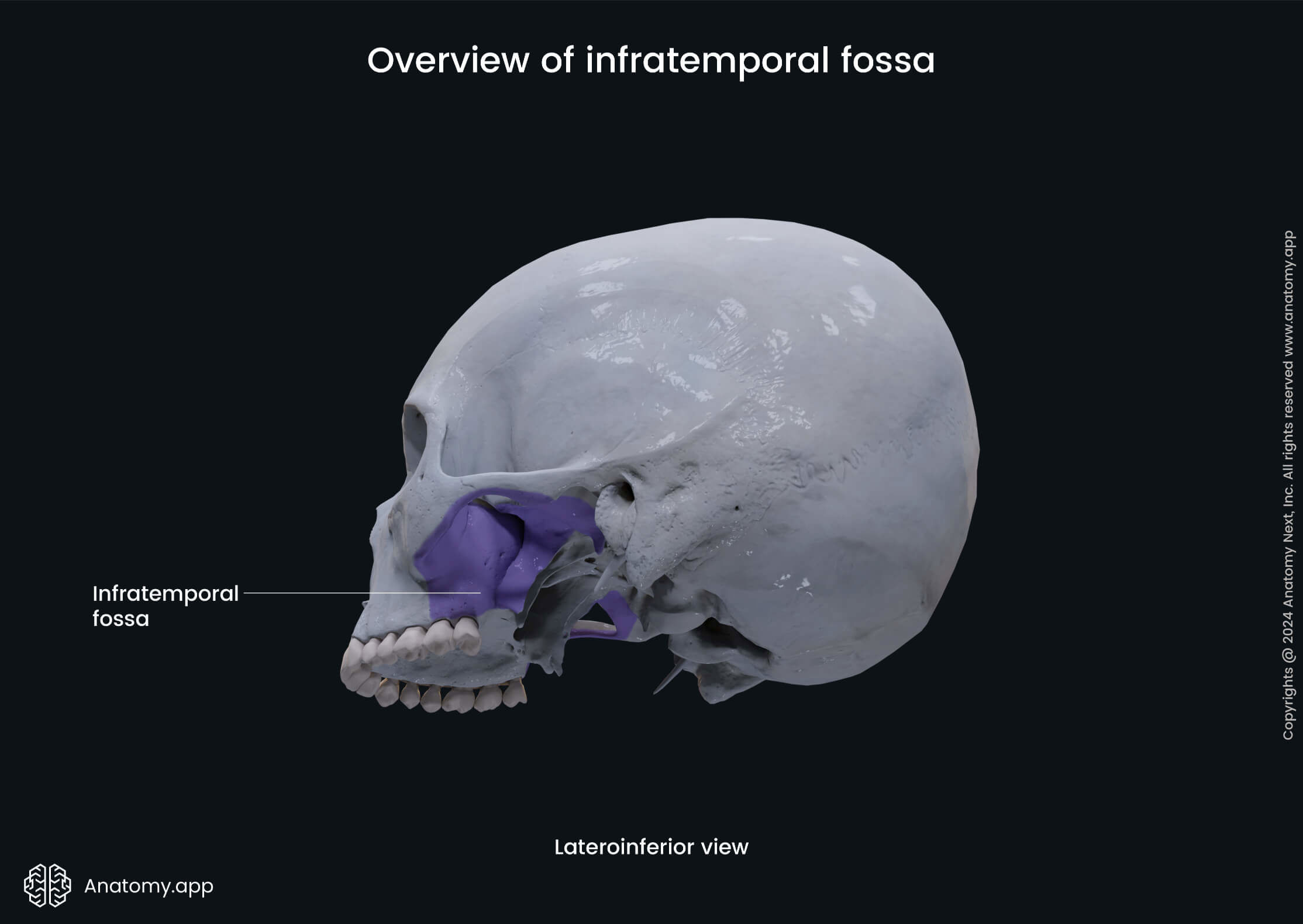
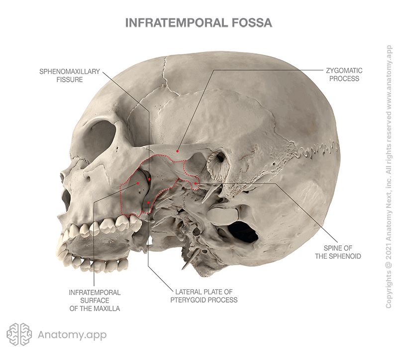
The infratemporal fossa is an irregular-shaped space situated on each side of the skull below the temporal fossa and deep to the ramus of the mandible. It is located between the base of the skull, lateral pharyngeal wall, and ramus of the mandible. The infratemporal fossa communicates with the temporal fossa via space to the zygomatic arch. It also communicates with the pterygopalatine fossa via the pterygomaxillary fissure.
| Infratemporal fossa | |||||||||
| Superiorly - infratemporal surfaces of the temporal bone and the greater wing of the sphenoid Inferiorly - medial pterygoid muscle Laterally - zygomatic arch, medial surface and condylar process of the mandibular ramus Medially - lateral plate of the pterygoid process, tensor veli palatini and superior pharyngeal constrictor muscles Posteriorly - carotid sheath Anteriorly - posterior border of the maxillary sinus | ||||||||
| Anterior - infratemporal surface of the maxilla Superior - infratemporal surface of the greater wing of the sphenoid bone Lateral - zygomatic arch and ramus of the mandible | ||||||||
| Major branches of the mandibular nerve (CN V3) Otic ganglion Middle meningeal vein Tendon of the temporal muscle | ||||||||
| |||||||||
Pterygopalatine fossa
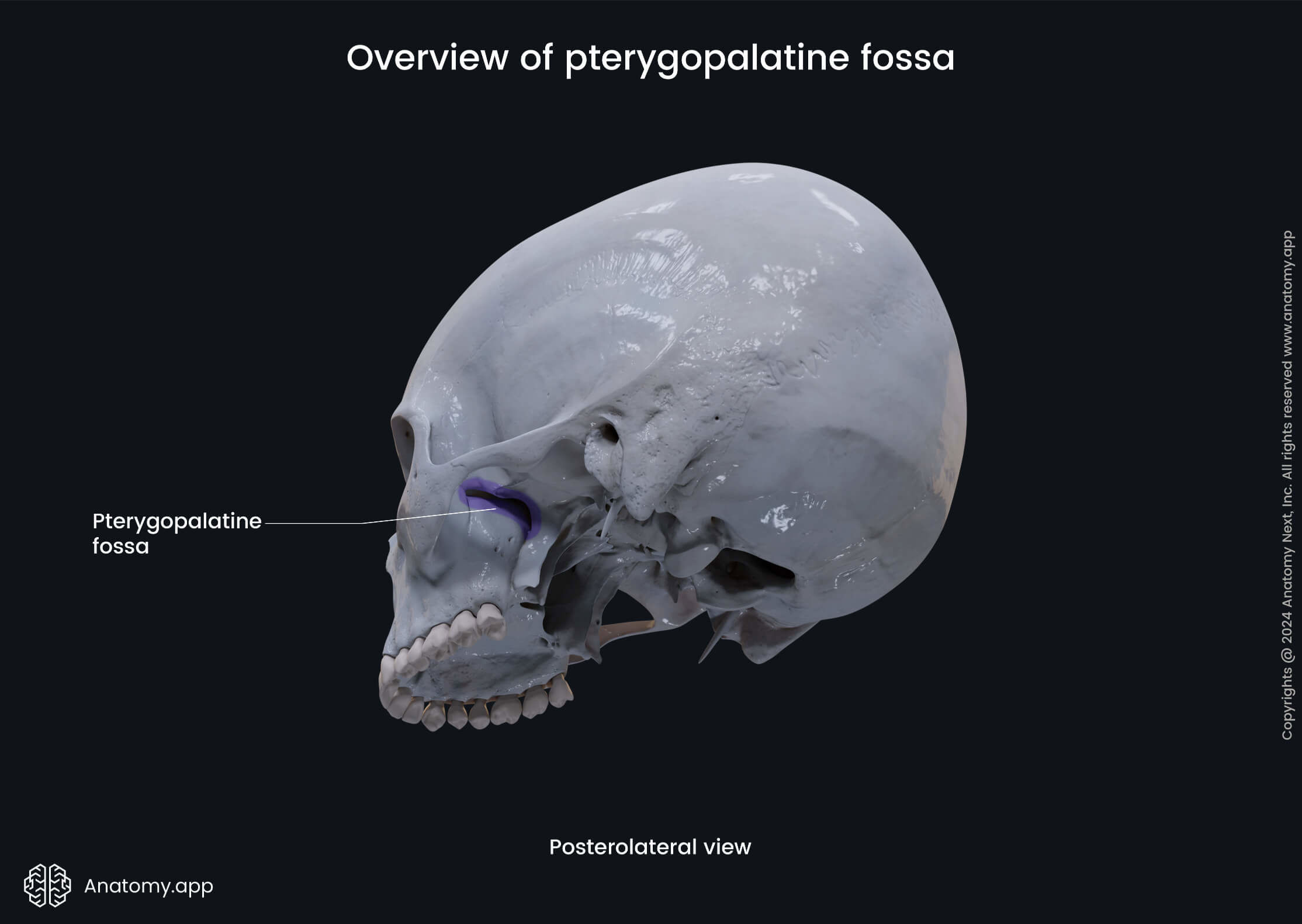
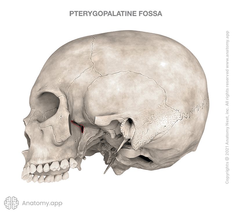
The pterygopalatine fossa, also known as the sphenopalatine fossa, is a cone-shaped, bilateral depression below the apex of the orbit on the lateral side of the skull. It is located between the maxilla anteriorly, the sphenoid bone posteriorly, and the palatine bones.
| Pterygopalatine fossa | |||||||||
| Anteriorly - superomedial part of the infratemporal surface of the maxilla Posteriorly - root of the pterygoid process and the adjacent anterior surface of the greater wing of the sphenoid bone Inferiorly - palatine bone Superiorly - maxillary surface of the greater wing of the sphenoid bone, inferior orbital fissure Medially - perpendicular plate of the palatine bone, orbital and sphenoidal processes of the palatine bone Laterally - pterygomaxillary fissure | ||||||||
| Pterygopalatine ganglion Posterior superior alveolar nerve Part of the maxillary artery | ||||||||
| Pharyngeal canal (palatovaginal canal) | ||||||||
Orbit
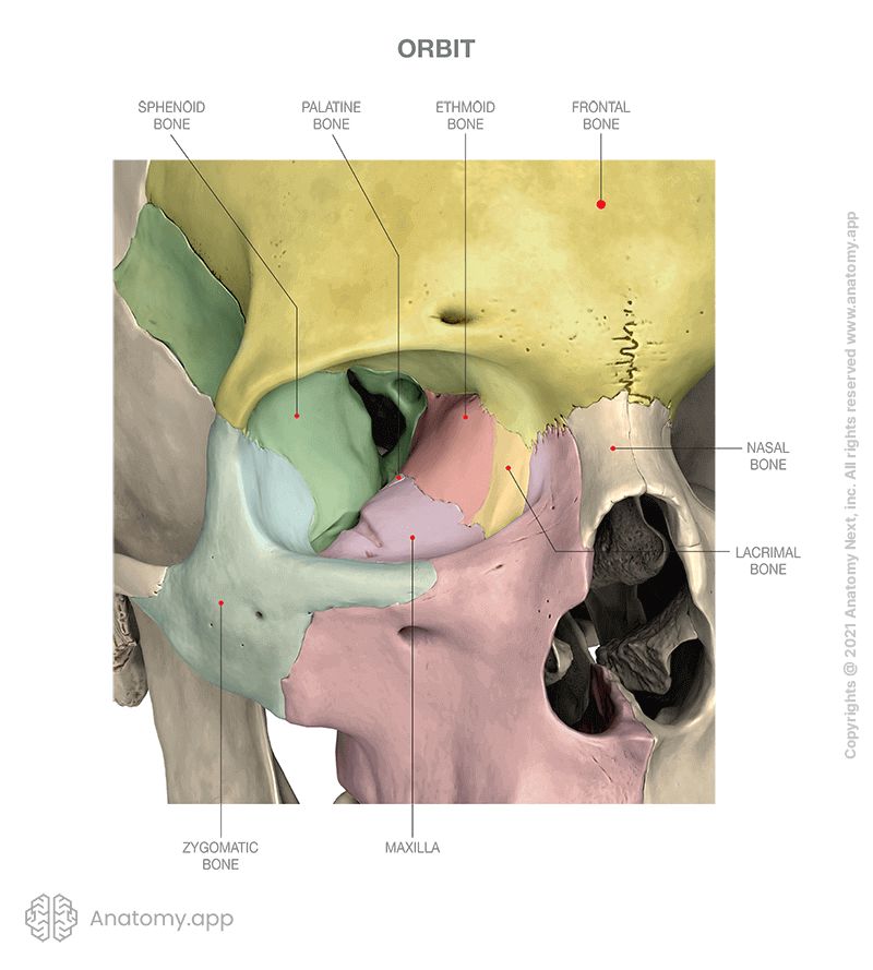
The orbit or orbital compartment is a pyramid-shaped paired skeletal cavity located in the skull on either side of the root of the nose. Its base opens anteriorly in the central part of the face and next to the midline. Its apex is pointed backward. The orbit is a bony cavity housing and protecting the eye, its appendages, and its accessory structures.
| Orbit | |||||||||
| Supraorbital - frontal bone Medial - frontal process of the maxilla Lateral - zygomatic processes of the frontal and zygomatic bones Infraorbital - zygomatic process of the maxilla and zygomatic bone | ||||||||
| Roof of the orbit (superior wall) - supraorbital margin and orbital surface of the frontal bone (anteriorly), orbital plate of the lesser wing of the sphenoid (posteriorly) Floor of the orbit (inferior wall) - orbital surface of the maxilla (anteromedially), orbital surface of the zygomatic bone, palatine bone (anterolaterally) Medial - lacrimal bone, orbital plate of the ethmoid, lesser wing of the sphenoid, frontal process of the maxilla Lateral - frontal process of the zygomatic bone (anteriorly), orbital surface of the greater wing of the sphenoid (posteriorly) | ||||||||
| Superior and inferior ophthalmic veins Orbital fascia Orbital fat | ||||||||
| |||||||||
Nasal cavity
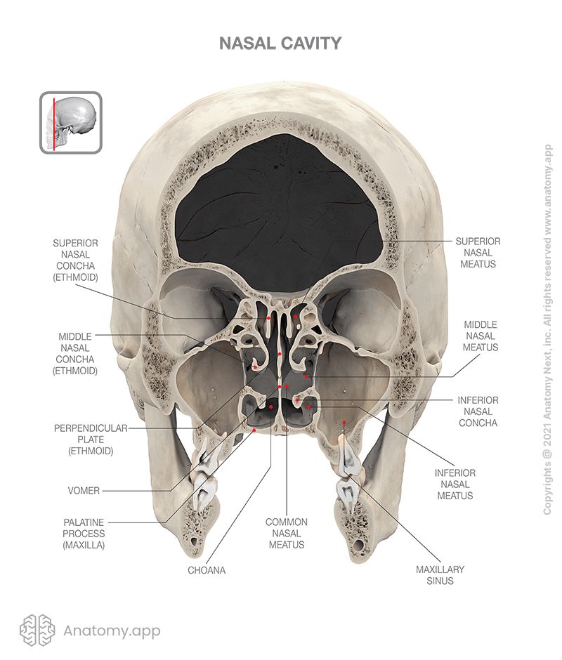
The nasal cavity or cavity of the nose is an irregular, bilateral air-filled space located within the skull above the roof of the mouth, forming the internal part of the nose. The nasal cavity serves as the initial part of the respiratory tract. It is lined with a mucous membrane, which also houses olfactory receptors. Bones forming the nasal cavity are the sphenoid, palatine, lacrimal, ethmoid, nasal bones, vomer, and maxilla.
Walls of nasal cavity
The nasal cavity is a bony compartment that is split into two symmetric compartments by the nasal septum. It has a roof, a floor, and four walls - all formed by bones of the skull. The roof of the nasal cavity is formed by the cribriform plate of the ethmoid and the anterior aspect of the body of sphenoid. The floor is formed by the bony palate. The bony palate is made by the palatine processes of the maxillae and horizontal plate of the palatine bone.
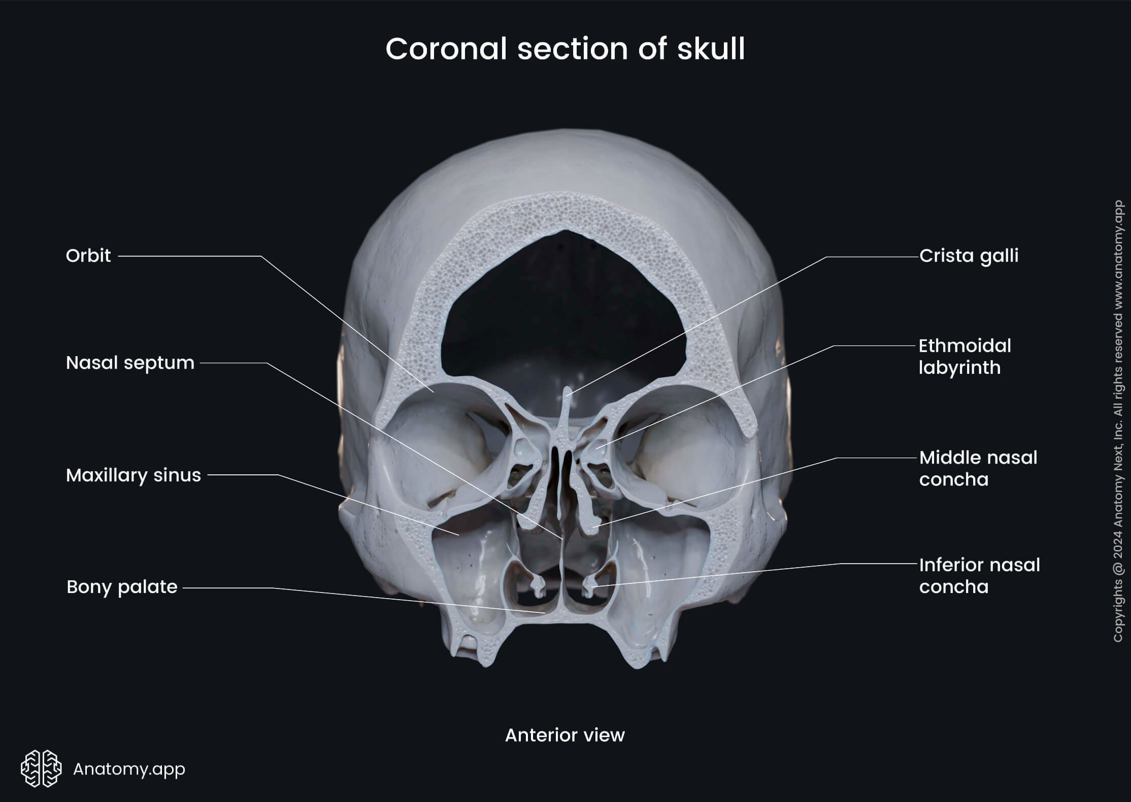
The medial wall of the nasal cavity is represented by the bony nasal septum composed of the vomer and the perpendicular plate of the ethmoid. The vomer is a small, thin bone forming the posterior part of the nasal septum. The vomer is attached to the rostrum of the sphenoid. The following structures form each lateral wall of the nasal cavity:
- Nasal surface of the maxilla
- Perpendicular plate of the palatine bone
- Ethmoidal labyrinth and superior and middle nasal conchae
- Lacrimal bone
- Medial plate of the pterygoid process of the sphenoid
- Inferior nasal concha
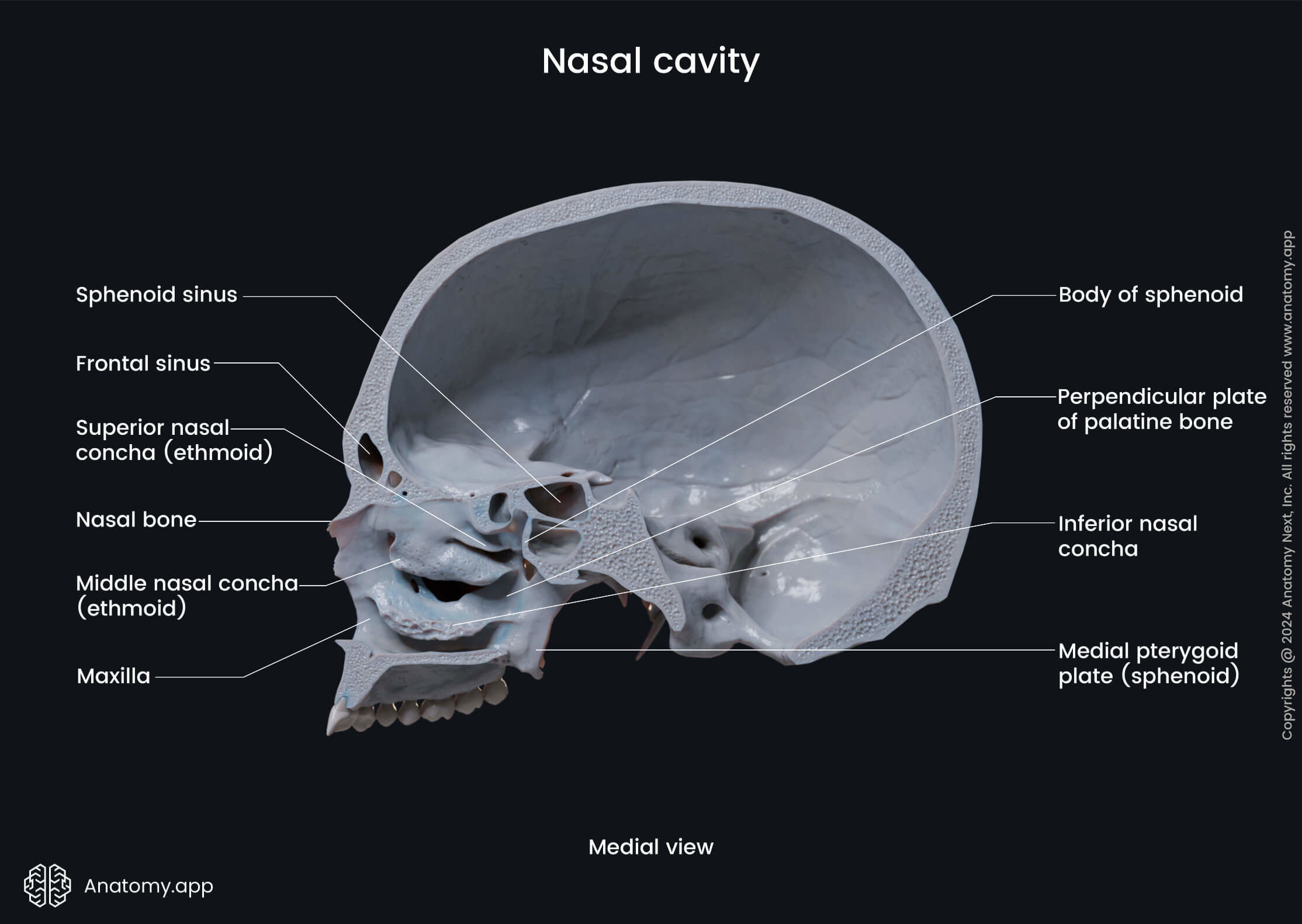
The nasal bones form the anterior wall. On each side, it opens with an anterior nasal aperture. Finally, the posterior wall of the nasal cavity is formed by the anterior surface of the body of the sphenoid, and it presents a pair of openings called the choanae.
Nasal conchae
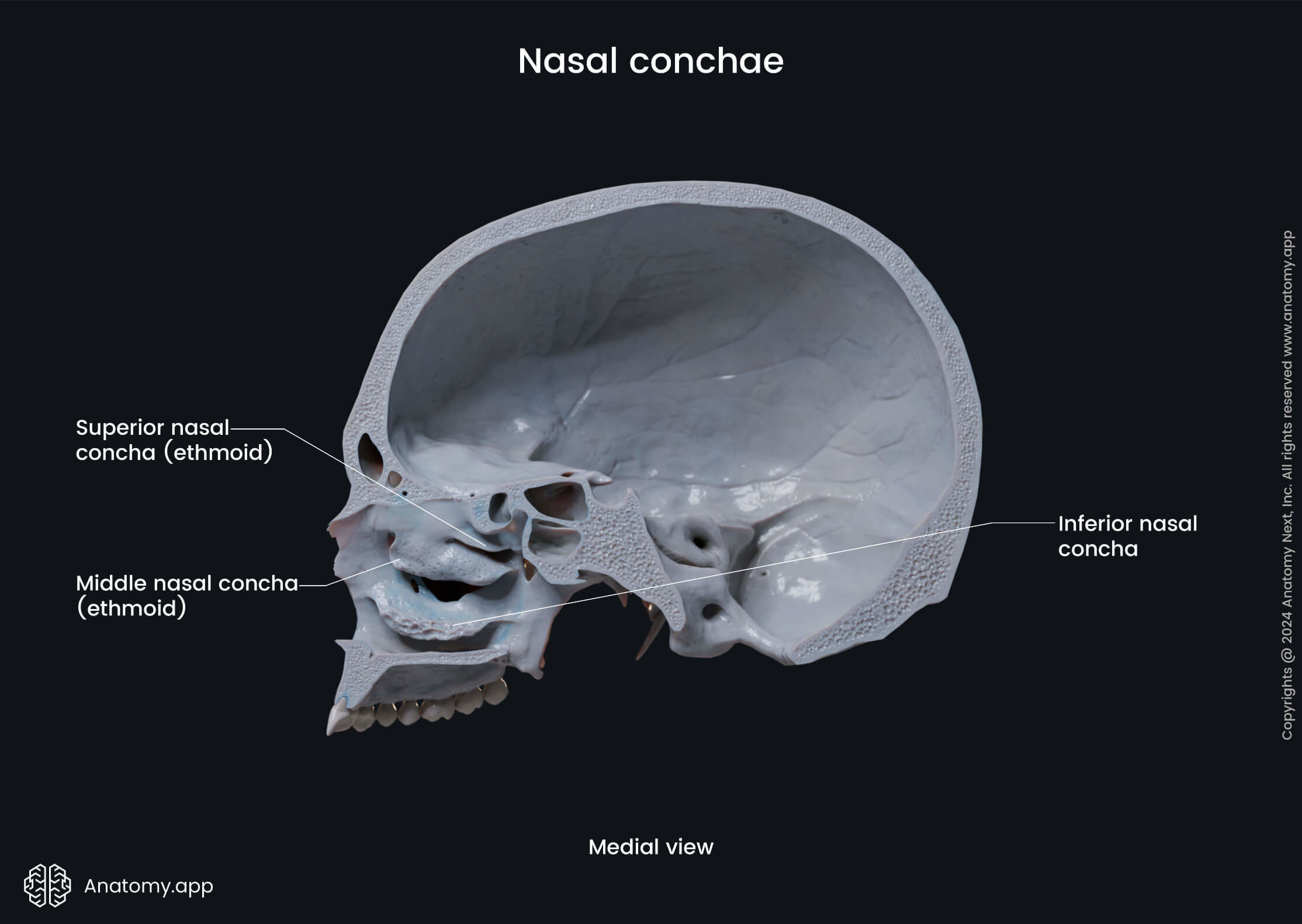
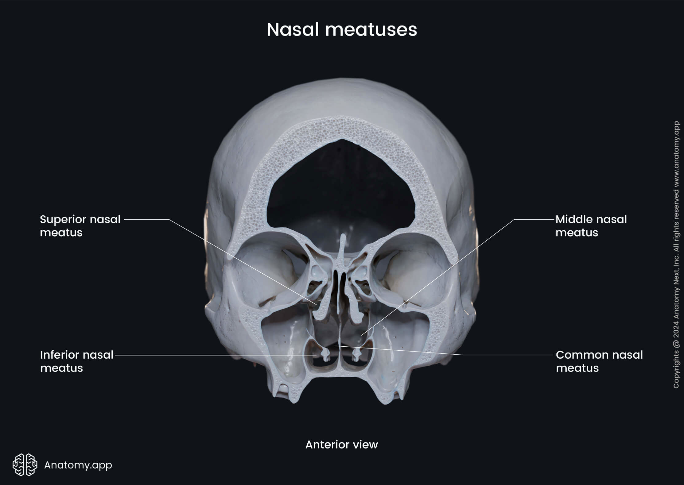
There are three bony formations in each lateral wall of the nasal cavity that resemble curved shelves called the nasal conchae - superior, middle, and inferior. They project into the nasal cavity creating five pathways called meatuses. These are the superior, middle, inferior, and common nasal meatuses, and the nasopharyngeal meatus.
Content and openings of nasal cavity
Like other skull regions, the nasal cavity serves as a passageway for several nerves and blood vessels of the head. The nasal cavity presents many openings in its bony walls, such as the following:
- Foramina of the cribriform plate
- Sphenopalatine foramen
- Incisive canal
- Opening of the nasolacrimal canal
The nasal cavity is also connected with the paranasal sinuses with openings to the frontal, maxillary and sphenoidal sinuses, and the ethmoidal air cells.
Periosteum
The periosteum is a layer of dense connective tissue covering the surfaces of bones, and that includes the skull. The periosteum that covers the skull bones is also known as the pericranium. The periosteum has a significant role in bone repair and growth and impacts the blood supply of the bone. It consists of two layers:
- Outer fibrous layer
- Inner layer (also known as the cambium)
The outer layer of the periosteum is a tough fibrous layer formed by collagen fibers that can be subdivided into a superficial and deep portion. Both contain collagen and elastic fibers, but the deep portion is is known as the fibroelastic layer as it has many elastic fibers and is highly collagenous. This layer also contains many nerve endings - therefore, pain sensation can be triggered by the damage to this layer.
The inner layer contains many blood vessels and cells and is highly vascular and cellular. The periosteum of the skull is also the innermost layer of the scalp. The layers of the scalp from the most superficial to the innermost are the:
- Epidermis of the skin with hair follicles and sebaceous glands;
- Dermis containing dense connective tissue with nerves, lymphatics, blood vessels;
- Epicranial aponeurosis (also known as the galea aponeurotica) - immobile connective tissue layer;
- Loose connective tissue;
- Periosteum, also known as the pericranium in the skull.
Functions of the skull
The primary function of the skull is to provide protection for the brain and the sensory organs connected with it. It also incorporates the upper part of the digestive and respiratory tracts. The skull supports the soft tissue of the head and facial anatomical structures. The skull also provides such functions as:
- Head movement
- Formation of the face framework
- Passage for cranial nerves and vessels
- Housing organs that provide special senses (vision, hearing, taste, smell)
- Supporting the muscles moving various parts of the head and providing facial expressions
- Attaching with the inner surfaces to the meninges (that stabilize the brain, blood vessels, and nerves)
- Protection of the eyeballs and attachment for muscles moving them
- Chewing and digestion (provided by the maxilla and mandible; these bones also contain alveolar processes with sockets that house the teeth)
- Storage of calcium
- Hematopoiesis