- Anatomical terminology
- Skeletal system
- Joints
- Muscles
- Heart
- Blood vessels
- Lymphatic system
- Nervous system
- Respiratory system
- Digestive system
- Urinary system
- Female reproductive system
- Male reproductive system
- Endocrine glands
- Eye
- Ear
Anatomical terminology
Medical students and healthcare professionals worldwide use universal anatomical terminology to facilitate effective communication, precise descriptions, and accurate documentation of patient information. This standardized language, rooted in ancient Greek and Latin, serves as a common reference point, significantly enhancing precision and minimizing the risk of medical errors.
Embarking on the study of anatomy may initially seem like delving into a new language, and indeed, it can be challenging. Massive amount of new terms and very little time. However, mastering basic anatomical terms is the key to learning and understanding anatomy faster.
Think of anatomical terminology as the foundation of a home - an essential framework without which understanding the human body becomes challenging. Anatomy terminology is the foundation and the very beginning of medical studies.
In this article, we will describe what anatomical position means, explore regional terminology, anatomical planes, anatomical lines and directional terms, as well as discuss the various cavities and serous membranes found within the human body. Additionally, we will navigate through the abdominal region, describing its quadrants and regions. And finally, we will review anatomical terms that are associated with the movements.
Anatomical position
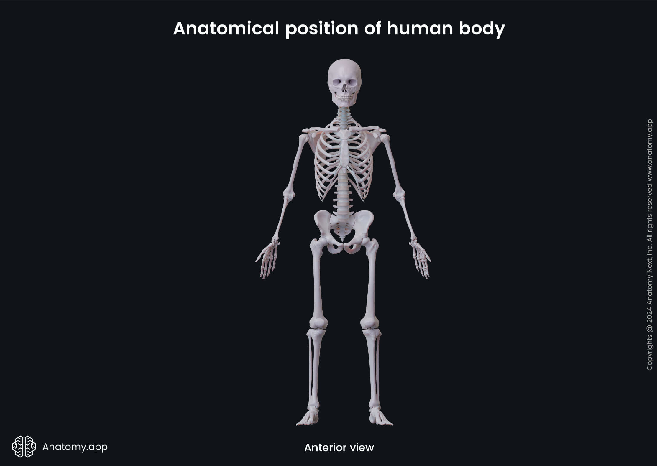
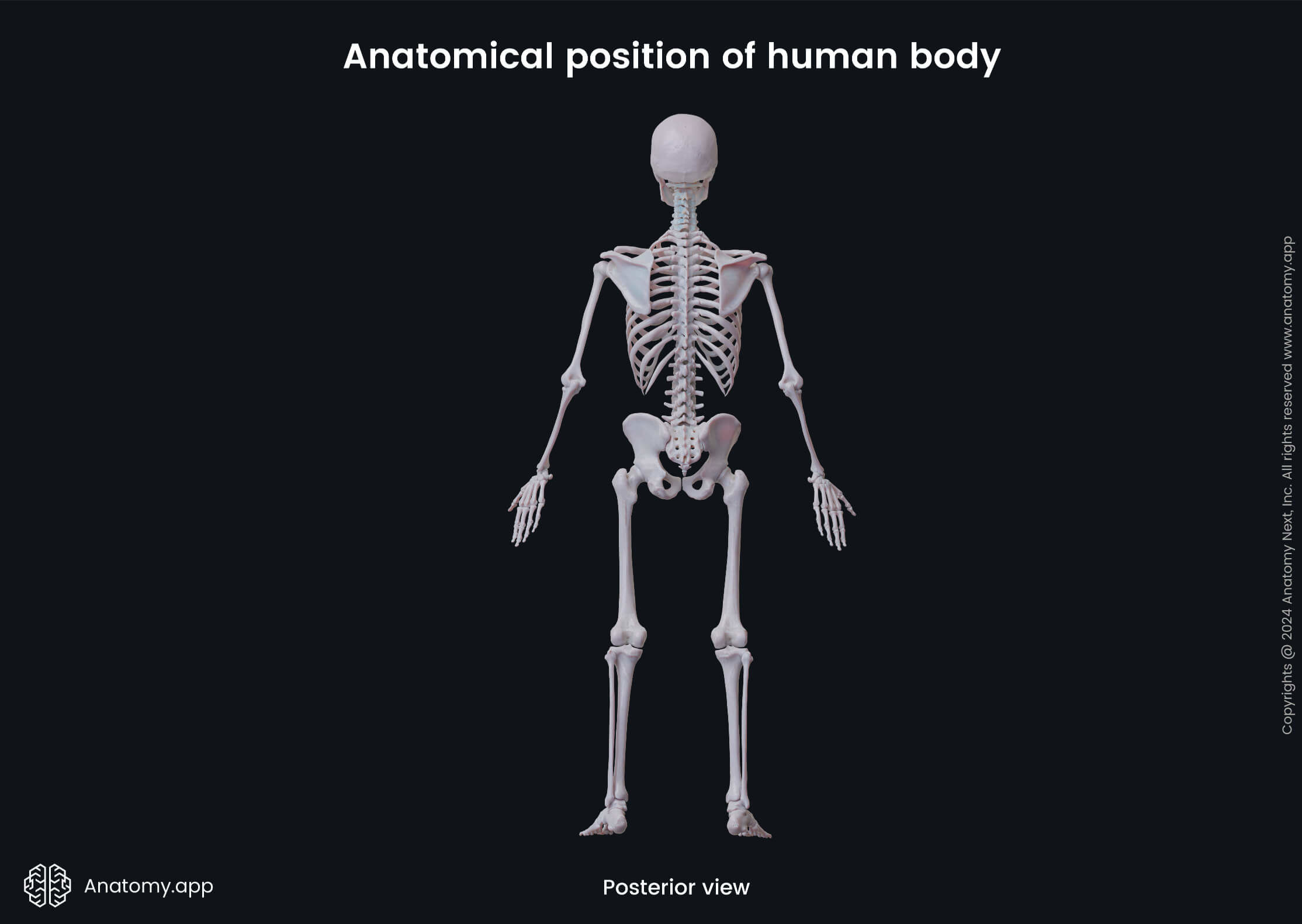
The anatomical position is the very first term one must understand in human anatomy. It can be described as the foundational reference point, providing a standard orientation or position of the human body. When describing any orientation, direction or movement, the reference point is always the anatomical position.
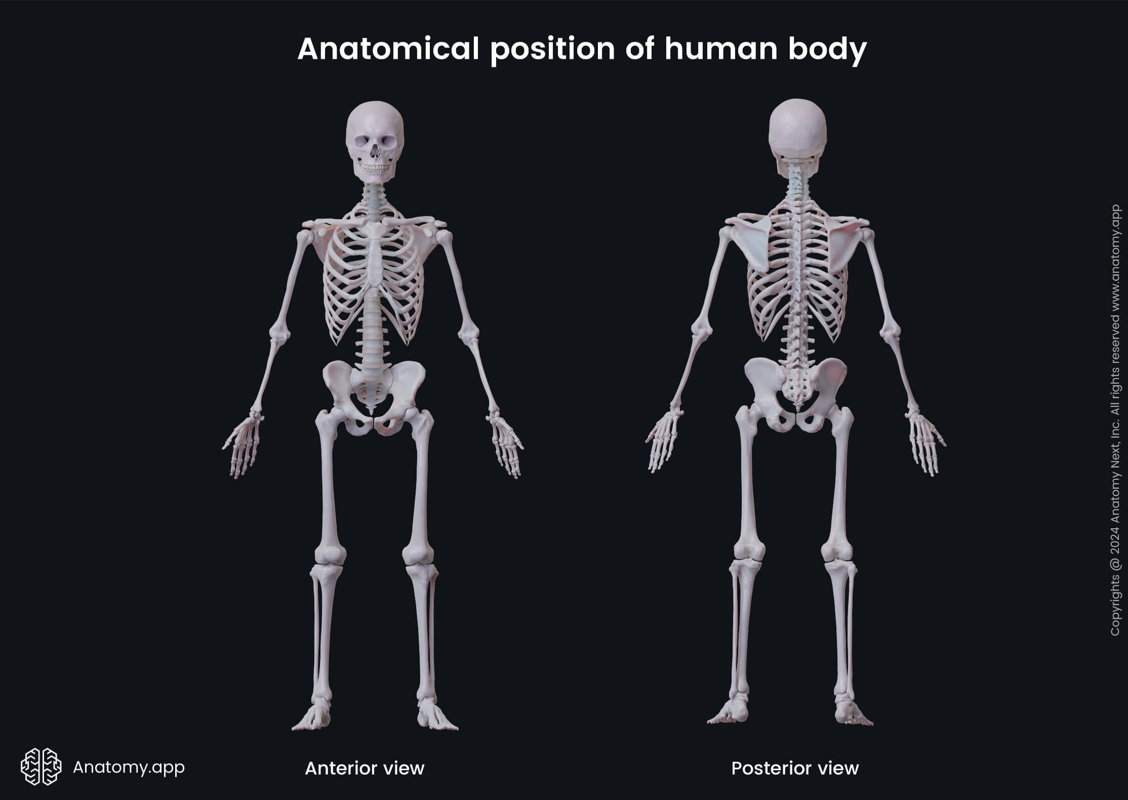
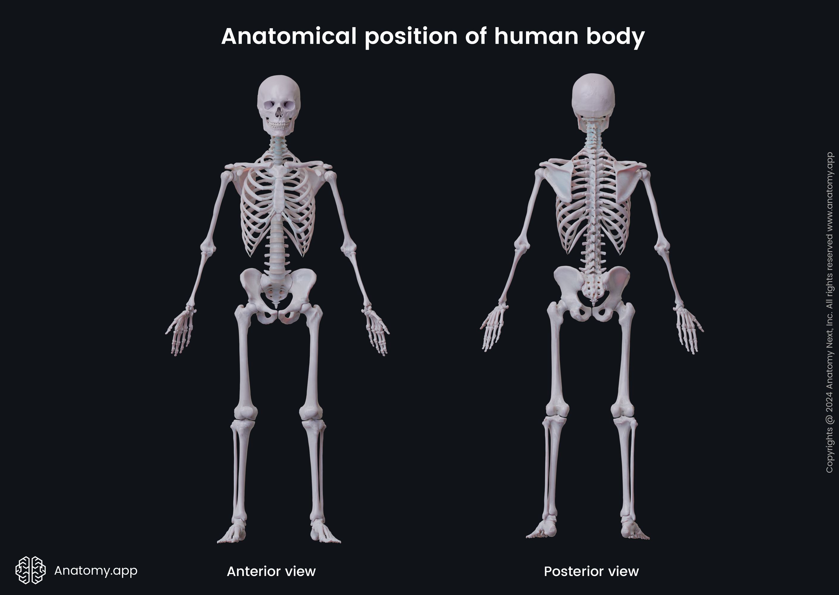
When a person is in the anatomical position, it means that he/she is standing upright and facing forward, the mouth is closed, and the facial expression is neutral. The eyes are focused on the distance. The arms hang straight by the sides, and the hands are held by the hips with palms facing forward and thumbs pointing away from the body. The legs are straight, with the feet standing parallel on the ground at shoulder width, and the toes are pointing forward.
This standardized position reduces confusion when describing any orientation, movement, location, and direction. Anatomical terms are consistently used as if the body is in the anatomical position, even if it lies differently.
Note: Sometimes, two more terms are used during surgical procedures or specific physical examinations - prone position and supine position. These terms refer to the body that is lying down. The prone position is used when the body is in a face-down orientation, while the supine position describes a face-up position of the human body.
Regional terminology
The human body can be perceived as a unified whole. However, it can also be divided into several smaller regions, such as the head, neck, thorax, abdomen, pelvis, spine and back and the upper and lower limbs. All these regions can be further subdivided into even smaller areas based on the anatomical contents.
Overall, countless terms are used to describe regions of the human body to help increase precision. For example, the lumbar and sacral regions together define the lower back. However, each separately represents a more precise location of the lower back.
Students learn most of the terms when studying a particular region. However, here is the summary of the most commonly used regional terms:
- Cephalon (adj. cephalic) - refers to the entire head region;
- Cranium (adj. cranial) - used to describe the skull;
- Frons (adj. frontal) - refers to the frontal part of the head or the forehead;
- Occiput (adj. occipital) - region at the back of the head or the base of the skull;
- Facies (adj. facial) - a term that describes the face;
- Orbit (adj. orbital) - applies to the eye socket;
- Oculus (adj. ocular) - defines the eyes;
- Nasus (adj. nasal) - applies to the nose;
- Oris (adj. oral) - refers to the mouth region;
- Mentis (adj. mental) - a term that is used to describe the chin area;
- Bucca (adj. buccal) - represents the cheek region;
- Auris (adj. otic) - refers to the ear region;
- Cervicis (adj. cervical) - applies to the neck;
- Shoulder (adj. acromial) - used to describe the shoulder region;
- Axilla (adj. axillary) - applies to the armpit region;
- Brachium (adj. brachial) - defines the upper arm;
- Antecubitis (adj. antecubital) - defines the front of the elbow;
- Olecranon (adj. olecranal) - applies to the back of the elbow;
- Antebrachium (adj. antebrachial) - refers to the forearm or the lower arm;
- Carpus (adj. carpal) - describes the wrist;
- Manus (adj. manual) - defines the hand;
- Palma (adj. palmar) - used to describe the palm;
- Pollex - refers to the thumb of the hand;
- Thoracis/thorax (adj. thoracic) - represents the entire chest region between the neck and the abdomen;
- Sternum (adj. sternal) - describes the region in the center of the thorax that corresponds to the breastbone (sternum);
- Mamma (adj. mammary) - used to describe the breast region;
- Abdomen (adj. abdominal) - applies to the entire region of the belly;
- Umbilicus (adj. umbilical) - corresponds to the area of the navel or belly button;
- Coxa (adj. coxal) - describes the hip region;
- Pelvis (adj. pelvic) - represents the region between both hip bones;
- Pubis (adj. pubic) - an anteroinferior region of the pelvis;
- Gluteus (adj. gluteal) - a term that is used to describe the buttock;
- Inguen (adj. inguinal) - defines the groin region;
- Perineum (adj. perineal) - used to describe the region between the external genitalia and the anus;
- Dorsum (adj. dorsal) - represents the entire back region;
- Vertebra (adj. vertebral) - refers to the region that corresponds to the entire length of the spine;
- Lumbus (adj. lumbal) - describes the region along the lower part of the spine or the loin;
- Sacrum (adj. sacral) - triangular-shaped region at the base of the spine;
- Femur (adj. femoral) - refers to the thigh region;
- Patella (adj. patellar) - describes the kneecap region or the front of the knee;
- Popliteus (adj. popliteal) - defines the back of the knee;
- Crus (adj. crural) - describes the front of the lower leg between the knee and the ankle;
- Sura (adj. sural) - refers to the back of the lower leg or the calf;
- Pes (adj. pedal) - used to describe the entire foot region;
- Calcaneus (adj. calcaneal) - defines the heel of the foot;
- Tarsus (adj. tarsal) - applies to the ankle region;
- Planta (adj. plantar) - a term that is used to describe the sole of the foot;
- Digits (adj. digital or phalangeal) - describe the fingers of the hand or toes of the foot;
- Hallux - refers to the great toe of the foot.
Anatomical planes
Anatomical planes are imaginary two-dimensional planes that intersect and pass through the body, creating various cuts of structures and organs. These planes are also used to describe the location and position of the structures in human anatomy.
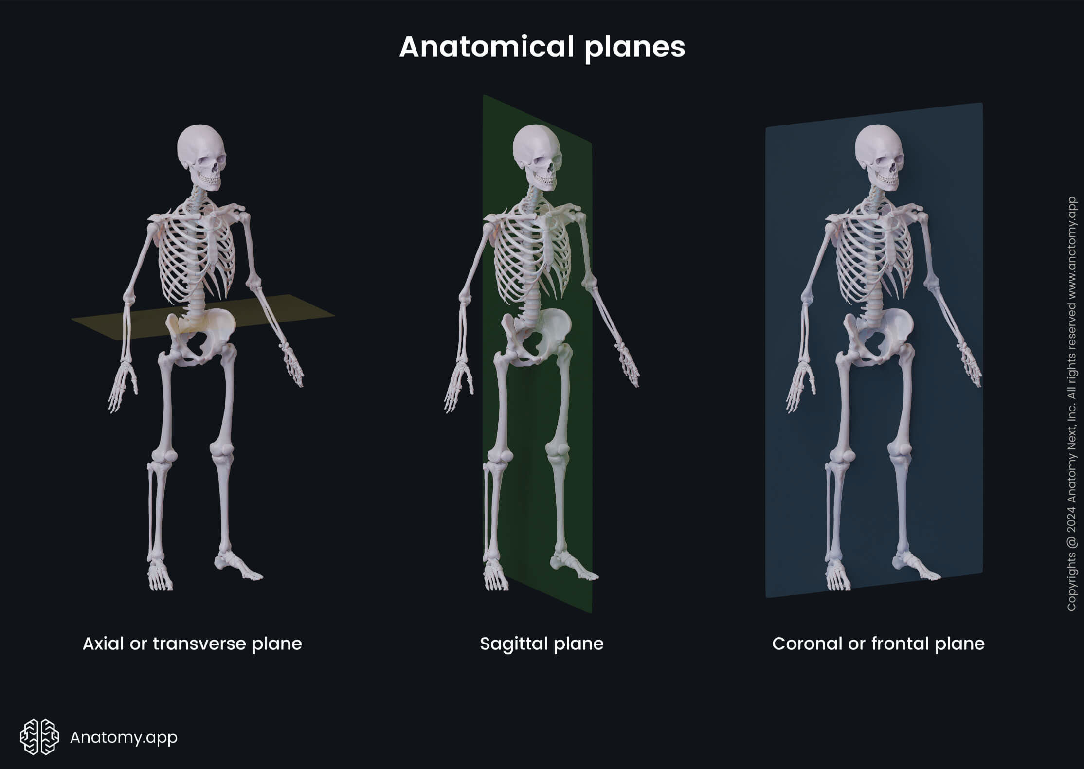
Anatomical planes are most commonly used in radiological studies, and they are applied to the human body that stands in the anatomical position. All planes can be classified as either longitudinal (vertical) planes or transverse (horizontal) planes.
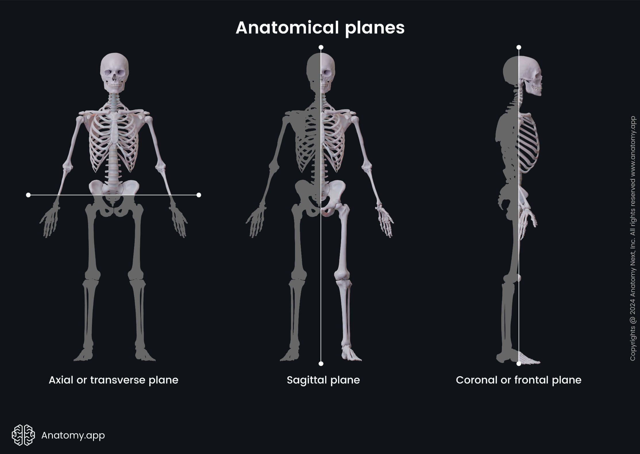
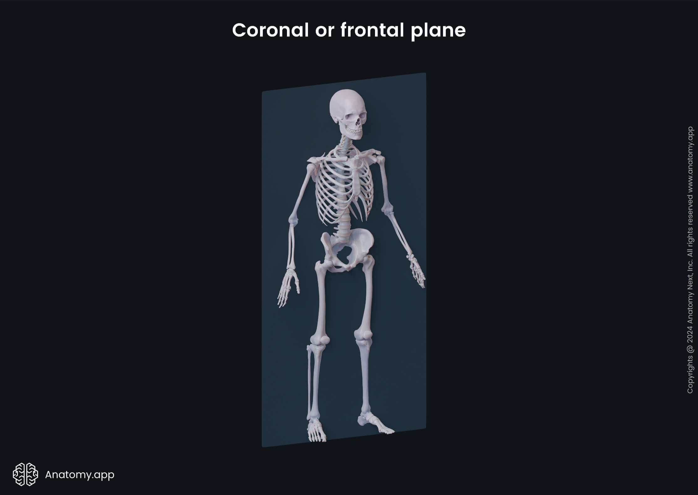
- Coronal or frontal plane - a vertical plane that goes longitudinally from one body side to another; it divides the body or any of its structures into the anterior and posterior portions;
- Sagittal plane - a vertical plane that runs longitudinally from the front to the back of the body; it goes perpendicular to the coronal plane; this plane divides the body or any of its structures into the left and right portions;
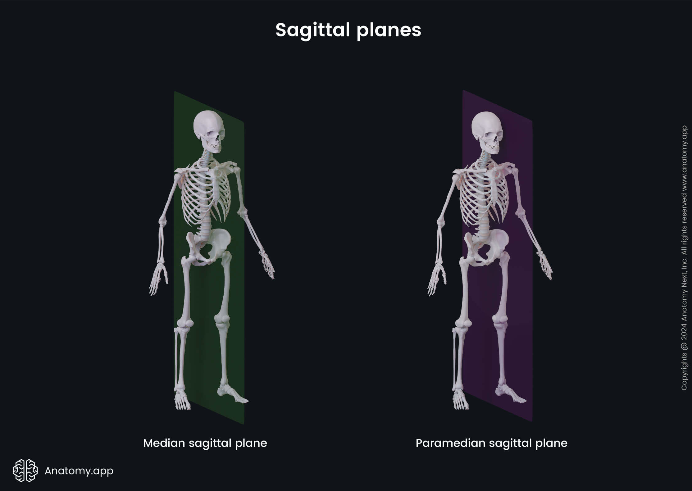
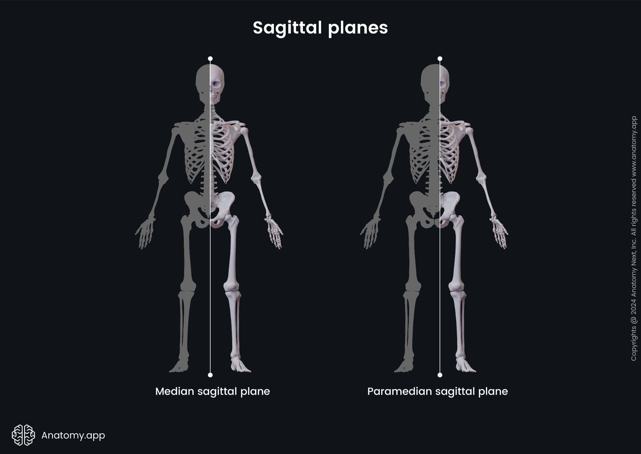
- Median, median sagittal or midsagittal plane - a sagittal plane that goes through the midline or the center of the body and evenly divides it into two equal halves;

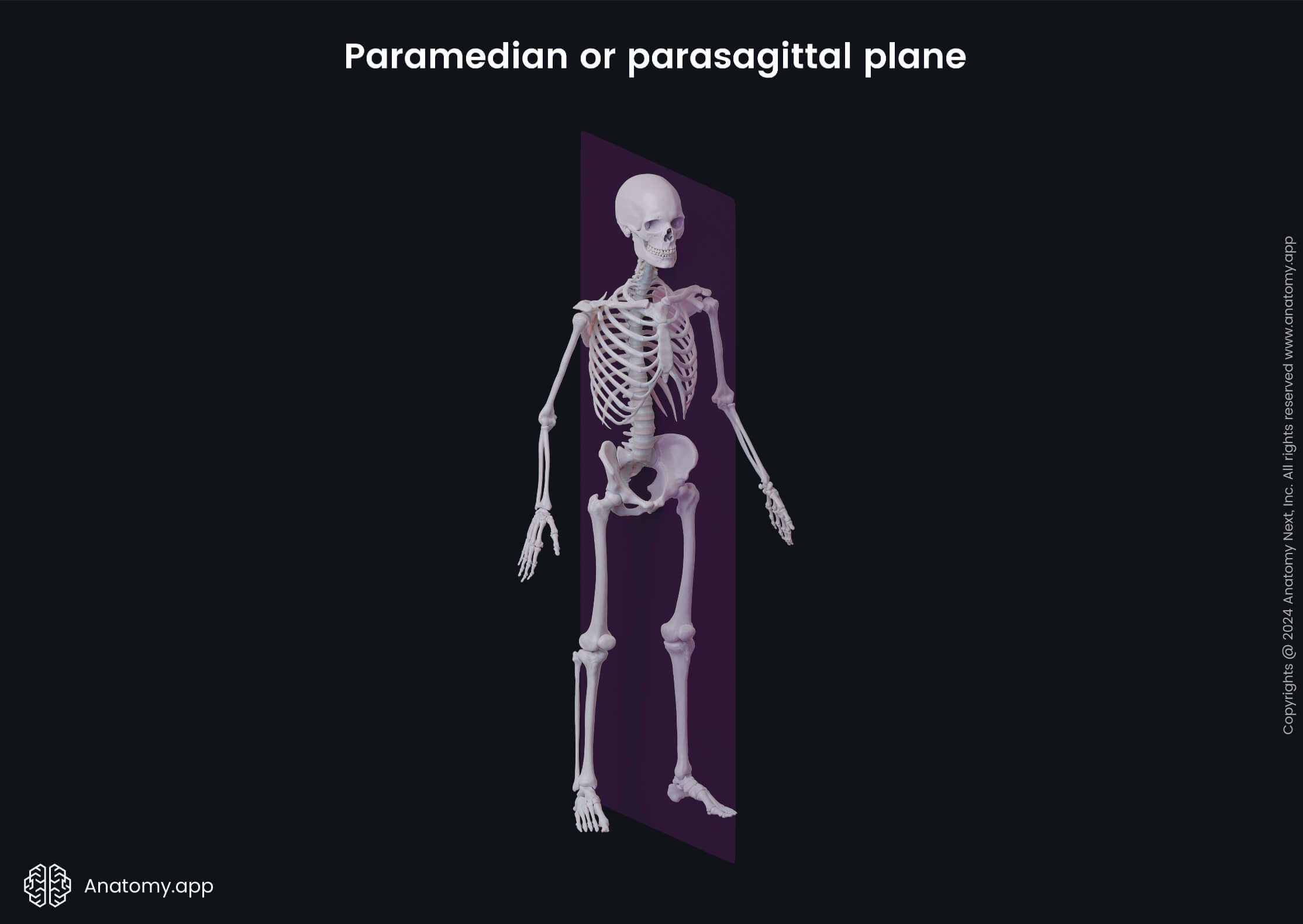
- Paramedian, parasagittal or paramedian sagittal plane - a vertical plane that goes parallel to the midline of the body and divides the body or any of its structures into unequal right and left portions;
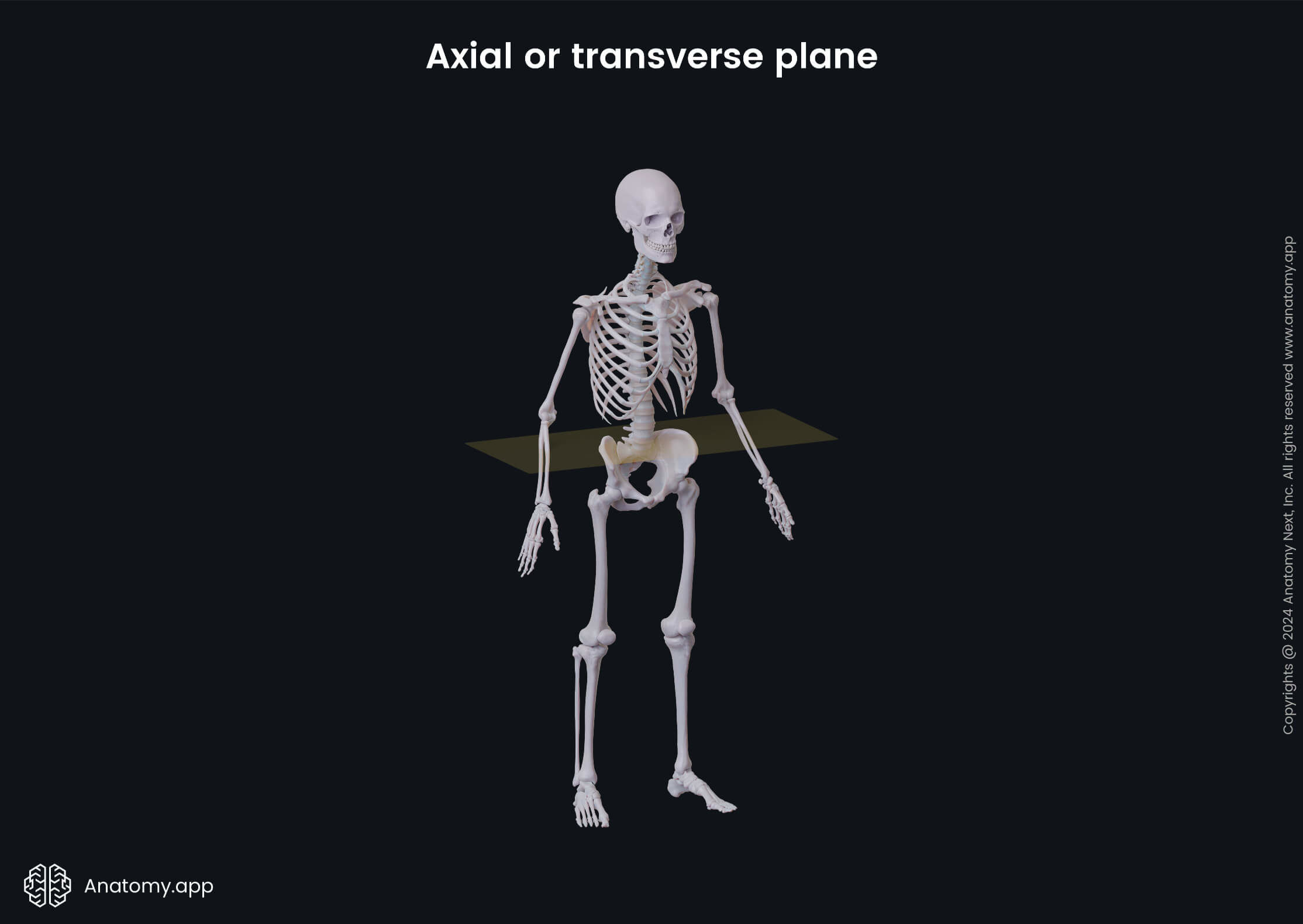
- Axial or transverse plane - a horizontal plane that divides the body or any of its structures into the upper and lower parts; it is perpendicular to all previously described planes, and it goes parallel to the ground; in radiology, the transverse planes produce images that are referred to as cross-sectional images;
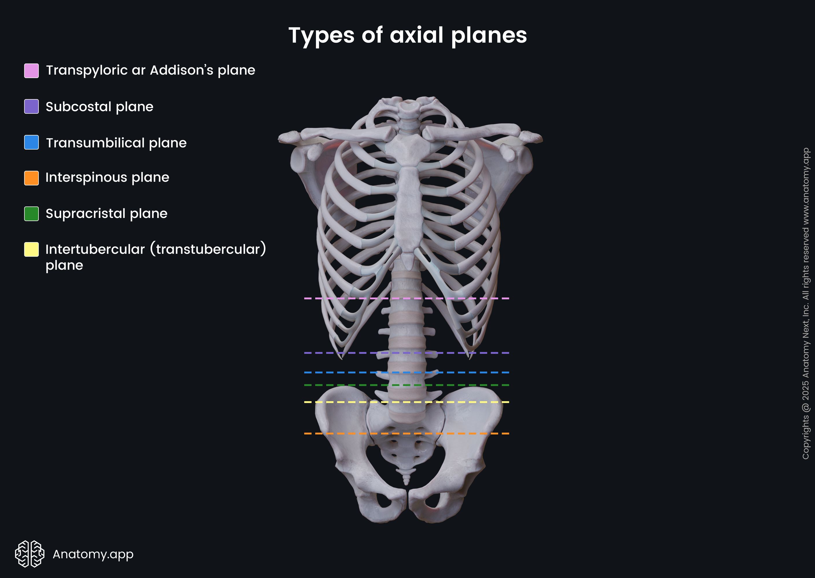
- Transpyloric or Addison's plane - a horizontal plane that goes through the body midway between the superior margin of the manubrium of the sternum and the pubic symphysis;
- Subcostal plane - a horizontal plane that goes through the body at the lowest level of the costal cartilage (at the level of the tenth rib);
- Transumbilical plane - a horizontal plane that goes through the body at the level of the navel or umbilicus;
- Supracristal plane - a horizontal plane that goes through the summits of the iliac crests and the fourth lumbar vertebra;
- Intertubercular (transtubercular) plane - a horizontal plane that goes through the body at the level of the iliac tubercles;
- Interspinous plane - a horizontal plane that goes through the anterior superior iliac spines.
Note: The term section is applied to a two-dimensional surface of a three-dimensional structure that has been cut. In radiology, they are called scans.
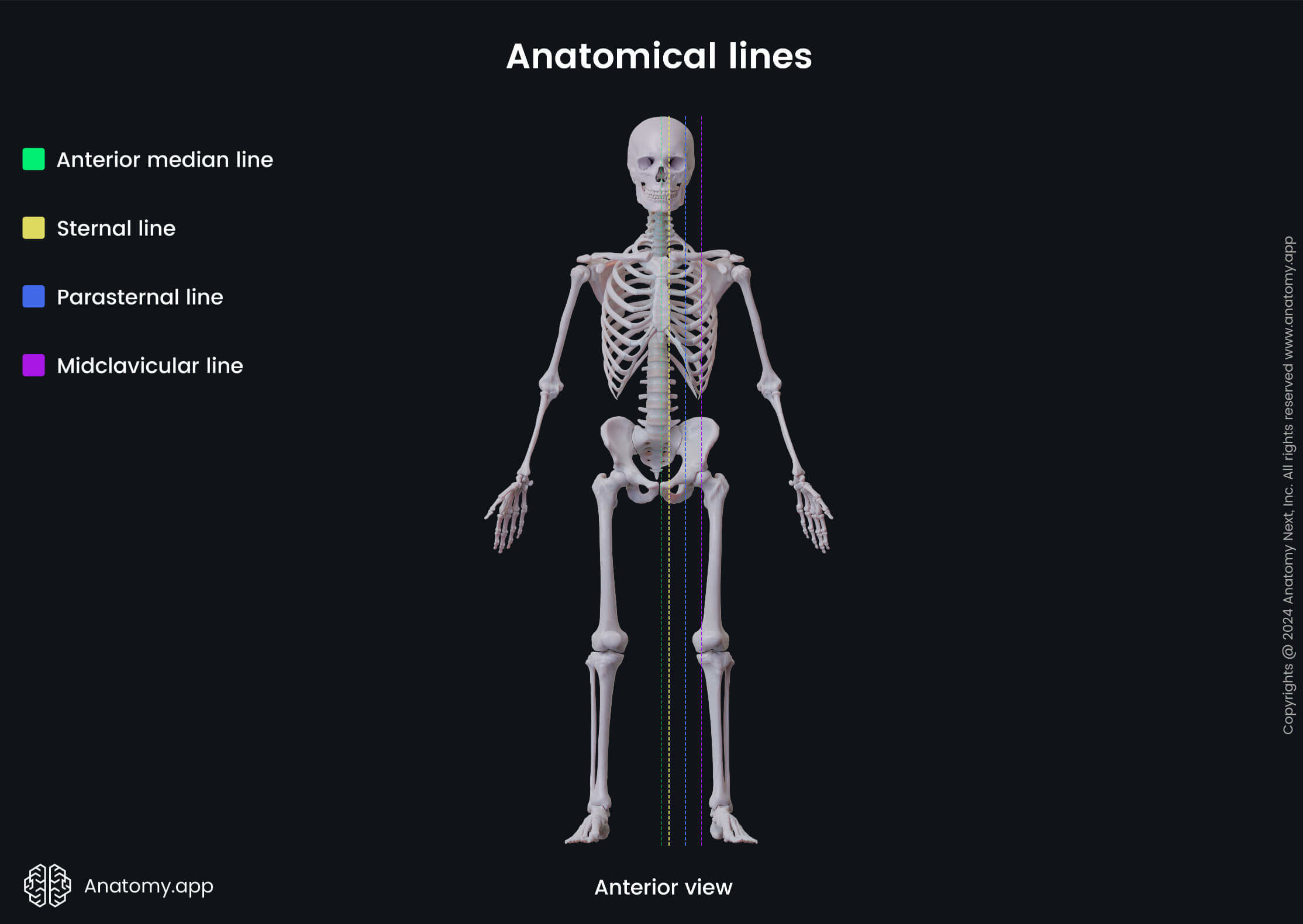
Anatomical lines
The term anatomical lines is applied to theoretical lines that are drawn through various structures on the anterior walls of the chest and abdomen, as well as on the back. They serve as reference lines and help in localizing structures. These lines are often used in surgery and during propedeutics rotation.
- Anterior median line - a vertical line that goes through the anterior aspect of the center of the body;
- Sternal line - a vertical line that goes along the lateral margin of the sternum;
- Parasternal line - a vertical line that is drawn midway between the sternal and midclavicular lines;
- Midclavicular line - a vertical line that is drawn from the middle of the clavicle;
- Mamillary or nipple line - a vertical line that goes through the nipple; in some individuals, it can match with the midclavicular line;
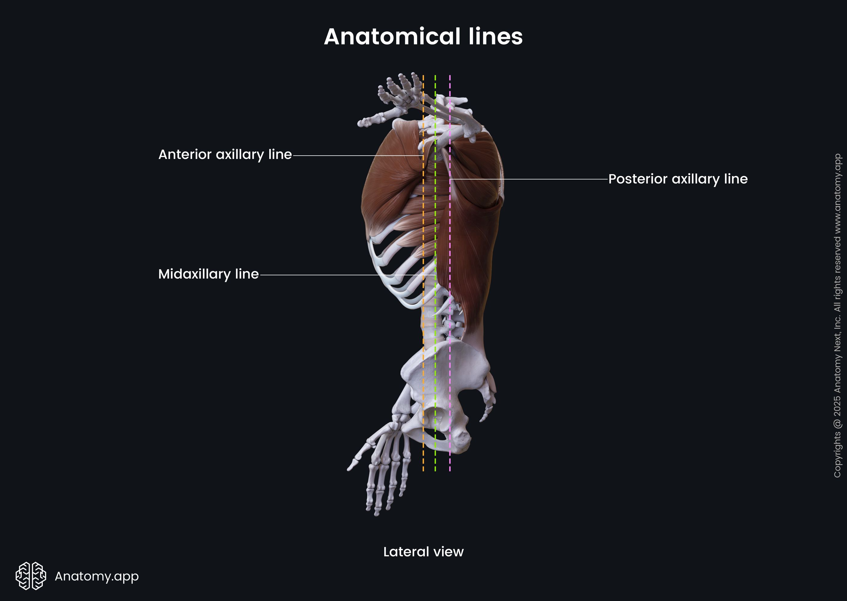
- Anterior axillary line - a vertical line that goes along the anterior axillary fold;
- Midaxillary line - a vertical line that is drawn from the middle of the armpit between the anterior and posterior axillary lines;
- Posterior axillary line - a vertical line that goes along the posterior axillary fold;
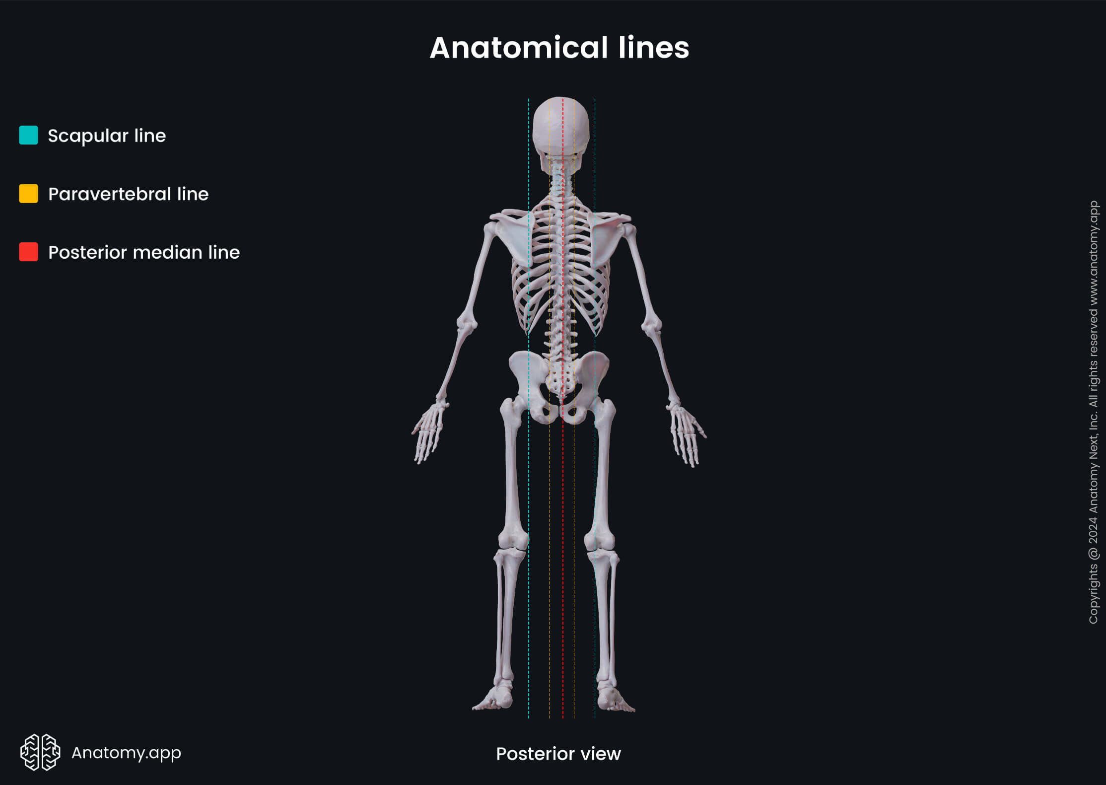
- Scapular line - a vertical line that goes through the inferior angle of the scapula;
- Paravertebral line - a vertical line that goes along the spine; it is drawn through the tips of the transverse processes of the vertebrae;
- Posterior median line - a vertical line that goes through the posterior aspect of the center of the body (it is drawn through the spinous processes of the vertebrae).
Directional terminology
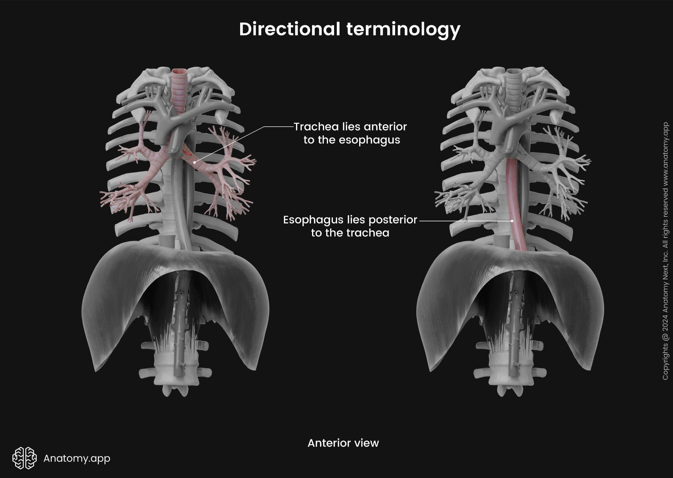
Directional terminology in anatomy is used to describe the position and location between various relative structures. Overall, these terms compare the position of adjacent structures and explain how far or close they are to one another. Here are the main directional terms:
- Anterior or ventral - describes the front or direction toward the front of the body; i.e., the trachea lies anterior to the esophagus;
- Posterior or dorsal - refers to the back or direction toward the back of the body; i.e., the heart is found posterior to the sternum;
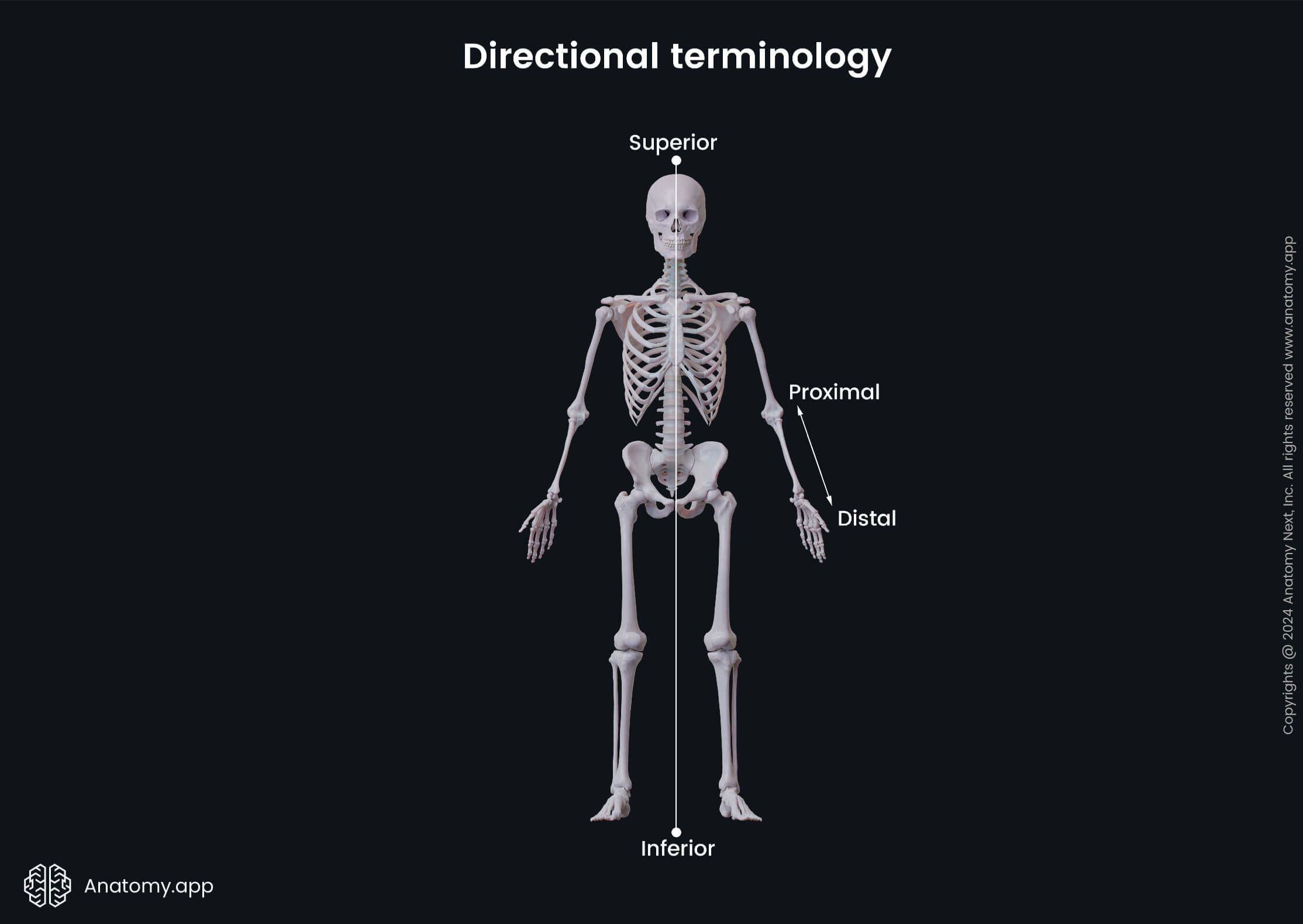
- Superior - defines the position that is above the other structure or toward the top of the head; i.e., the sixth cervical vertebra is superior to the seventh cervical vertebra;
- Inferior - refers to the position that is below the other structure or toward the feet; i.e., the stomach lies inferior to the diaphragm;

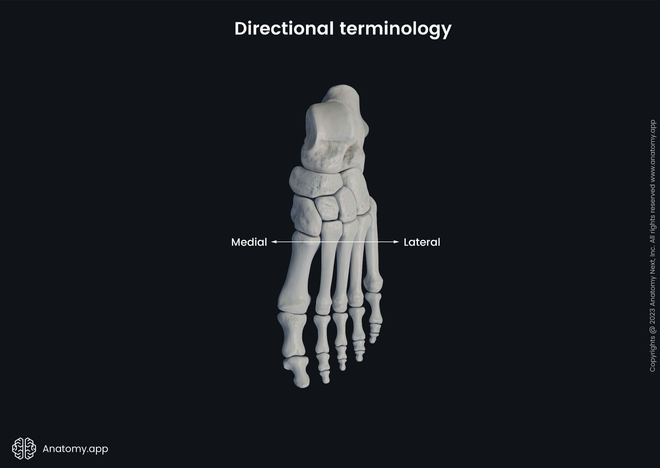
- Lateral - defines the structure that is found farther from the midline of the body; describes the direction away from the midline of the body; i.e., the little toe is located on the lateral side of the foot;
- Medial - refers to the structure that is found closer to the midline of the body; describes the direction toward the midline of the body; i.e., the hallux is located on the medial side of the foot;
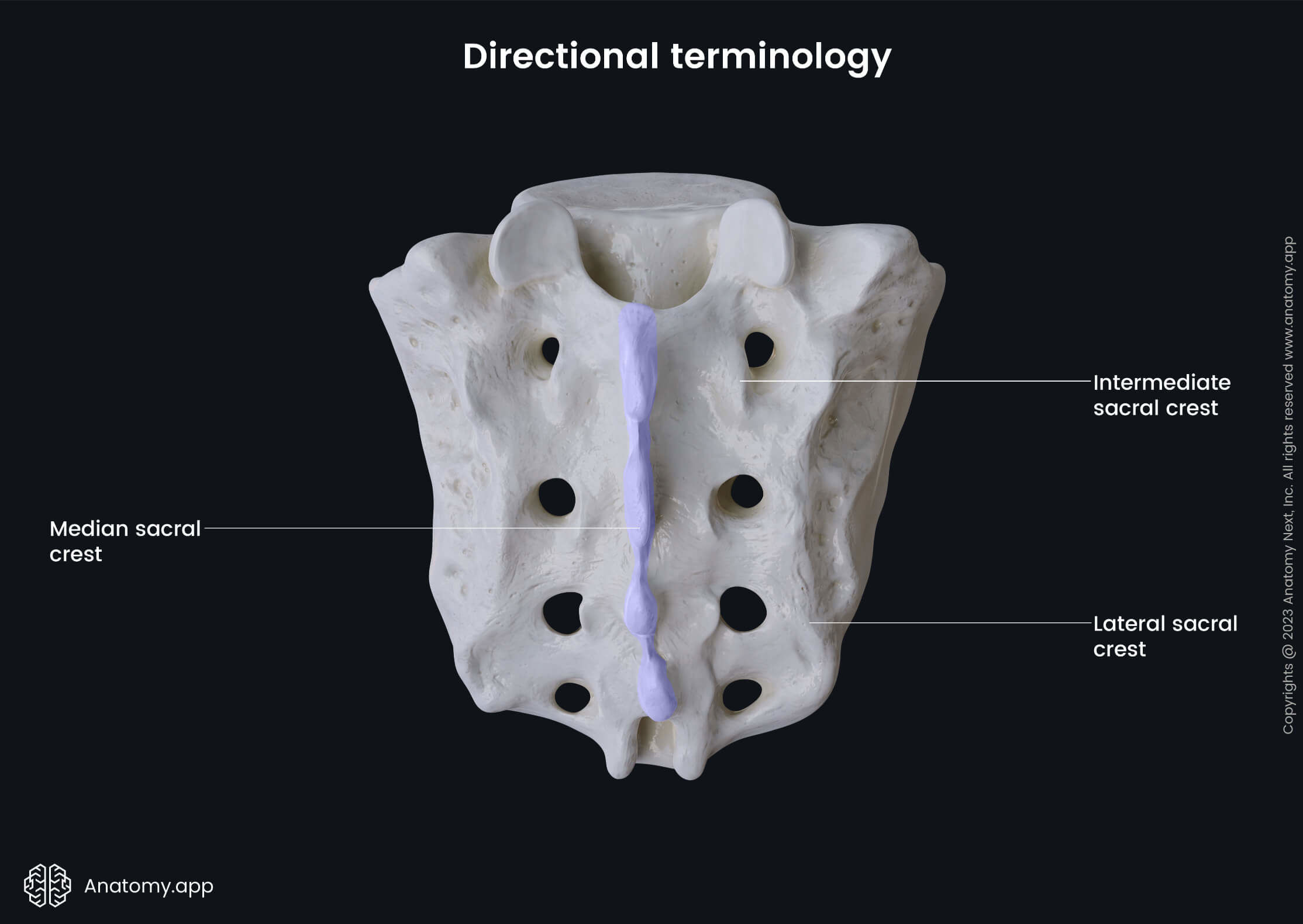
- Median - refers to the midline of the body; it matches the midline of the body; i.e., the sternum is a median structure;
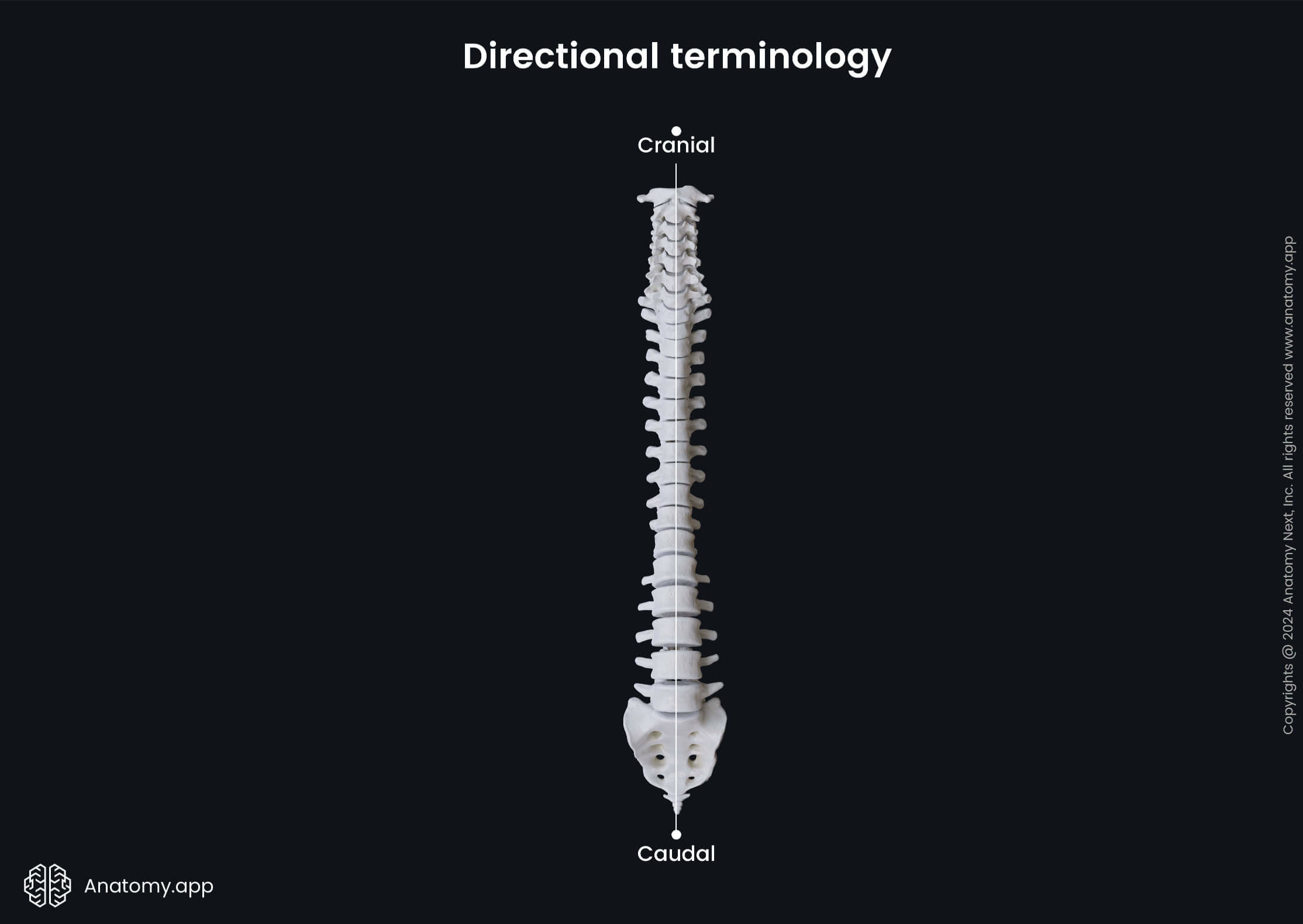
- Cranial - describes the structure that is directed toward the head; i.e., the cranial end of the spinal cord connects with the medulla oblongata;
- Caudal - refers to the structure that is positioned away from the head and toward the tail (in humans, the tail refers to the final part of the spine (tailbone or coccyx)); i.e., the caudal end of the spinal cord features the cauda equina;
- Proximal - defines the structure that is found toward or nearest the trunk of the body or the point of origin of the body part; i.e., the proximal end of the femur articulates with the hip bone;
- Distal - describes the structure that is found away or farthest from the trunk or the point of origin of the body part; i.e., the distal end of the femur articulates with the tibia;
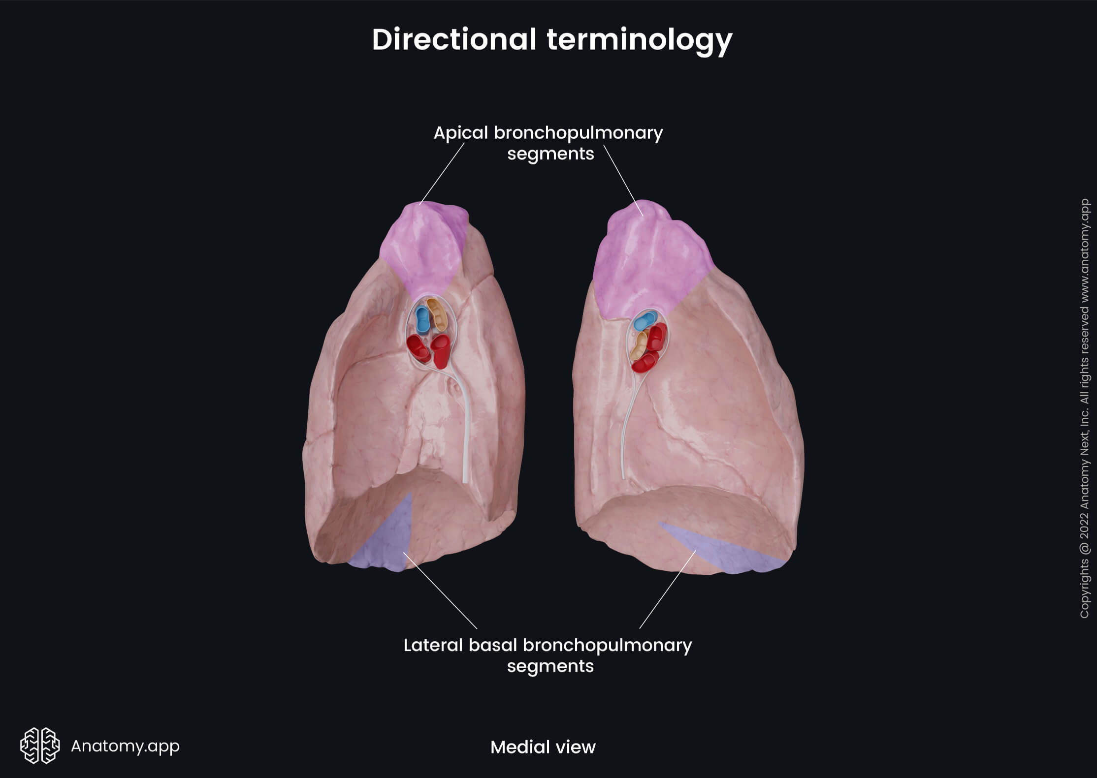
- Apical - a term that describes the direction/location toward the apex of a particular structure; i.e., apical bronchopulmonary segment of the right lung;
- Basal - a term that describes the direction/location toward the base of a particular structure; i.e., lateral basal bronchopulmonary segment of the right lung;
- Luminal - inner space within a tubular structure or cavity; i.e., the luminal surface of the small intestine is lined with villi;
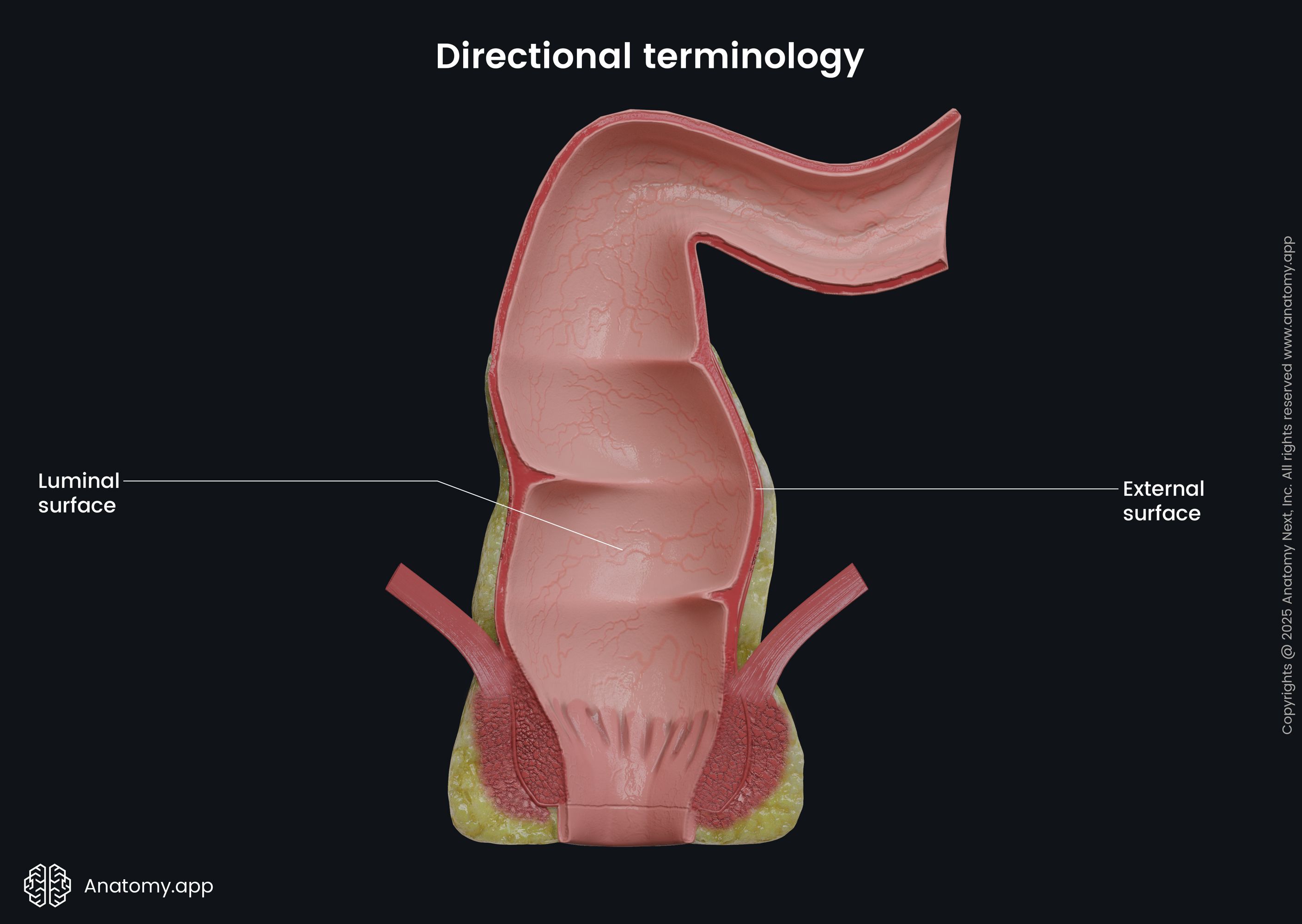
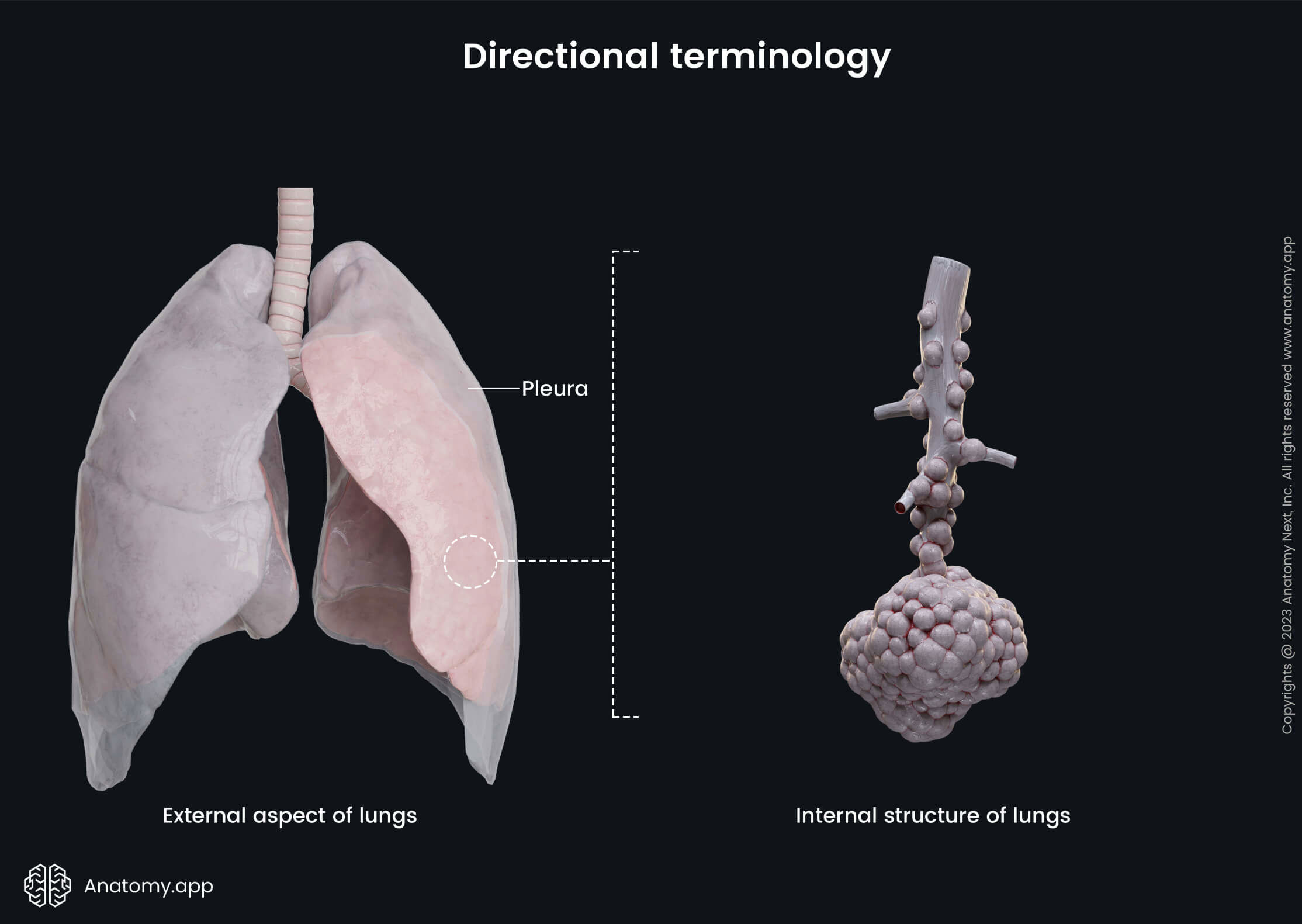
- External - refers to the structure that is closest to the surface of the body; also describes the outer aspect of a structure; i.e., the external surfaces of the lungs are covered by the pleura;
- Internal - defines the structure that is found away from the surface of the body; also describes the inner aspect of a structure; i.e., internally, the lungs are composed of alveoli;
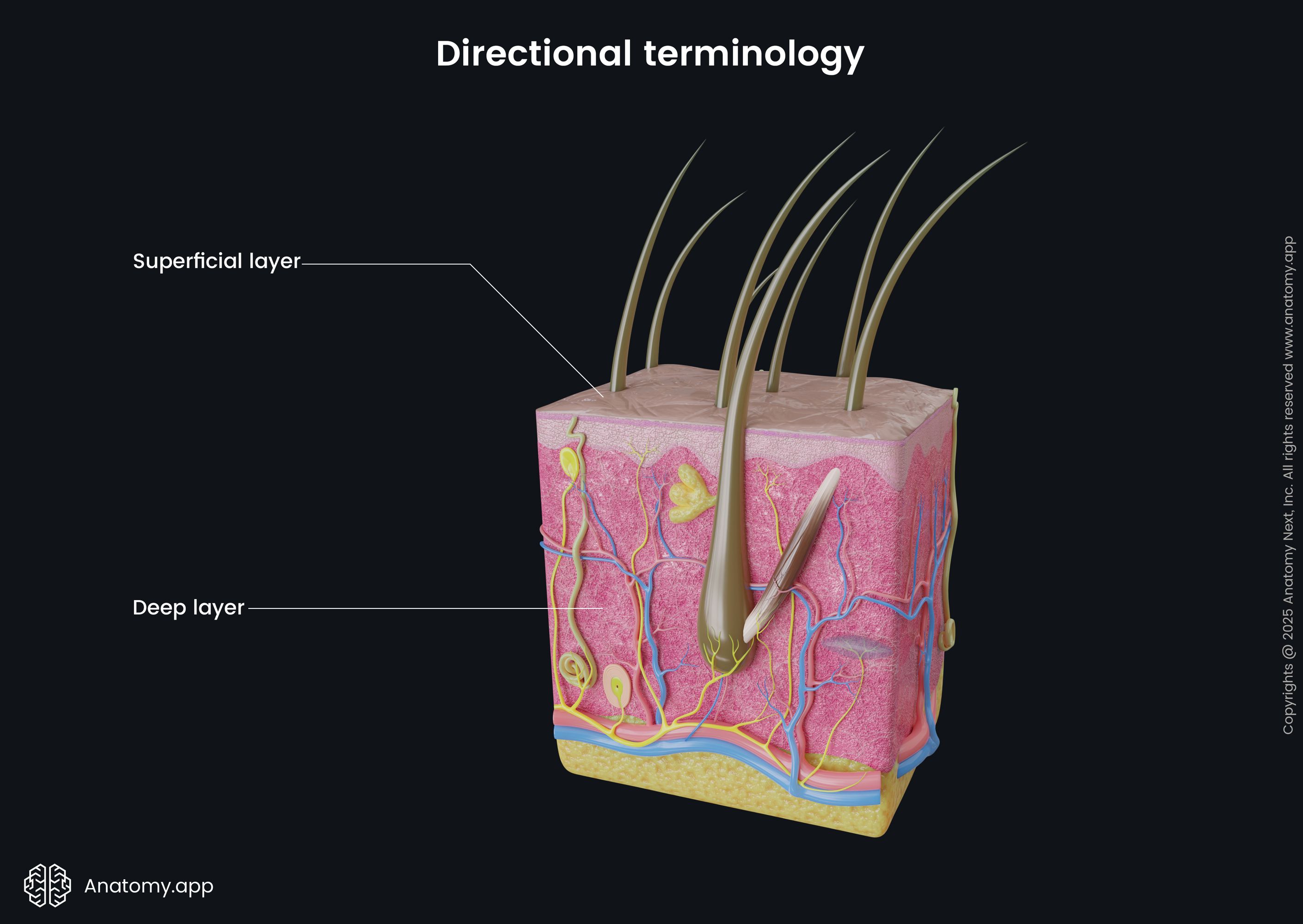
- Superficial - describes the position that is nearer and closer to the surface of the body; i.e., the skin is superficial to the bones;
- Deep - defines the position that is farther from the surface of the body; i.e., the ovaries lie deep within the pelvis;
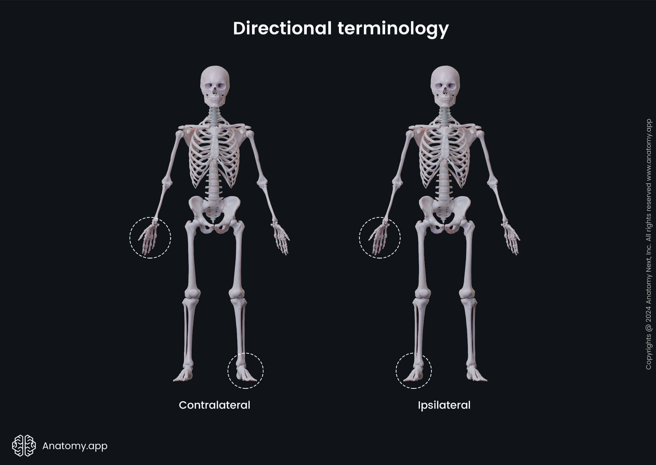
- Contralateral - this term is applied to the structure that is located on the opposite side of the midline of the body; i.e., the contralateral leg of the right side is the left leg;
- Ipsilateral - this term describes the structure that is positioned on the same side of the midline of the body; i.e., the ipsilateral arm of the right side is the right arm;
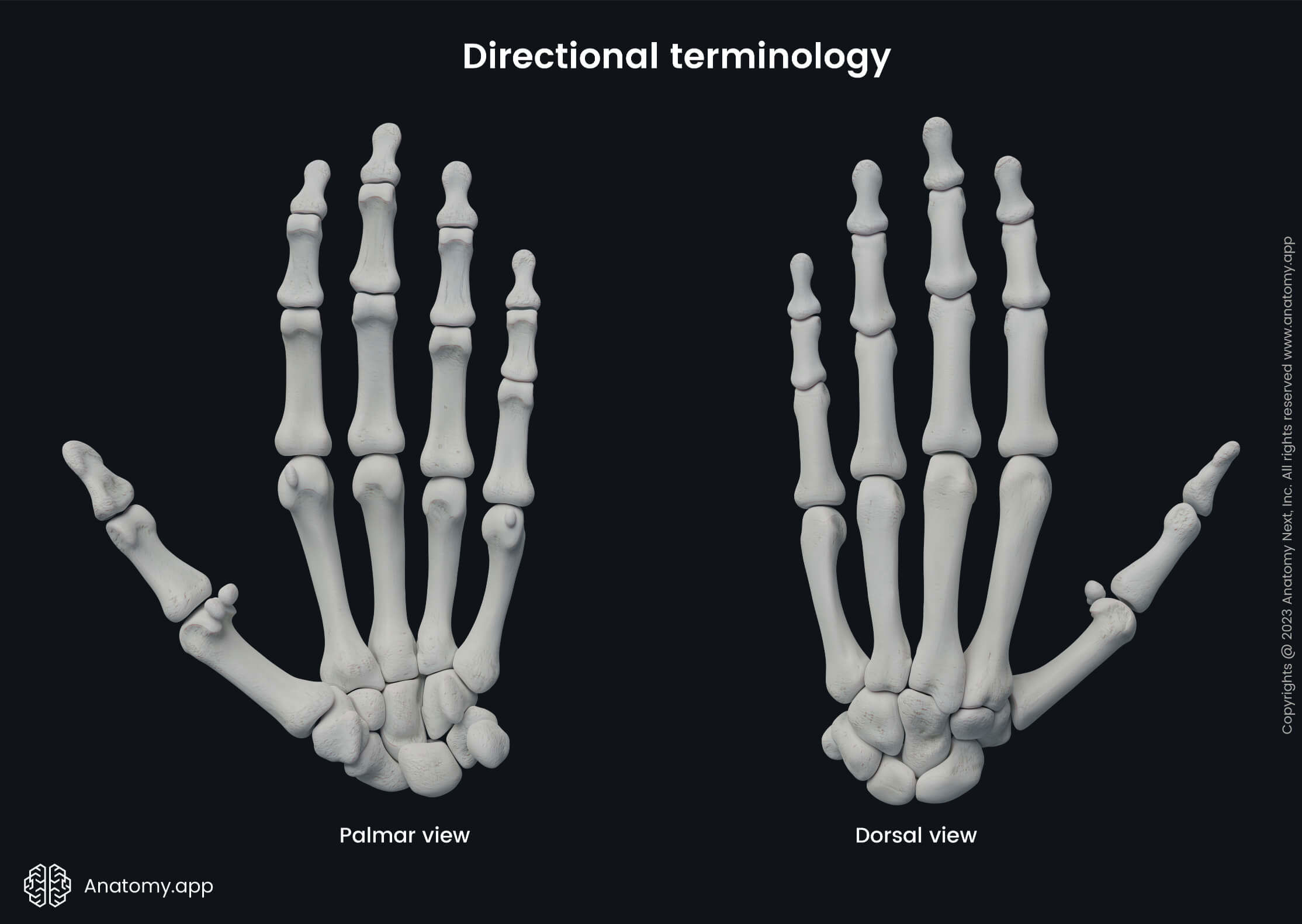
- Palmar (of hand) - grasping or anterior side of the hand or the palm; i.e., the fingerprints are found on the skin on the palmar side of the hand;
- Dorsal (of hand) - describes the posterior side of the hand; i.e., the fingernails are found on the dorsal sides of the distal phalanges;
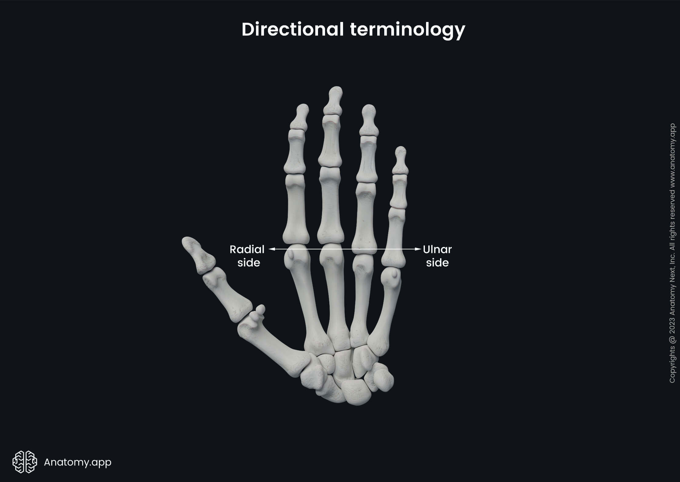
- Ulnar side (of hand) - refers to the little finger or medial side of the hand;
- Radial side (of hand) - describes the thumb or lateral side of the hand;
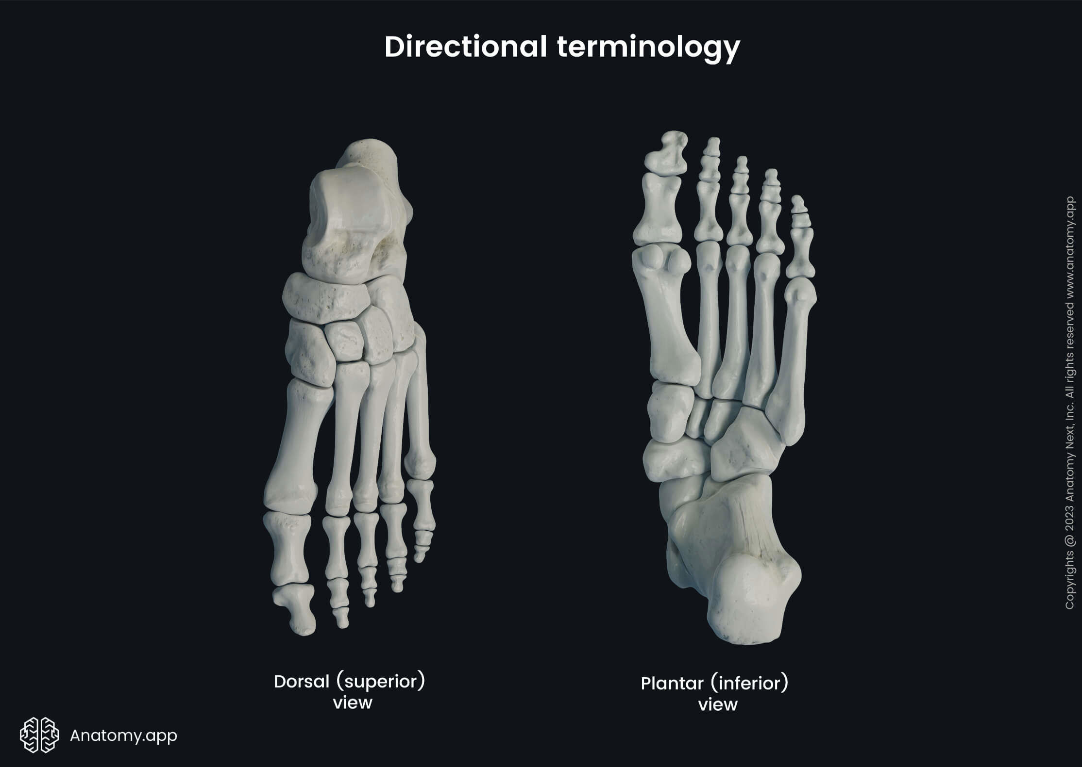
- Plantar (of foot) - applies to the inferior aspect of the foot or the sole; i.e., when a person stands upright, the ground touches the plantar sides of the feet;
- Dorsal (of foot) - used when describing the superior aspect of the foot; i.e., the fingernails are found on the dorsal sides of the distal phalanges of the feet.
Abdominal quadrants and regions
When a patient complains about abdominal pain, it is essential to understand in which region the pain is localized to better understand possibly involved structures. So, for the purpose of clear communication, the abdomen is typically divided into four quadrants or nine regions. Each quadrant and region contains different structures and organs.
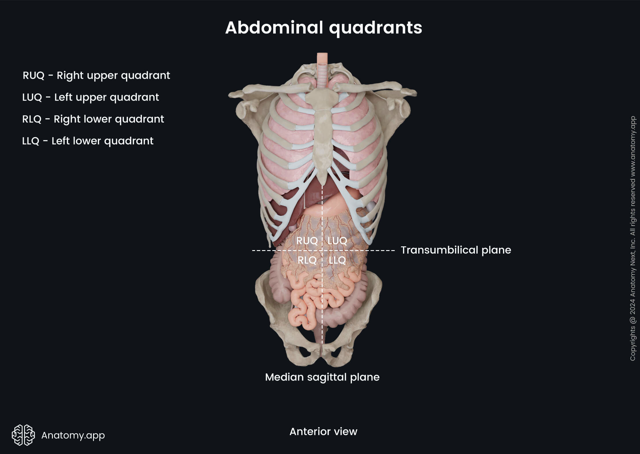
Abdominal quadrants
The first approach uses two planes - one is horizontal (transumbilical plane) and one is vertical (median sagittal plane). Both intersect at the umbilicus. This results in the formation of the following four abdominal quadrants:
- Right upper quadrant (RUQ)
- Left upper quadrant (LUQ)
- Right lower quadrant (RLQ)
- Left lower quadrant (LLQ)
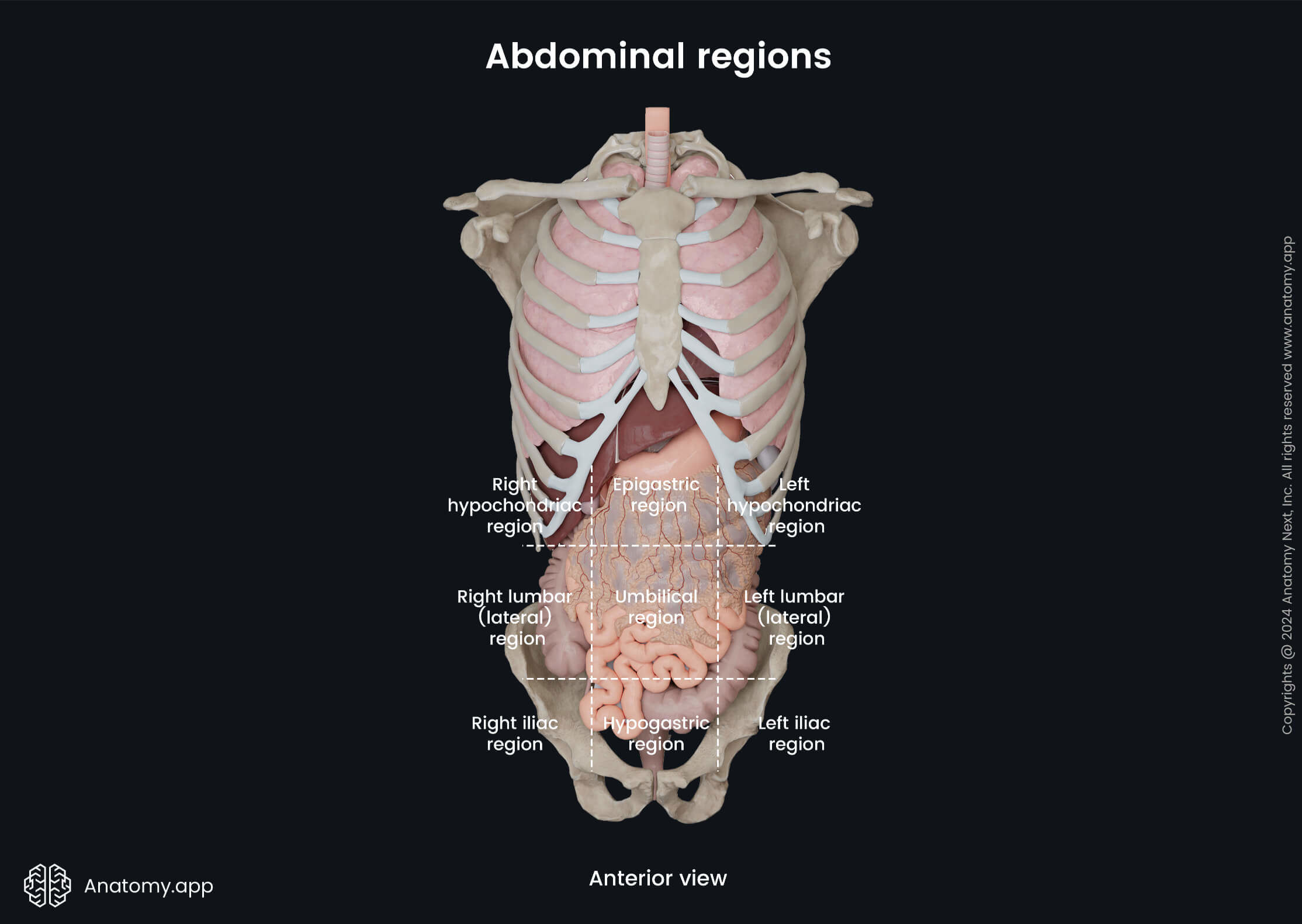
Abdominal regions
The second approach is a bit more complex as it uses four planes - two horizontal (subcostal and intertubercular planes) and two vertical planes (parasagittal planes). Both vertical planes are drawn from the midpoints of each clavicle. The upper horizontal plane connects both inferior aspects of the thorax, while the lower horizontal plane connects the iliac crests and transverses both iliac tubercles. As a result, the abdomen is subdivided into nine abdominal regions:
- Umbilical region - in the middle;
- Epigastric region - above the umbilical region;
- Hypogastric region - below the umbilical region;
- Right hypochondriac region - upper region of the right side;
- Right lumbar (lateral) region - below the right hypochondriac region;
- Right iliac region - below the right lumbar region;
- Left hypochondriac region - upper region of the left side;
- Left lumbar (lateral) region - below the left hypochondriac region;
- Left iliac region - below the left lumbar region.
Body cavities
Internally, the human body is organized into several spaces or cavities by various membranes, structures, and sheaths. Each cavity houses, lubricates, and protects various internal organs and structures. Moreover, they allow changes in the size and shape of the organs and structures found within them. For instance, the intestines can expand without disrupting the activity of nearby organs.
Overall, each person has two main spaces called the ventral (anterior) and dorsal (posterior) cavities. Each cavity can be further subdivided into even smaller compartments.
Ventral cavity
The ventral cavity is larger, and the diaphragm subdivides it into two portions - the thoracic and the abdominopelvic cavities.
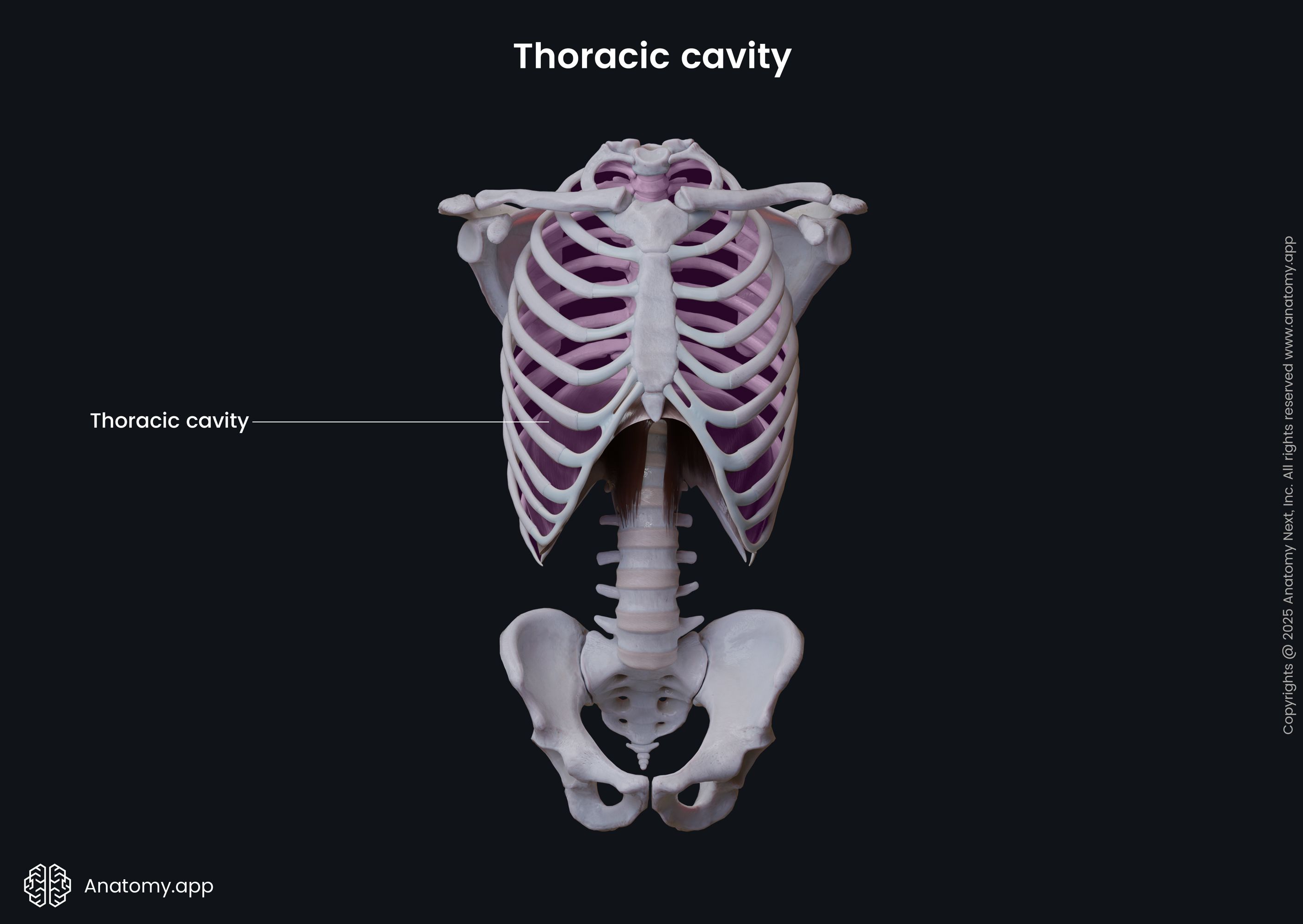
Thoracic cavity
The thoracic cavity is the upper subdivision of the ventral cavity. It is enclosed by the ribs and found within the thorax. Overall, the thoracic cavity houses the lungs, trachea, esophagus, heart, great vessels of the heart, pericardium, pleura, and a part of the bronchial tree. Additionally, between the lungs and the pleural cavity is the mediastinum.
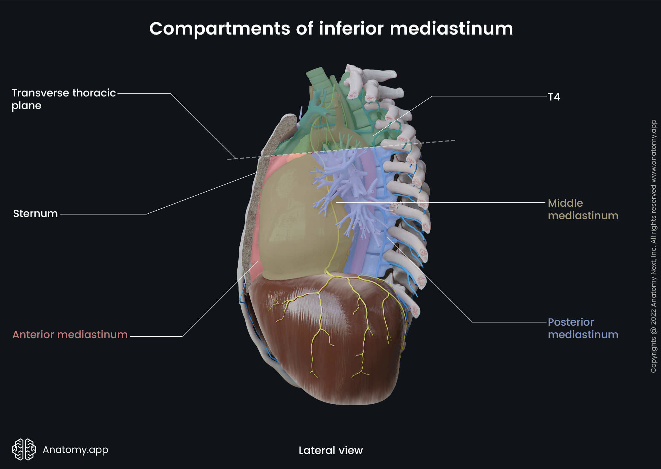
Mediastinum
The mediastinum is the central compartment of the thoracic cavity, located between the pleural sacs of the lungs. It extends from the superior thoracic aperture superiorly to the superior surface of the diaphragm inferiorly and from the sternum anteriorly to the bodies of the thoracic vertebrae posteriorly.
For organizational purposes, the mediastinum is typically further subdivided into two smaller sections by a horizontal plane known as the transverse thoracic plane (it goes through the sternal angle and the intervertebral disc between the fourth and fifth thoracic vertebrae):
- Superior mediastinum - found between the superior thoracic aperture and the transverse thoracic plane;
- Inferior mediastinum - extends between the transverse thoracic plane and the superior surface of the diaphragm.
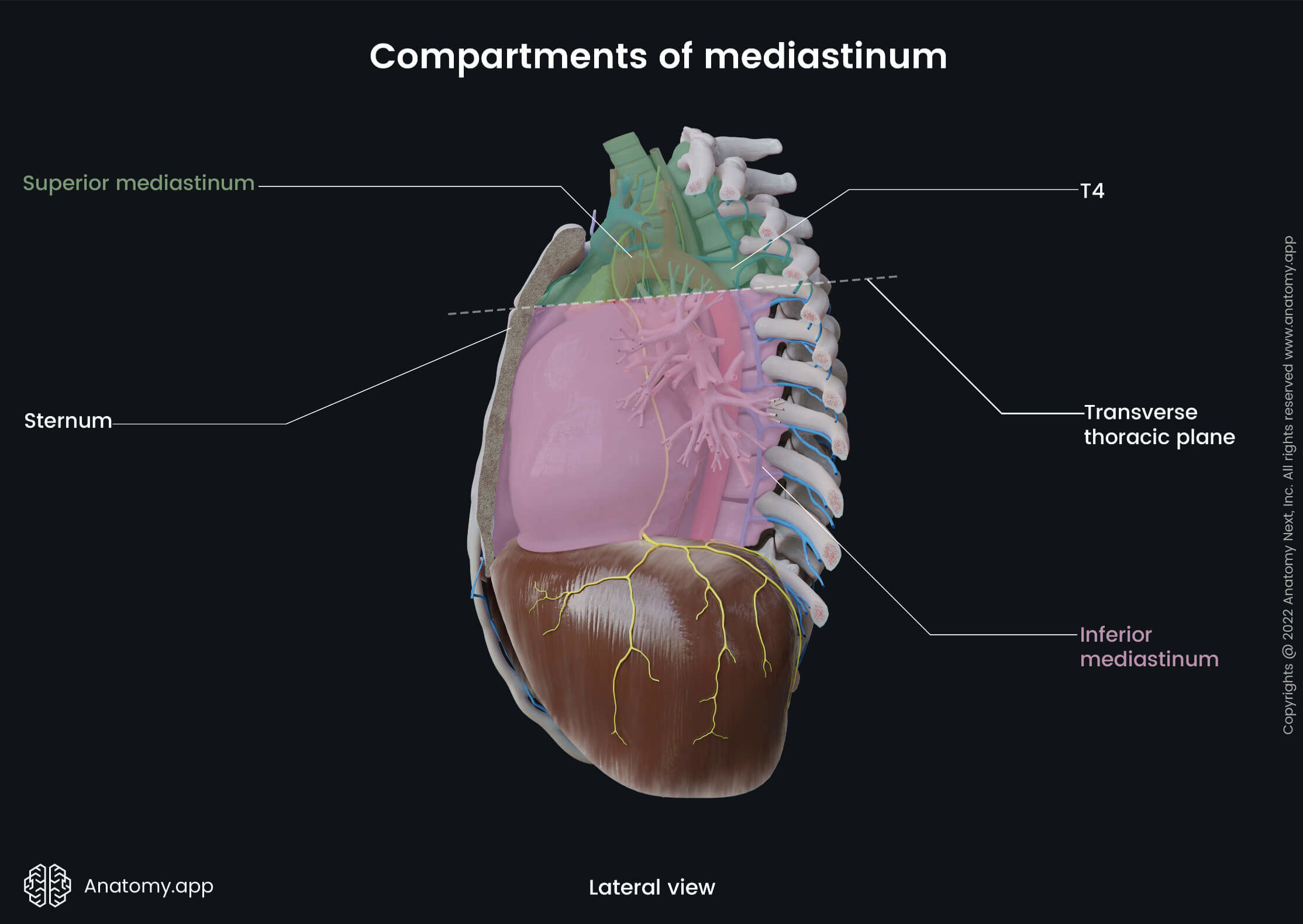
The inferior mediastinum is larger than the superior, and the pericardium further subdivides it into three even smaller compartments:
- Anterior mediastinum - lies between the sternum and pericardium;
- Middle mediastinum - contains the heart covered by the pericardium;
- Posterior mediastinum - lies between the pericardium and the thoracic vertebrae.
Every compartment of the mediastinum contains vital organs, as well as vascular and neural structures that are closely related to one another.
Abdominopelvic cavity
The abdominopelvic cavity is found below the diaphragm. It is not only the largest compartment of the ventral cavity but is also the largest cavity in the human body overall. For organizational purposes, the abdominopelvic cavity is usually subdivided into the abdominal and pelvic portions.
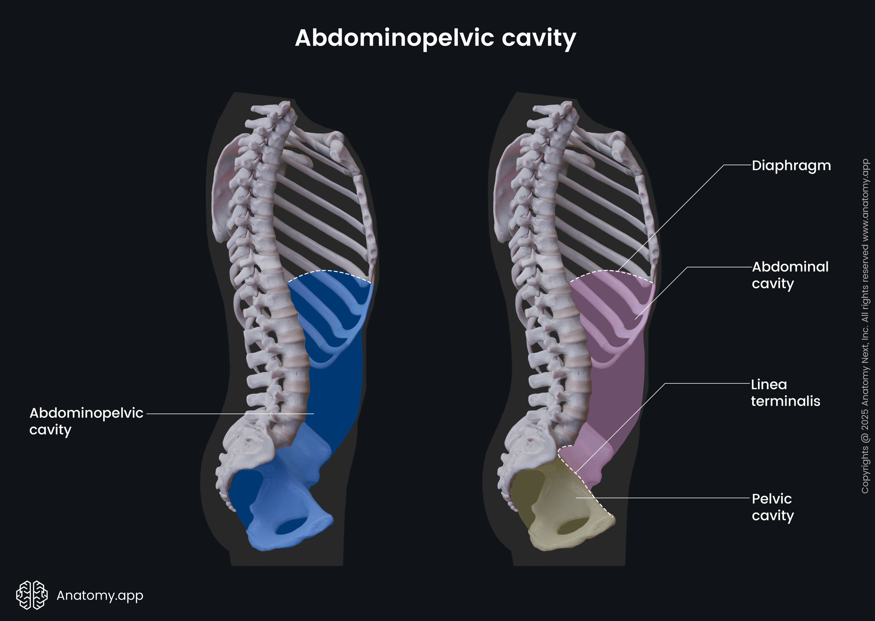
- The abdominal cavity is positioned between the pelvis and the diaphragm. It mainly contains the organs of the digestive system, as well as the kidneys, adrenal glands, and spleen.
- The pelvic cavity is found within the pelvis. In contrast to the abdominal cavity, the pelvic cavity houses the rest of the urinary system organs, organs of the reproductive system, and the rectum.
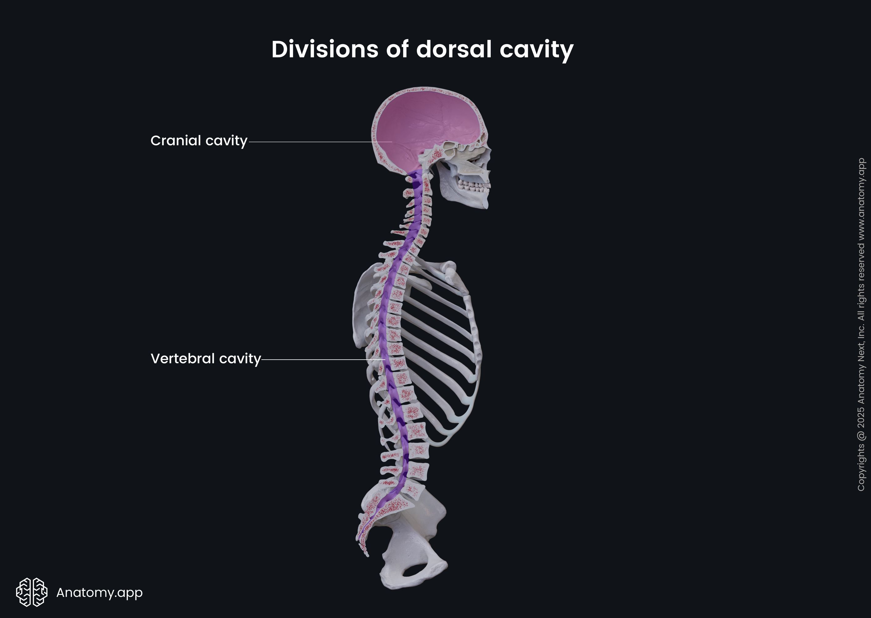
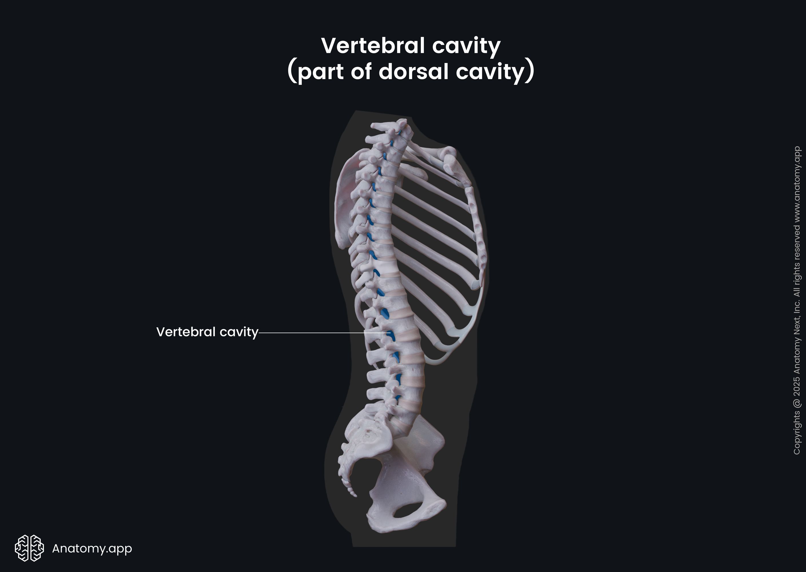
Dorsal cavity
The dorsal cavity is smaller than the ventral cavity. It contains organs that lie more posteriorly in the human body. Like the ventral cavity, it also can be subdivided into two smaller compartments - the cranial and the vertebral cavities.
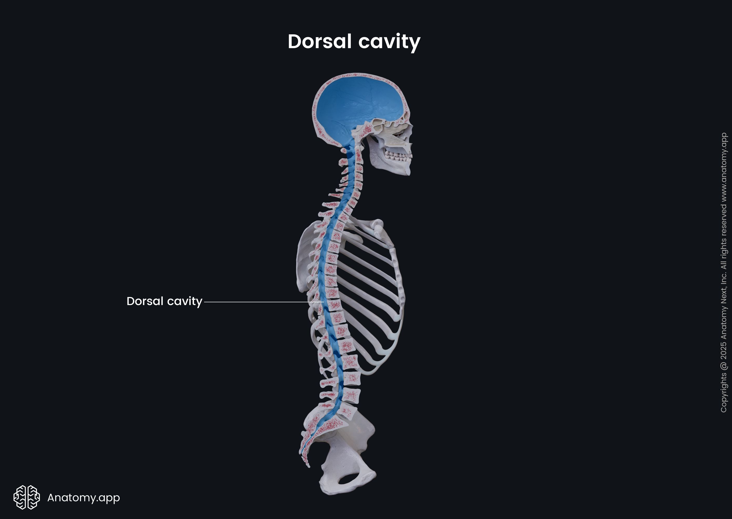
- The cranial cavity is the upper part of the dorsal cavity, containing the brain and meninges.
- In contrast, the vertebral cavity is the lower portion, and it houses the spinal cord and spinal meninges. The vertebral cavity is also referred to as the vertebral canal, spinal cavity, or spinal canal.
Each of these cavities and their structures are protected by the bones of the skull and vertebral column. Like the brain and spinal cord, the cranial and spinal cavities are continuous with each other. Additionally, the dorsal cavity contains cerebrospinal fluid, which nourishes the spinal cord and brain.
Serous membranes
Some parts of the ventral cavity, its walls and contents are lined by thin membranes referred to as serous membranes. Most of them have two layers - parietal and visceral. There is usually also a potential space between both layers that contains serous fluid produced by these membranes. It lubricates the layers and reduces friction between the walls of the cavities and the internal organs, allowing them to move. Overall, the serous membranes give additional protection to the organs found within the body cavities.
The three serous membranes of the human body are the pleura, pericardium, and peritoneum. They are all found within the ventral cavity.
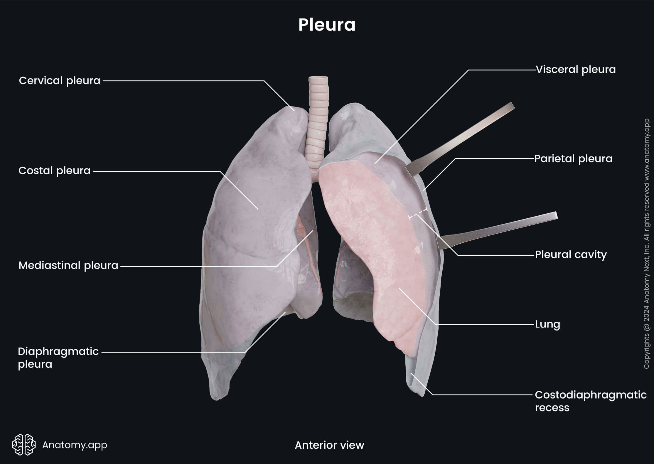
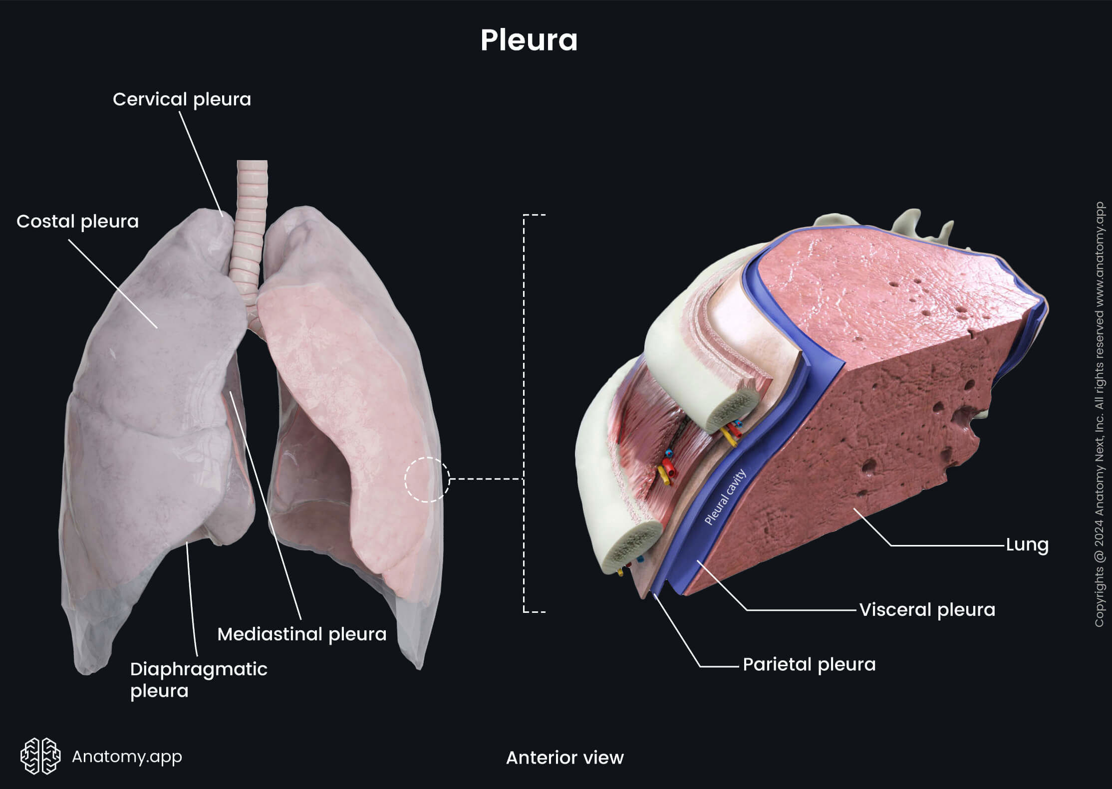
Pleura
The term pleura describes a thin serous membrane that covers each lung and lines the thoracic cavity. The parietal layer, or parietal pleura, covers the internal surface of the thoracic wall, while the visceral layer, or visceral pleura, surrounds the external surfaces of the lungs. Between both pleural layers is a potential space known as the pleural cavity.
Pericardium
One more serous membrane is found within the thoracic cavity - the pericardium. It is a fibroserous membrane that surrounds the heart from all sides and encloses the roots of the great vessels. It also defines the middle mediastinum, which is the middle compartment of the inferior mediastinum.
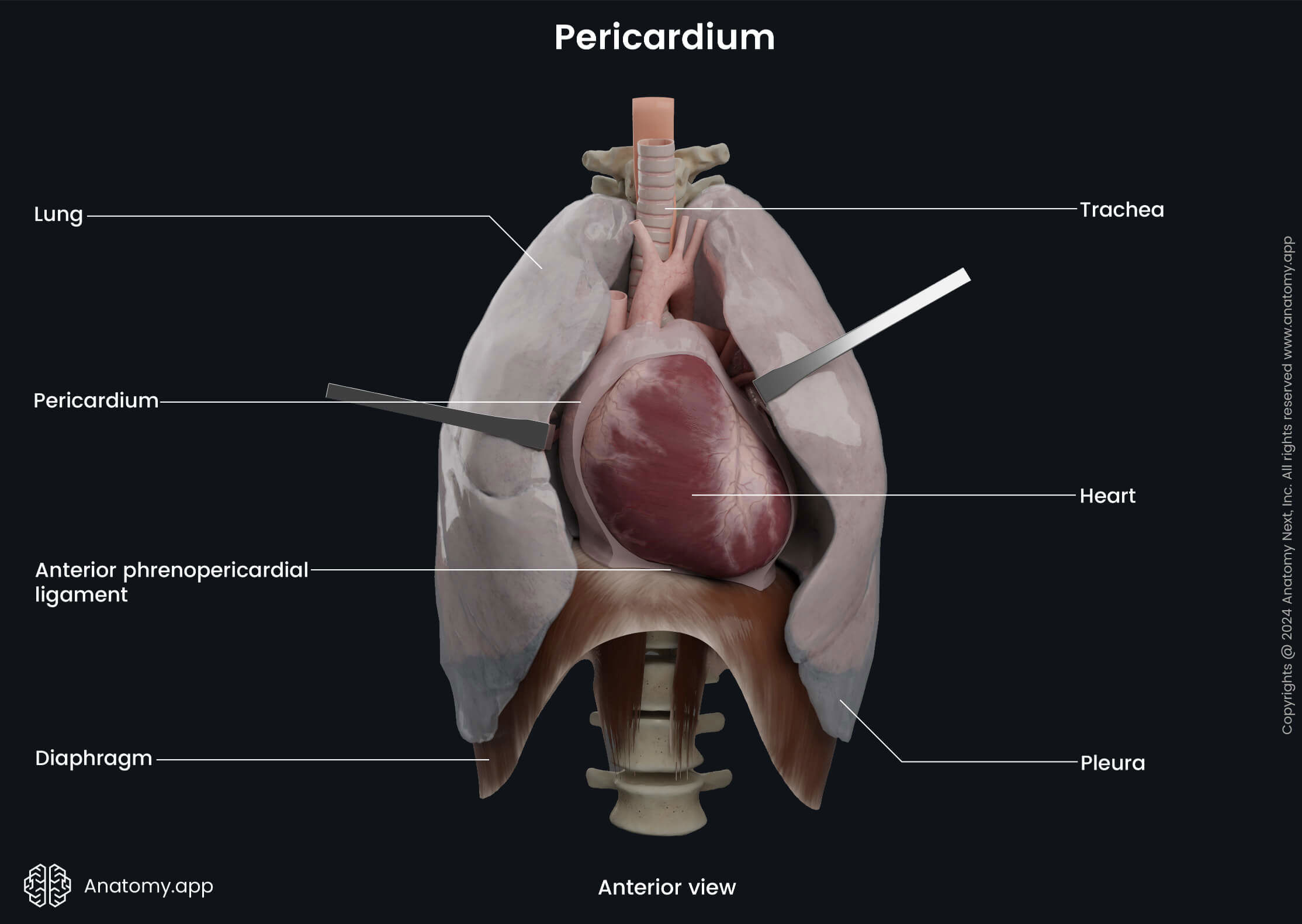
The pericardium consists of two layers - fibrous or the outermost layer and the serous or the innermost layer. The serous layer is a double layer, and it is composed of two sublayers (parietal and visceral).
Between both sublayers of the serous pericardium is a potential space known as the pericardial cavity. It contains a small amount (0.3 - 1.7 oz or 10 - 50 ml) of a serous fluid known as the pericardial fluid. Its main function is to lubricate both sublayers and reduce friction between them when the heart contracts.
Peritoneum
The peritoneum is a serous membrane that lines the abdominopelvic cavity and suspends and supports its organs. It consists of the parietal layer or parietal peritoneum, which lines the abdominopelvic cavity and the visceral layer or visceral peritoneum that surrounds the internal organs. The space between both layers is known as the peritoneal cavity. It is filled with 1.7 - 2.4 oz (50 - 70 ml) of serous fluid.
Anatomical terms of movements
The human body is composed of both - passive and active structures. A joint or articulation is a junction of two or more bones connecting and articulating with each other. Joints are supported and stabilized by various connective tissue structures. Muscles that cross the joints not only stabilize them but also activate their motions. In order to move, the muscles contract and, therefore, produce movements at joints in various directions and degrees of motion.
There are several anatomical terms that are used to describe a particular movement type around a certain axis in one of the anatomical planes. They are referred to as anatomical terms of movement. Most of these movements have opposite or antagonistic movements. Additionally, when describing movements, it is crucial to understand that the body is in an anatomical position and that the movement occurs from this position.
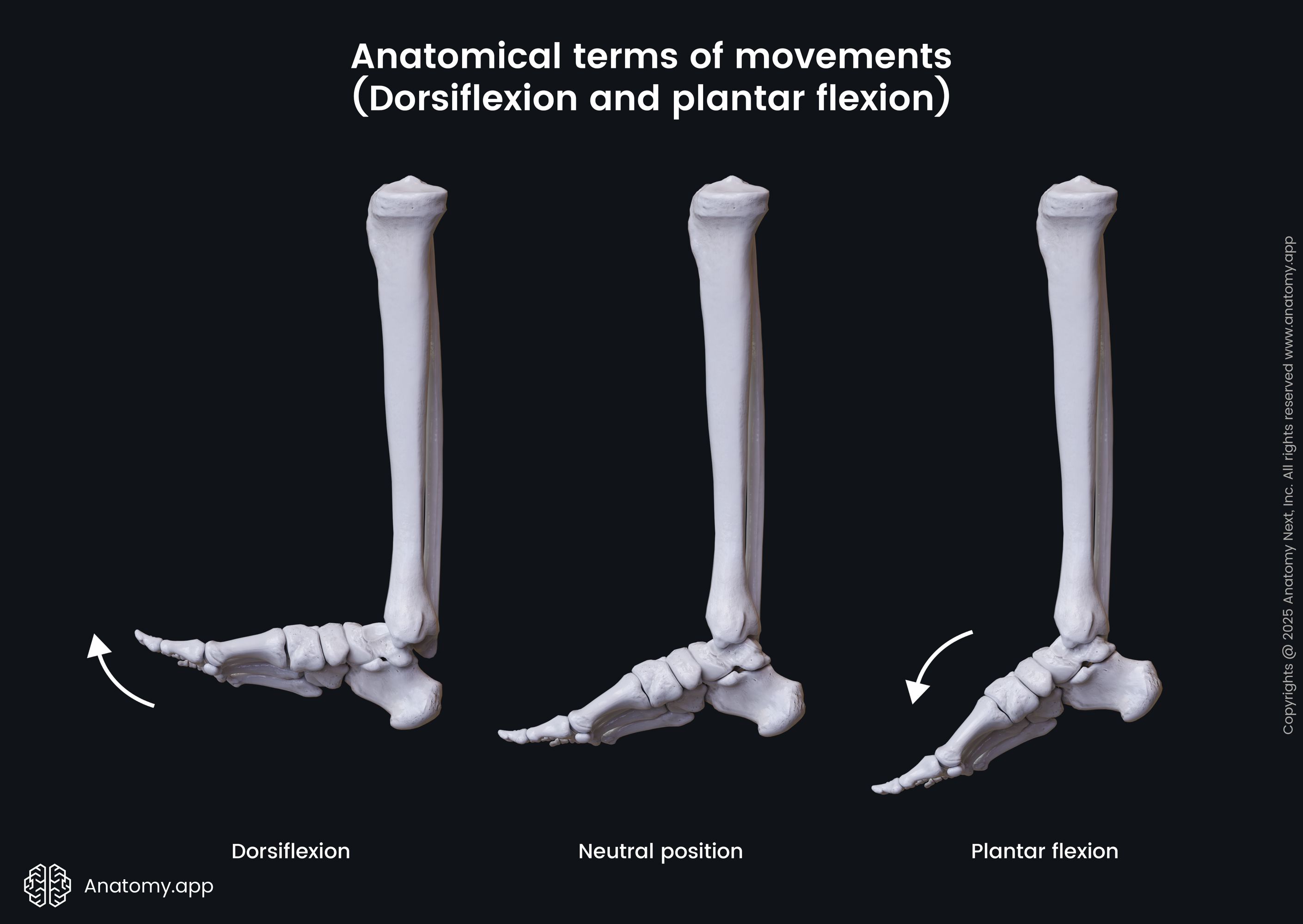
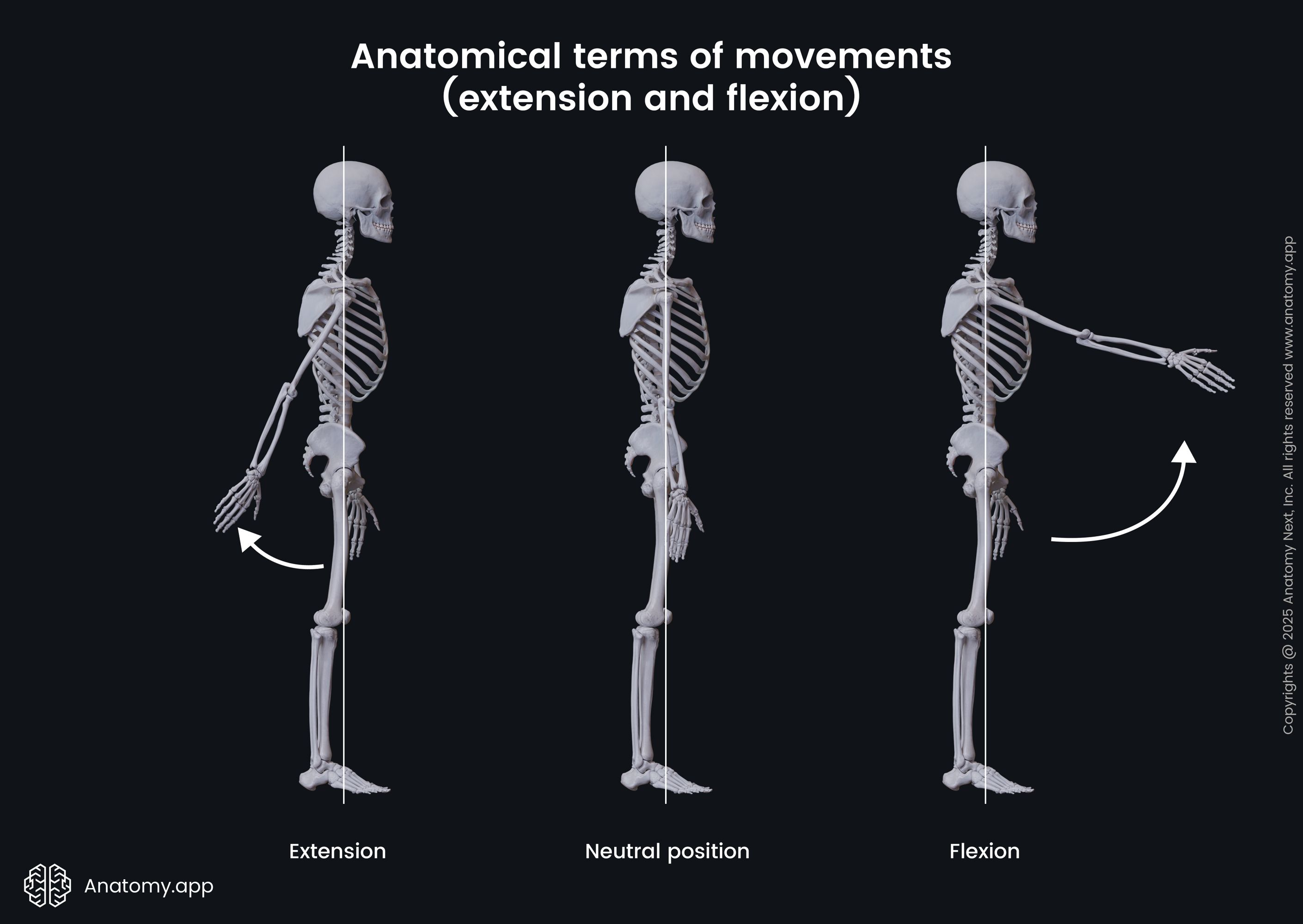
- Flexion - this movement occurs in the sagittal plane; it refers to the movement that decreases the angle between two body parts; i.e., flexion of the elbow brings the ulna and radius closer to the humerus; two specific terms are used when speaking of flexion that happens at the ankle joint:
- Dorsiflexion - an upward flexion of the foot; it can be described as the movement in which the toes are brought closer to the lower leg bones and the foot flexes towards the anterior leg; dorsiflexion decreases the angle between the lower leg and the dorsal surface of the foot;
- Plantar flexion - it can be described as the inferior displacement of the toes and superior displacement of the heel; the toes are pointing downward; this movement decreases the angle between the sole and the back of the leg;
- Extension - applied to the opposite movement of the flexion; it increases the angle between two body parts back to the anatomical position; i.e., extension of the elbow returns the radius, ulna, and humerus back to the anatomical position;
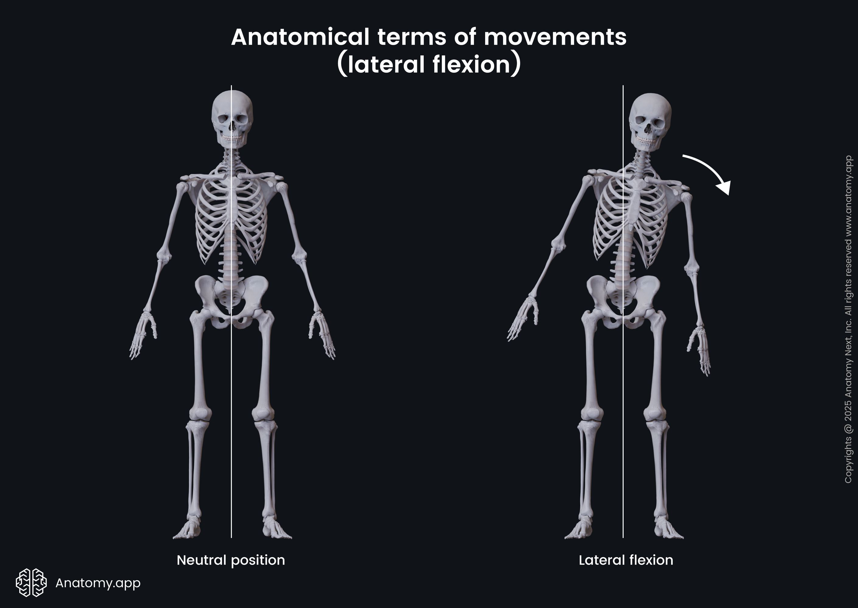
- Lateral flexion - refers to the bending or movement of the body sideways, typically to the left or right; it occurs in the frontal plane; i.e., lateral flexion of the trunk is bending to the side;
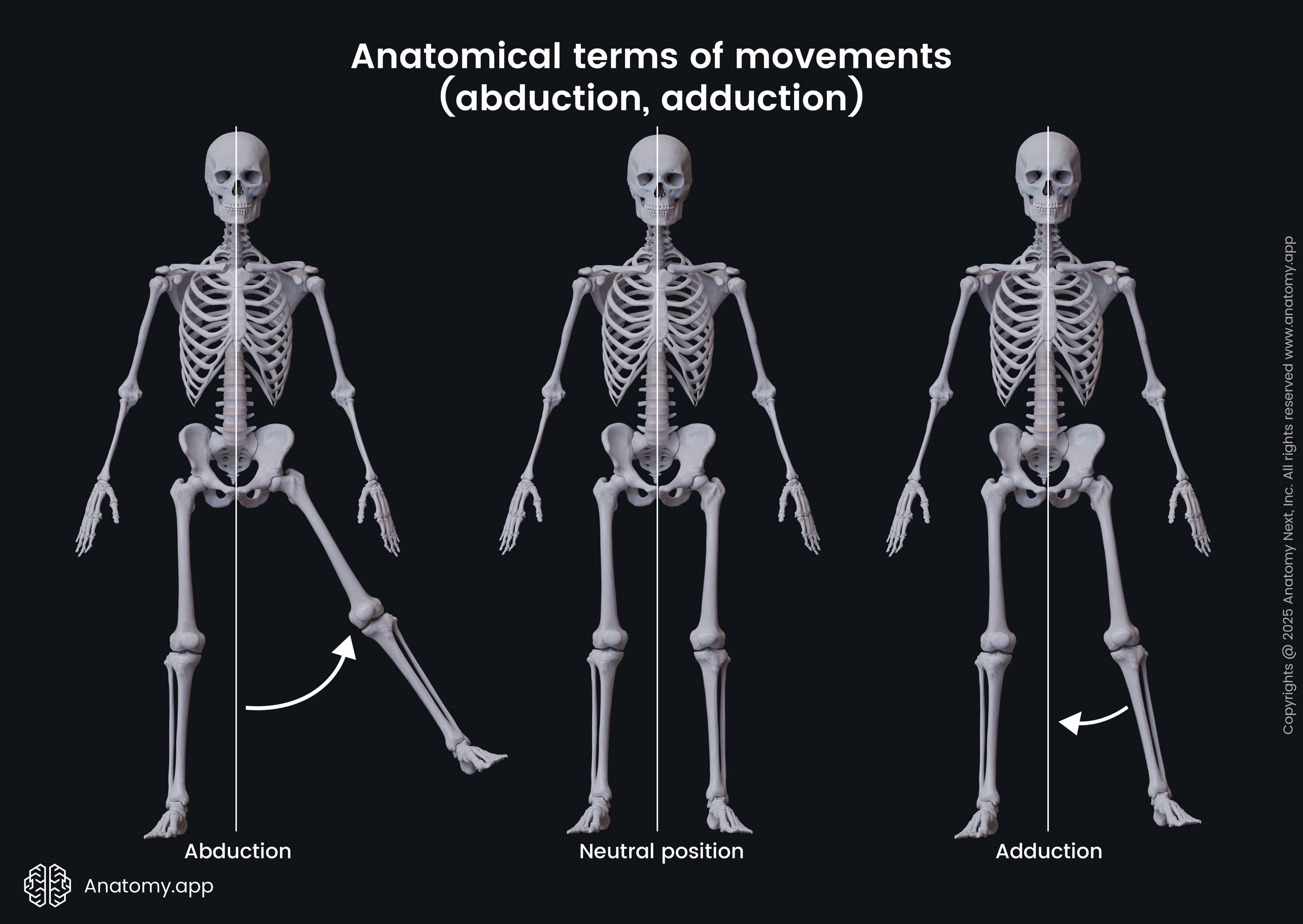
- Abduction - happens in the frontal plane; it is described as a movement away from the midline of the body; i.e., abduction at the hip joint causes one lower limb to move away from the other;
- Adduction - the opposite movement to abduction; it is described as a movement toward the midline of the body; i.e., adduction at the hip joint causes the lower limb to return to its anatomical position - it brings both lower limbs together again;
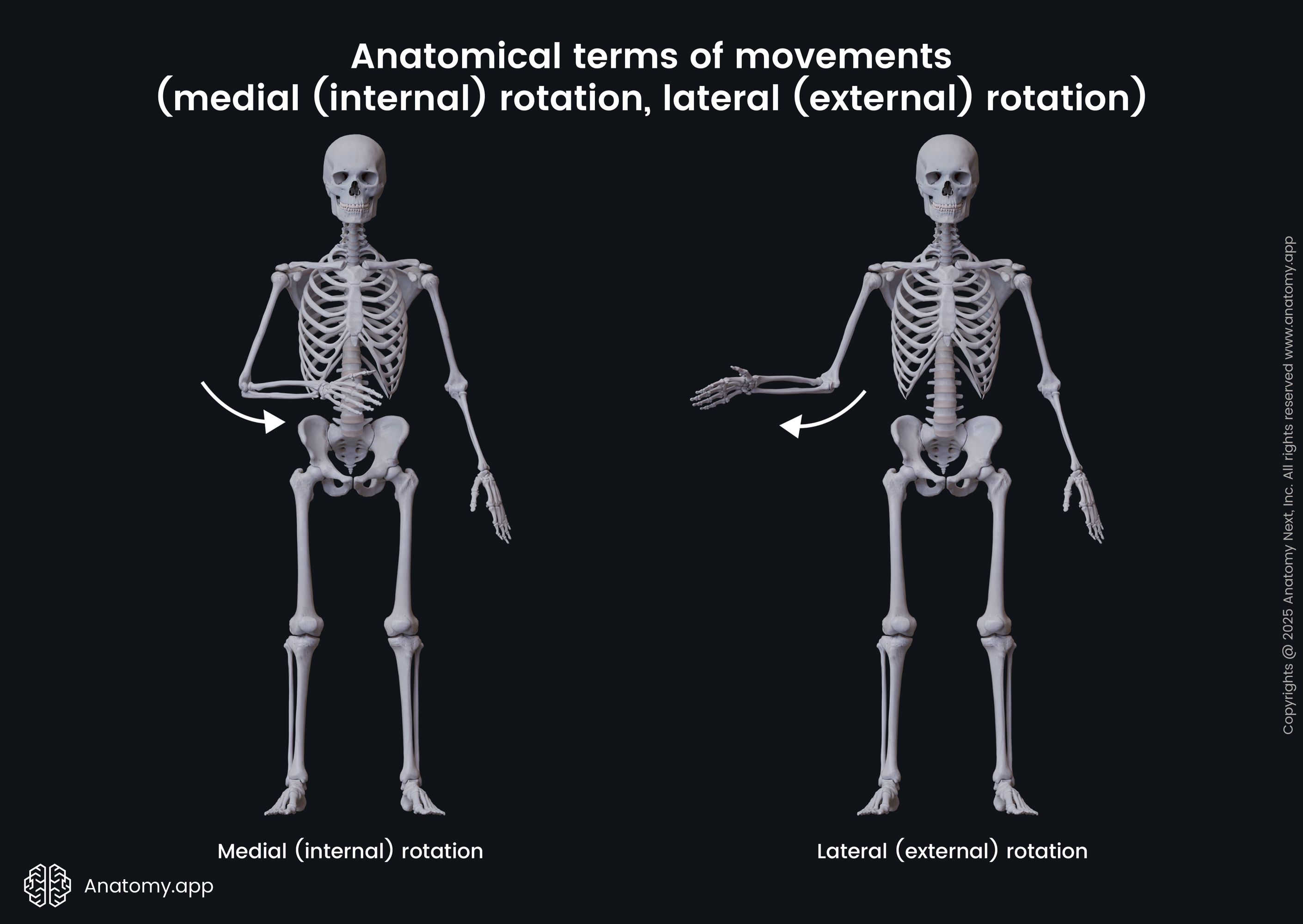
- Medial (internal) rotation - movement that happens around the longitudinal axis of the structure in the transverse plane; rotational movement toward the midline of the body; i.e., with the flexed elbow at 90 degrees, internal rotation at the shoulder causes the forearm and hand move toward the body;
- Lateral (external) rotation - the opposite movement to medial rotation; rotational movement away from the midline of the body; i.e., with the flexed elbow at 90 degrees, external rotation at the shoulder causes the forearm and hand to move away from the body;
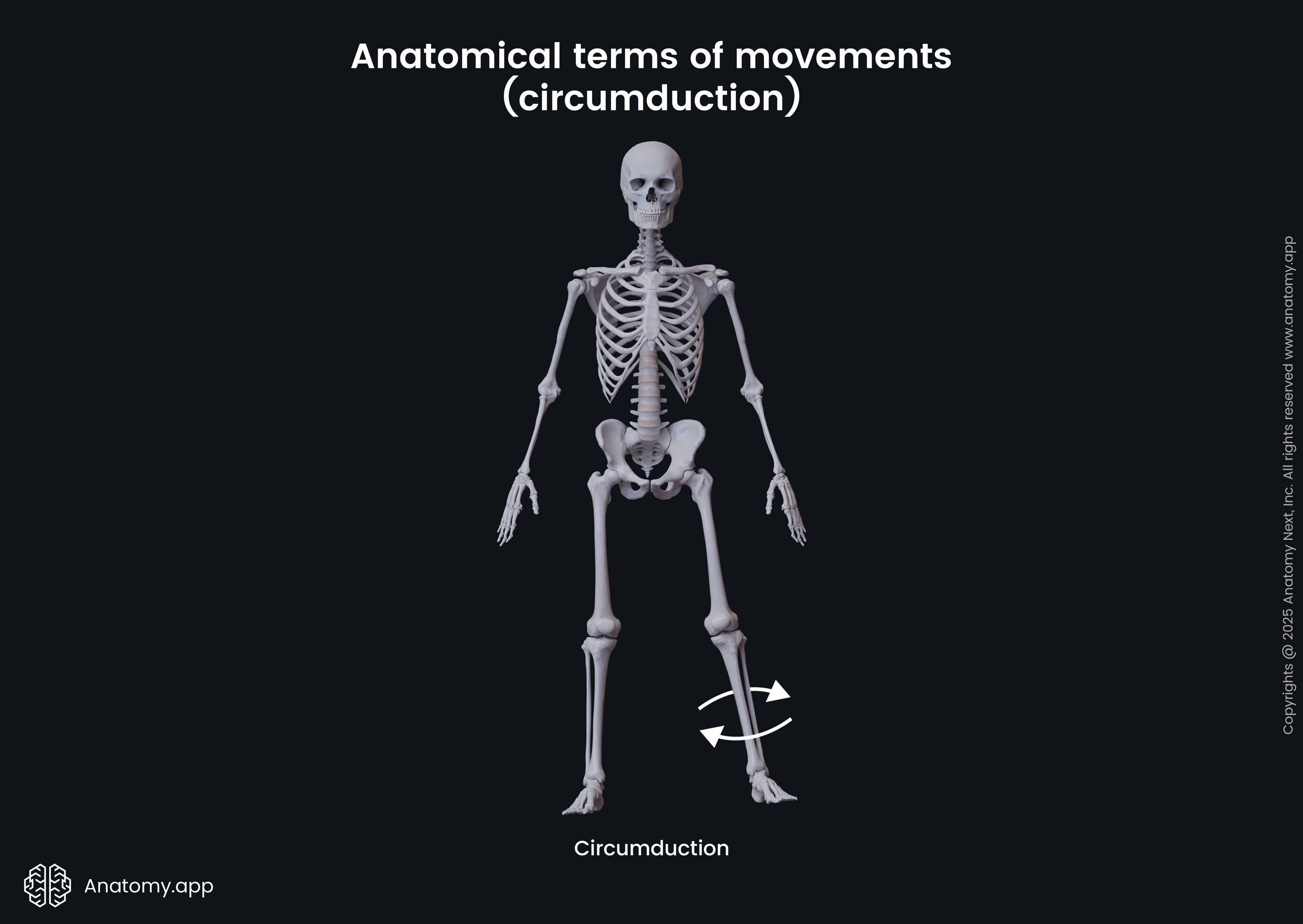
- Circumduction - it is a compound circular movement that occurs only at certain joint types (ball and socket joints); it combines flexion, extension, abduction, adduction, and rotation; it can be performed at shoulder and hip joints; it does not have an opposite movement;
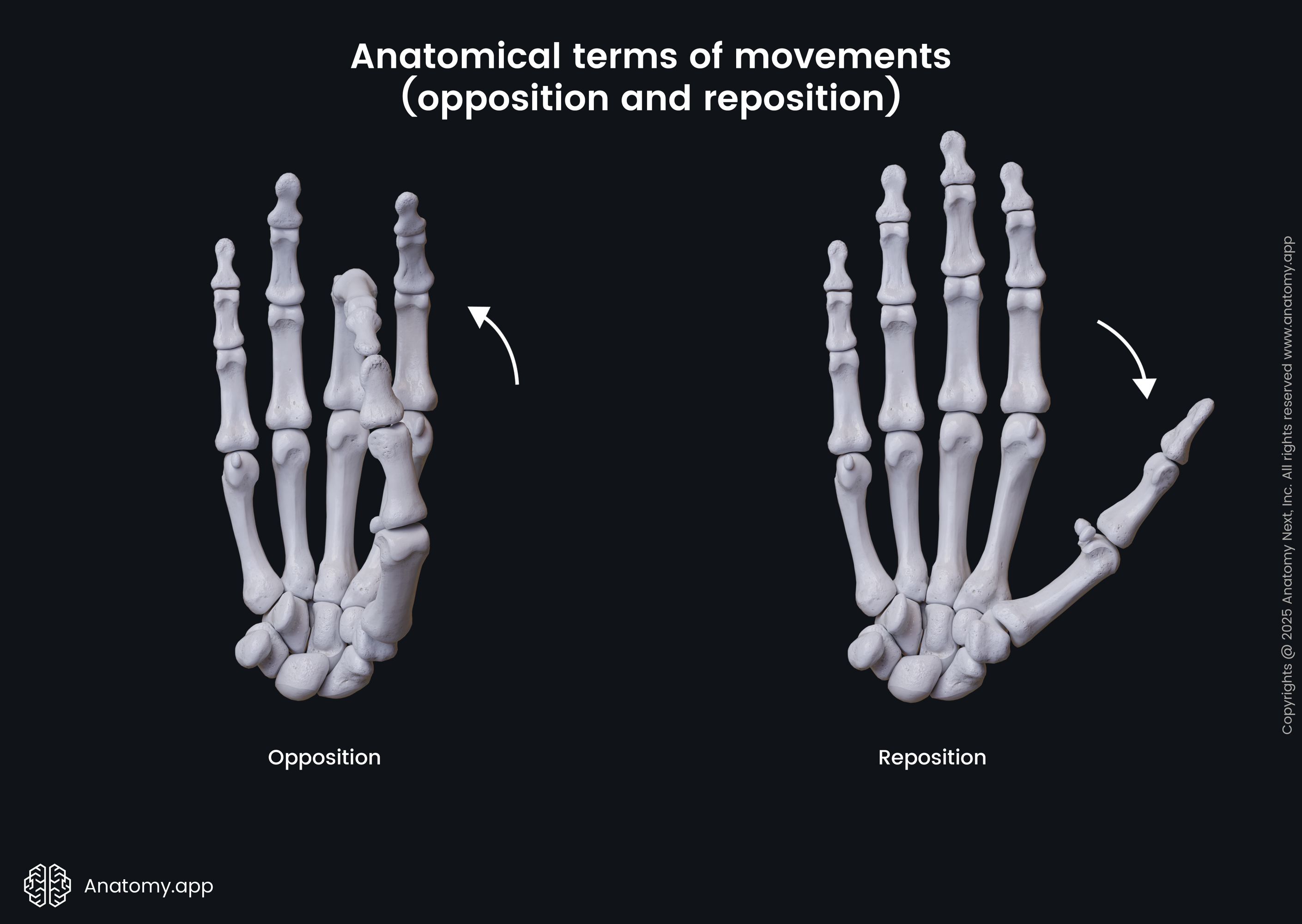
- Opposition - a special movement type that occurs in the hand and involves the first and other fingers; this movement helps to grasp objects; opposition brings the pads of the thumb and other fingers together in the midline of the hand;
- Reposition - antagonistic movement to opposition; it brings the other fingers and thumb away from each other back to the anatomical position;
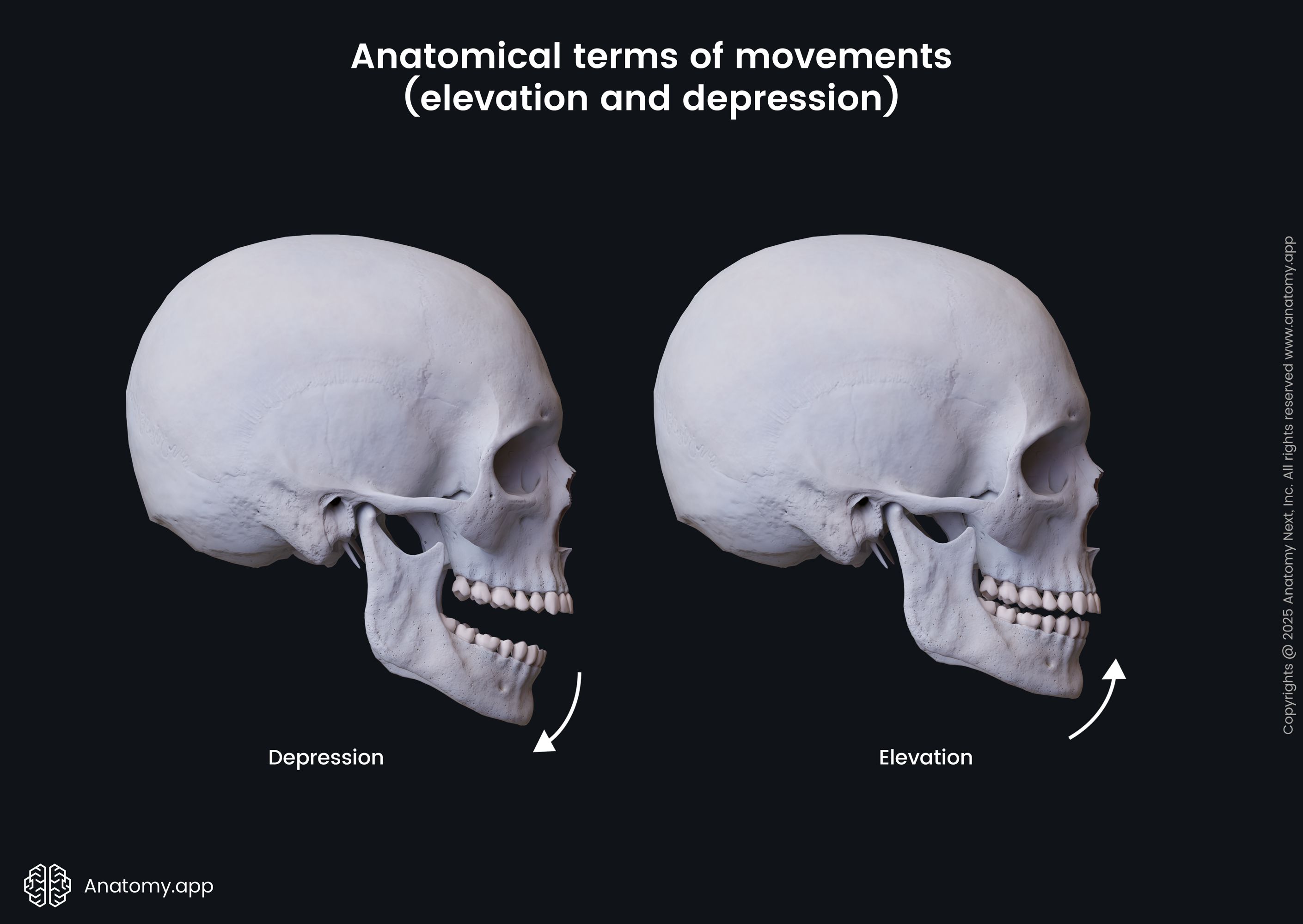
- Elevation - happens in the frontal plane; movement in the superior direction; refers to lifting a structure up; i.e., elevation of the mandible closes the mouth;
- Depression - the opposite movement to elevation; movement in the inferior direction; refers to lowering a structure down; i.e., depression of the mandible opens the mouth;
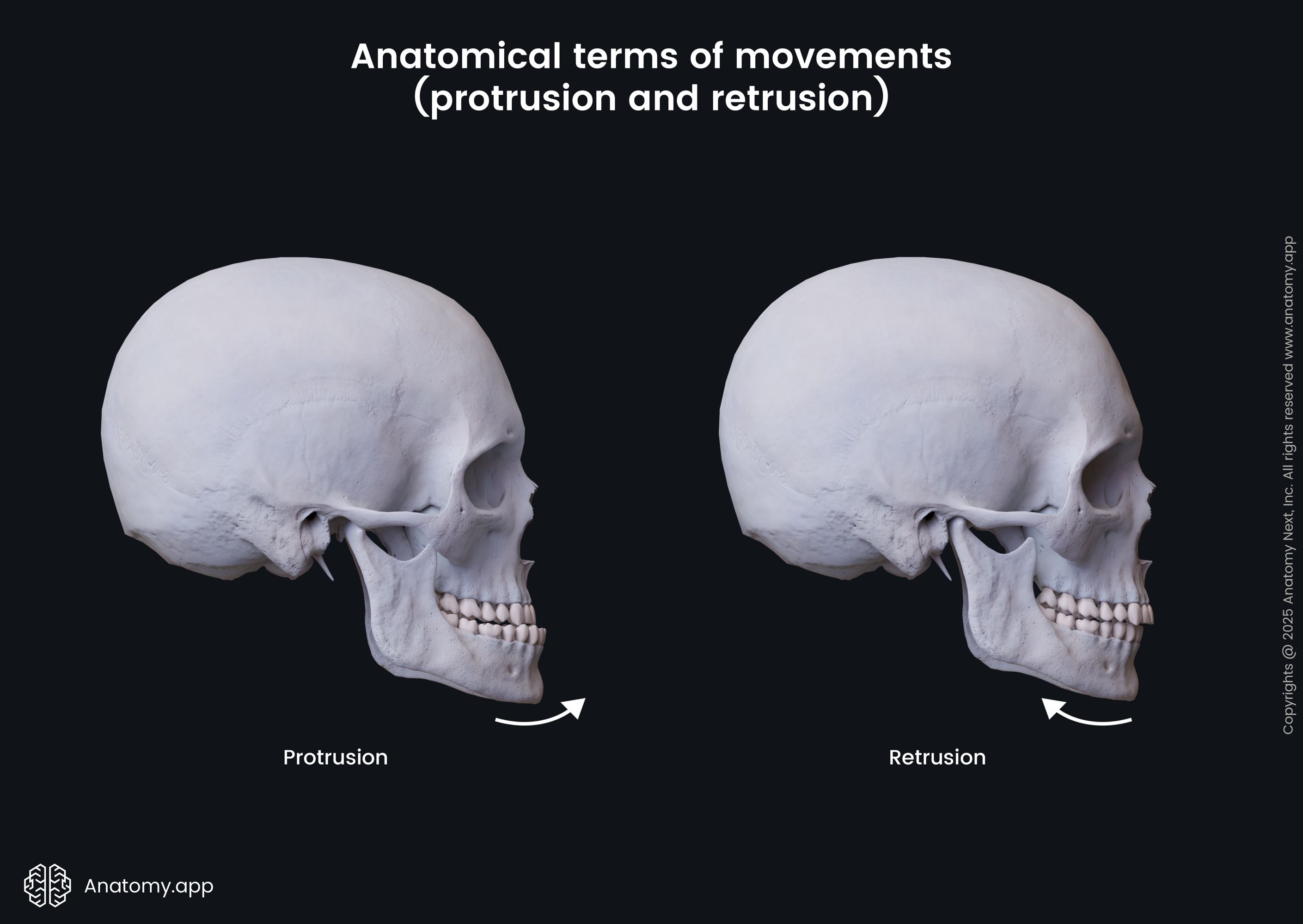
- Protrusion - movement that is performed in the anterior direction or forward; i.e., anterior movement of the mandible at the temporomandibular joints;
- Retrusion - the opposite movement to protrusion; it describes the backward or posterior movement; i.e., posterior movement of the mandible at the temporomandibular joints;
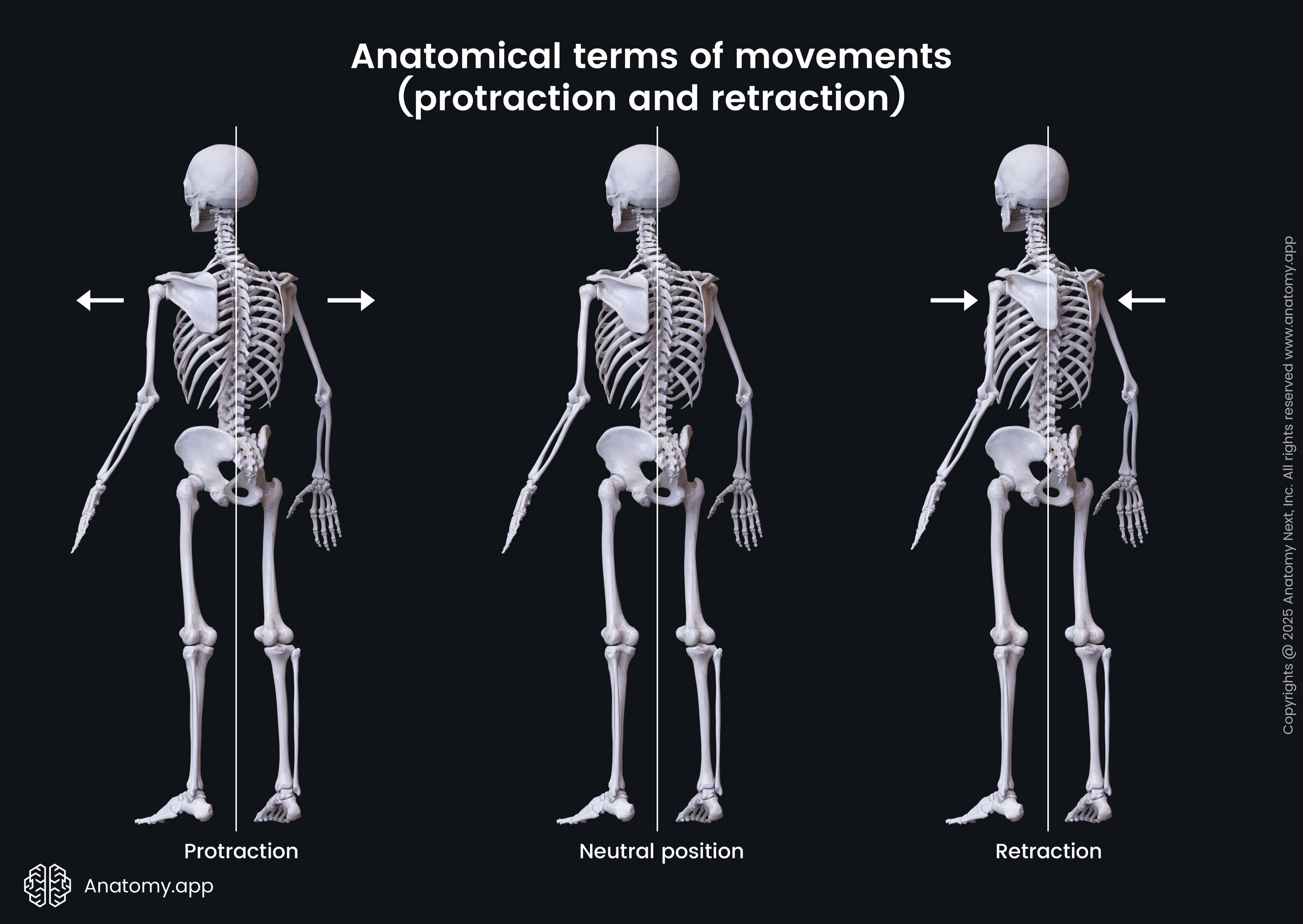
- Protraction - movement that happens in the anterior and lateral directions at the same time; this term is used when describing the anterolateral movement of the scapula on the thoracic wall when reaching out for something or hugging someone;
- Retraction - the opposite movement to protraction; movement that happens in the posterior and medial directions at the same time; it is also applied to scapular movement during which the scapula moves posteromedial; it happens when a person picks something up;
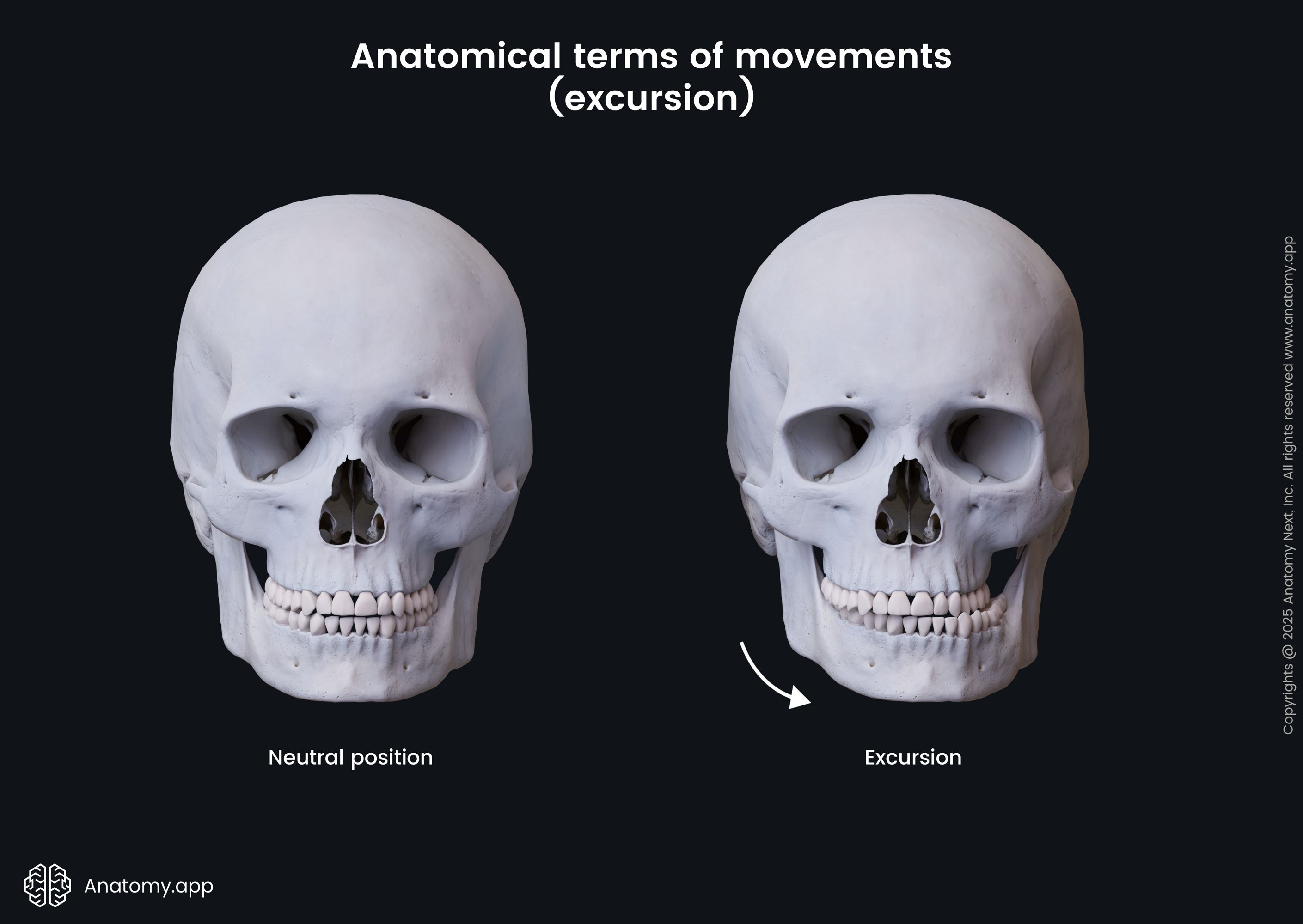
- Excursion - this term is used to describe side-to-side movements of the mandible; i.e., when the mandible moves away from the midline, it is called lateral excursion, while the opposite movement is referred to as medial excursion;
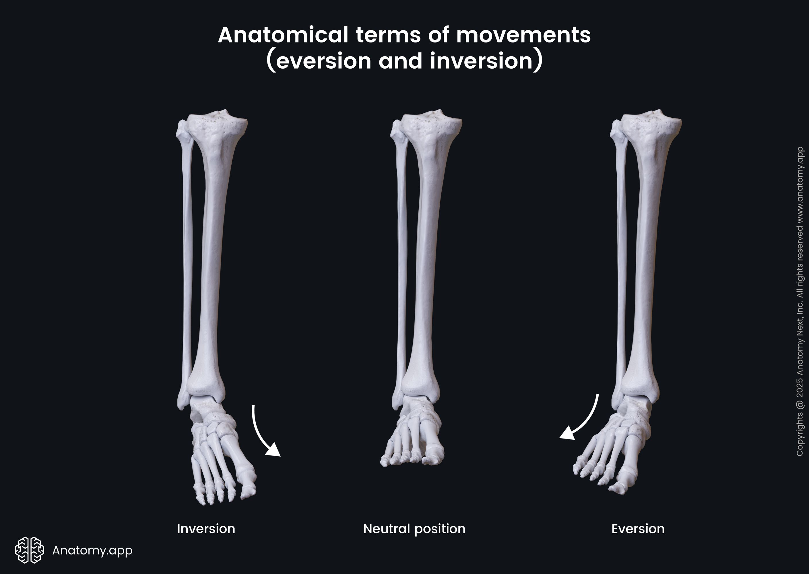
- Inversion - movement that occurs at the ankle joint in the frontal plane; inversion describes the movement that brings the sole toward the midline (the sole faces in the medial direction);
- Eversion - the opposite movement to inversion; it describes the movement that brings the sole away from the midline (the sole faces in the lateral direction);
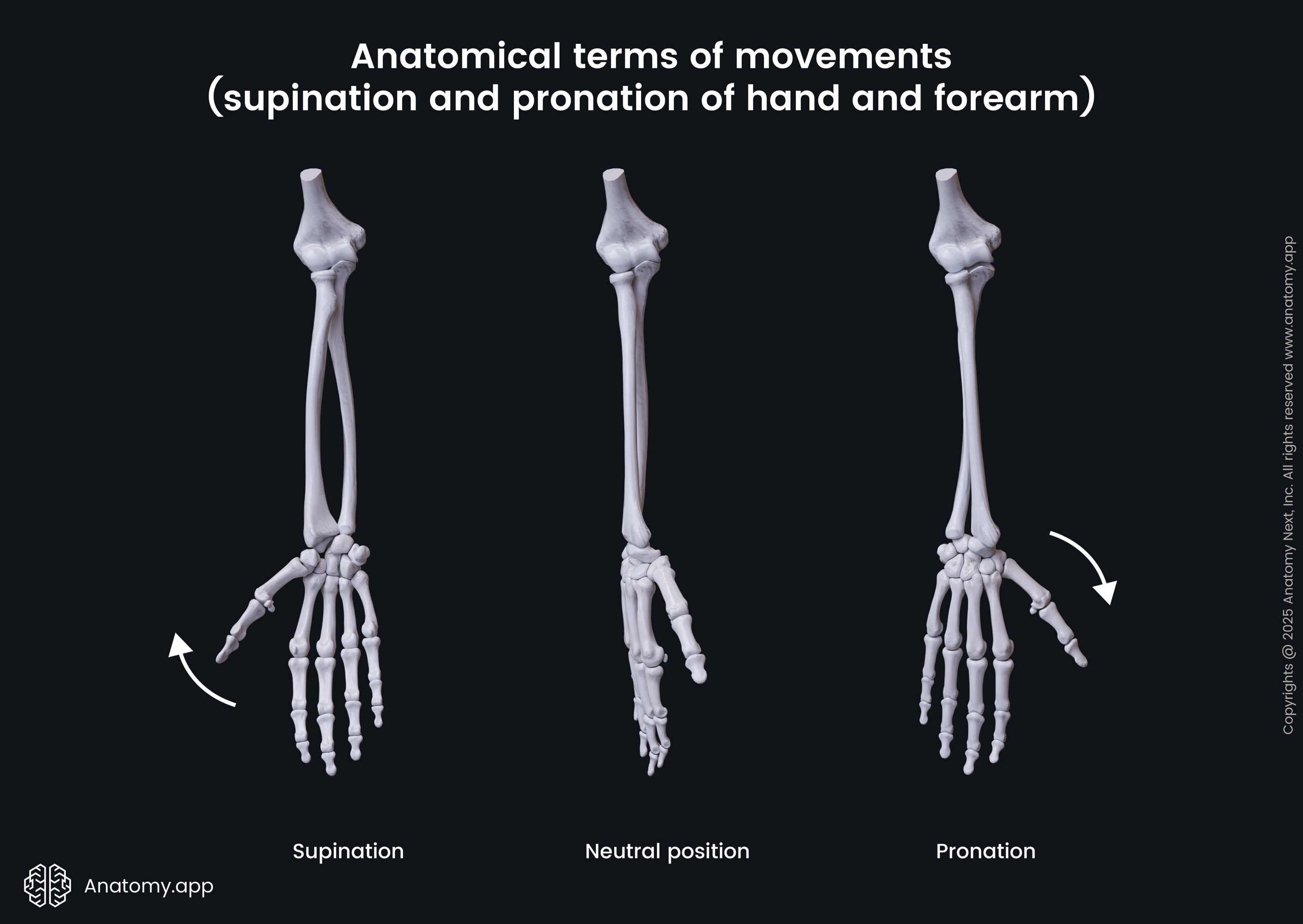
- Pronation of the hand and forearm - medial rotation of the radius; during pronation, the radial head rotates proximally, causing anteromedial movement of the distal radius; the distal radius crosses over the distal aspect of the ulna, allowing the hand and forearm to pronate; this movement results in the palm facing posteriorly if the body is in anatomical position or the palm faces the ground if the elbow is flexed;
- Supination of the hand and forearm - lateral rotation of the radius; opposite movement to pronation; this movement results in the palm facing anteriorly if the body is in anatomical position or the palm faces the sky if the elbow is flexed;
- Supination of the foot - a three-dimensional movement that combines eversion, abduction, and dorsiflexion; this combination causes the sole to face laterally;
- Pronation of the foot - an opposing movement to supination; it combines inversion, adduction, and plantarflexion, causing the sole to face medially.
Anatomy.app
Contact information
- For questions regarding business inquiries. Please contact:
- info@anatomy.app