- Anatomical terminology
- Skeletal system
- Joints
- Muscles
- Heart
- Blood vessels
- Lymphatic system
- Nervous system
- Respiratory system
- Digestive system
- Urinary system
- Female reproductive system
- Male reproductive system
- Endocrine glands
- Eye
- Ear
Brain
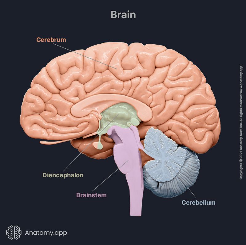
The brain (Latin: cerebrum) is the central anatomical part of the nervous system, and it is located in the cranial cavity of the skull. The brain is made up of the cerebrum, diencephalon, brainstem and cerebellum. It is a complex organ composed of neural tissue. Neural tissue is primarily made up of two types of cells: neurons – the functional units of the neural tissue, and supportive glial cells.
The human brain weighs on average 1.2 to 1.4 kg, comprising around 2% of the body's total weight. The brain is encased and protected by the neurocranium. Additional safety is ensured by three connective tissue layers known as the meninges that cover the brain. These are the dura mater, arachnoid mater, and pia mater.
The brain is a vital organ that integrates and coordinates sensory and motor information from the whole body, and it is involved in the organization and generation of voluntary and involuntary body functions, thoughts and feelings. The brain uses approximately 20% of the human body's total energy expenditure.
The brain and spinal cord are the main parts of the central nervous system. The brain connects to the spinal cord via the brainstem. Twelve pairs of cranial nerves are part of the peripheral nervous system. Eleven out of the twelve pairs of cranial nerves originate from the brainstem. The accessory nerve (CN XI) is the one exception.
The brain can be further divided into the cerebrum, diencephalon, cerebellum, and brainstem. The brain's subcortical structures are the diencephalon, pituitary gland, limbic structures and basal ganglia. Additional structures closely related to the brain are the brain's ventricles and meninges.
Brain parts and functions
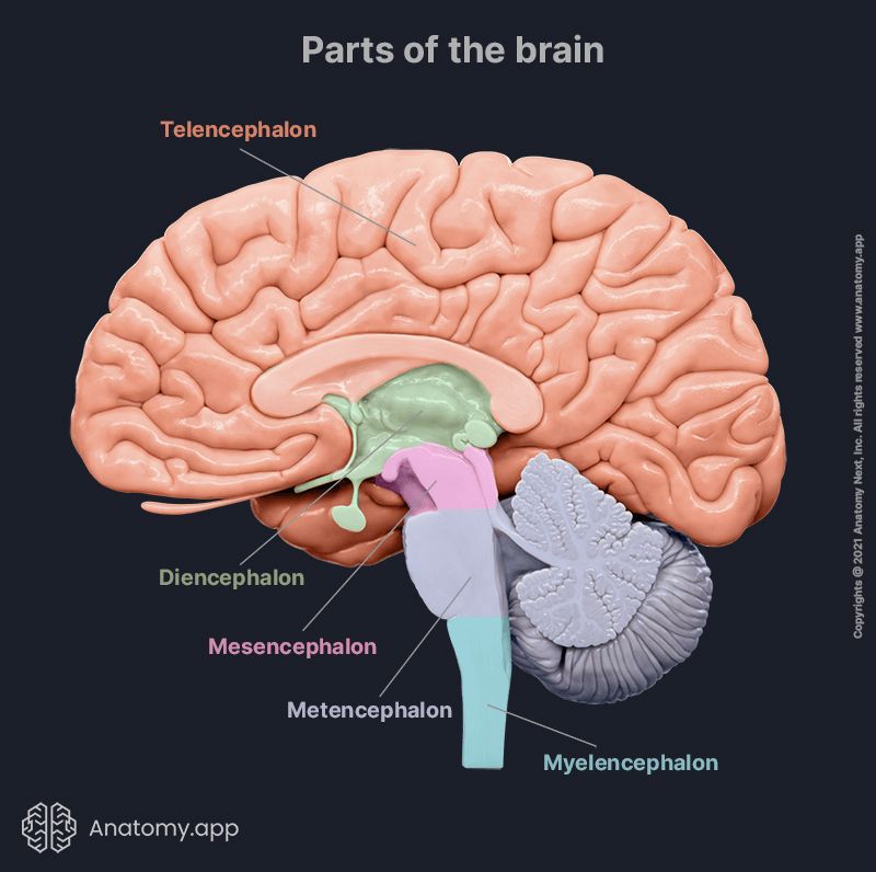
The major anatomical parts of the brain are the brainstem, diencephalon, cerebellum and cerebrum. There are also other commonly used subclassifications of the brain according to the embryonic development of the brain's structures. For instance: forebrain, midbrain and hindbrain. In addition to the preceding, the brain is also often divided into: telencephalon, diencephalon, mesencephalon, metencephalon and myelencephalon.
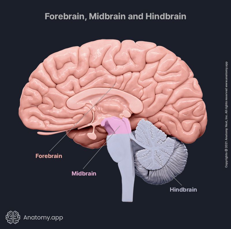
During embryonic development, there are three primary brain vesicles: prosencephalon (forebrain), mesencephalon (midbrain) and rhombencephalon (hindbrain). They further develop into secondary vesicles of the brain and further grow into the final brain parts. The prosencephalon further divides into the telencephalon (cerebrum) and diencephalon. The mesencephalon becomes the midbrain. The rhombencephalon splits into the metencephalon (pons and cerebellum) and the myelencephalon (medulla oblongata).
Brainstem
The brainstem is the distal part of the brain. It connects the cerebrum to the spinal cord and the cerebellum. The brainstem is the place of origin for ten of the twelve cranial nerves, contains many nuclei, tracts and neural pathways. Hence, it is involved in regulating heart rate, respiration, blood pressure, and other essential functions of the body.
The brainstem consists of three parts:
Another important component of the brainstem is the reticular formation. Although anatomically not well defined, the reticular formation is an interconnected network of nuclei of the brainstem. These nuclei are divided into three groups: lateral, medial, and median. The reticular formation is the site of integration and relay of vital information from the cerebrum, cerebellum and spinal cord.
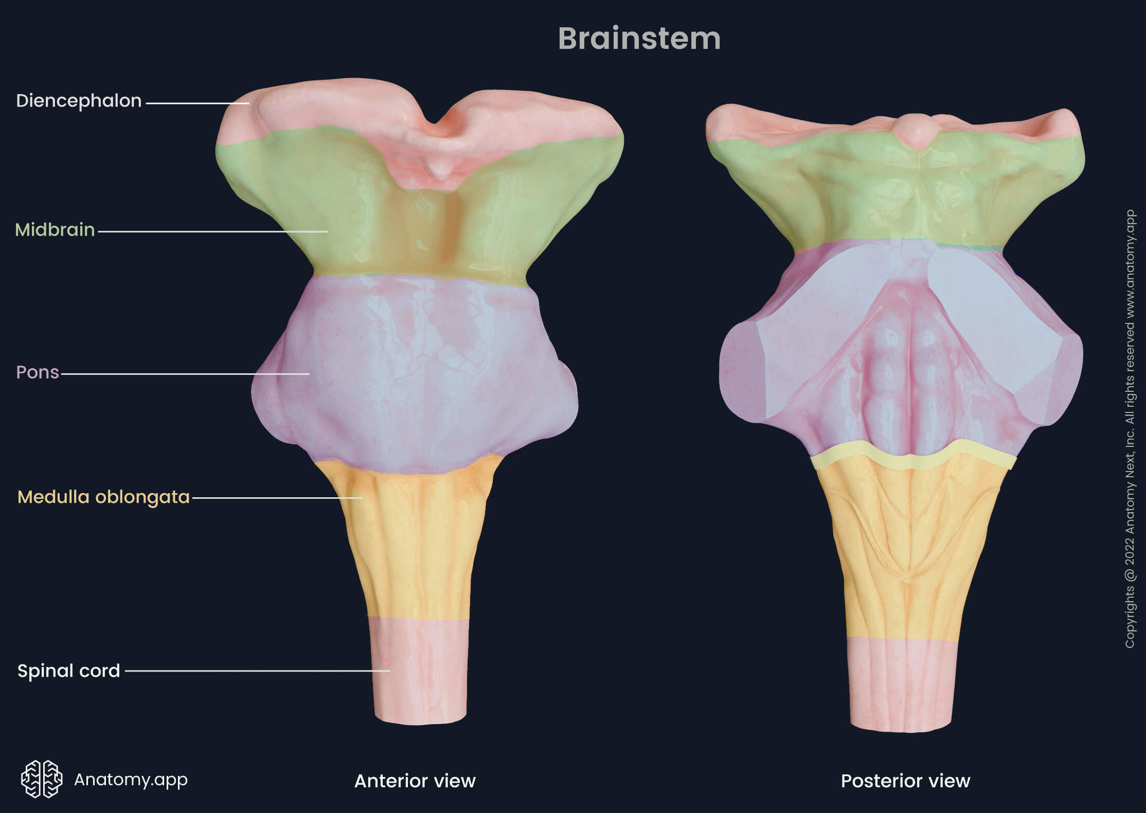
Medulla oblongata
The medulla oblongata is a stem-like structure located in the posterior cranial fossa. It is also known as the myelencephalon. The upper part of the medulla oblongata connects to the pons, and the lower part continues as the spinal cord. Besides being a conduit for fibers running between the spinal cord and higher brain regions, the medulla oblongata contains control centers for involuntary functions such as blood pressure, breathing and swallowing.
The medulla oblongata slightly decreases in width as it descends. It has two surfaces: the ventral (anterior) and dorsal (posterior) surface. The ventral surface of the medulla faces the basilar part of the occipital bone's clivus and the dens of the axis (C2). The upper rear part of the medulla oblongata gives rise to the lower part of the fourth ventricle. The central canal is a continuation of the fourth ventricle and continues into the spinal cord.
Along the midline of the ventral surface, five grooves are distinguished. The anterior median fissure runs along the midline of the ventral surface, between the two eminences called pyramids. On both lateral sides of the pyramid are grooves called the anterolateral sulci. Further laterally are additional grooves called the posterolateral sulci, which run parallel along both posterolateral sides of the medulla oblongata.
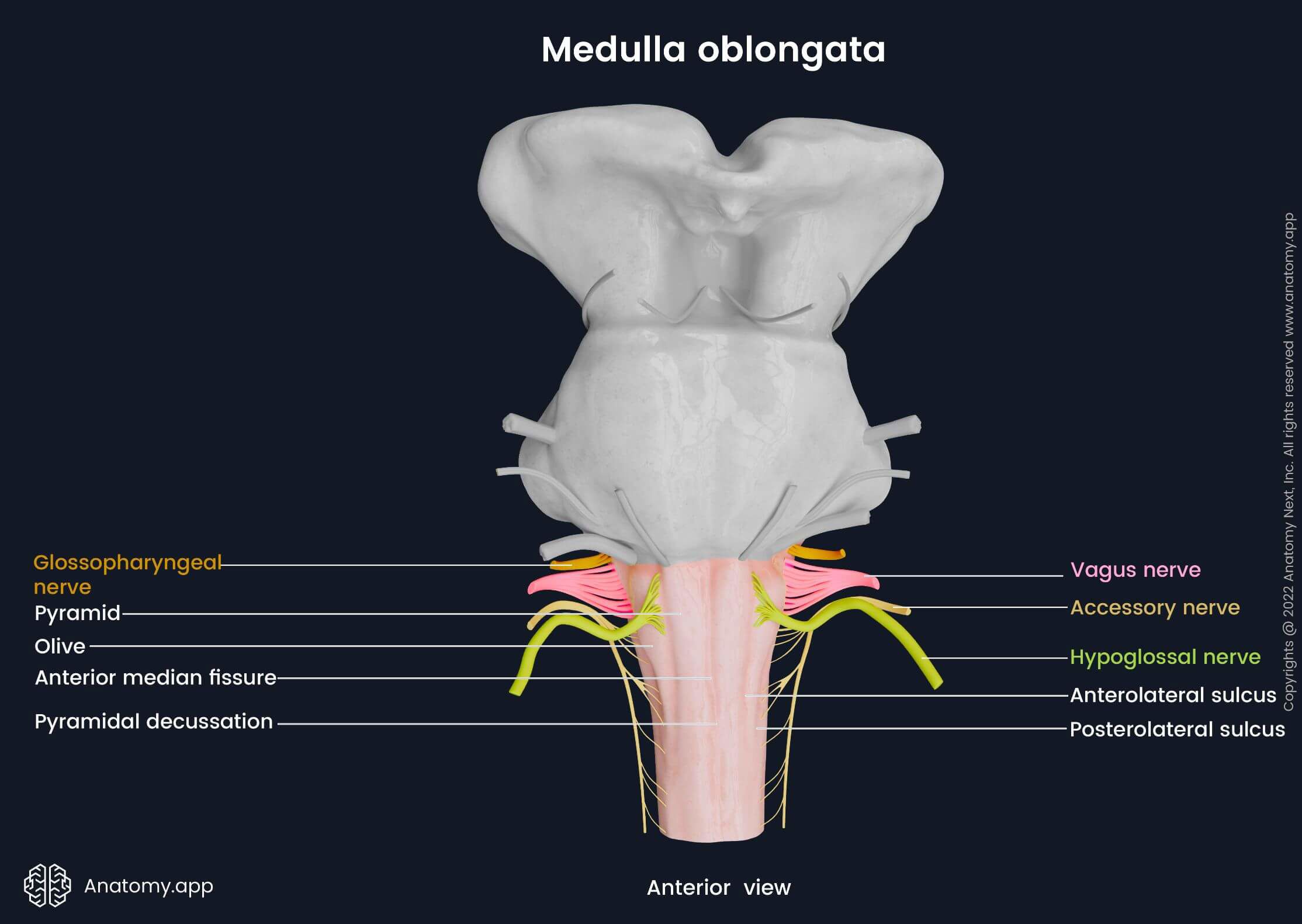
The pyramids are located anteriorly on both sides between the anterior median fissure and the anterolateral sulcus. The corticospinal tracts pass through the pyramids. Near the junction of the medulla oblongata and the spinal cord, the motor fibers of the corticospinal tracts cross in the midline, forming the pyramidal decussation. More superiorly, on the ventral surface of the medulla oblongata are two eminences called olives. They are located laterally between the anterolateral and posterolateral sulci and contain the superior and inferior olivary nuclei.
Four cranial nerves originate on the ventral surface of the medulla oblongata. The hypoglossal nerves (CN XII) arise from the anterolateral sulci. The glossopharyngeal nerves (CN IX), vagus nerves (CN X), and accessory nerves (CN XI) exit from the posterolateral sulci. Nuclei of the previously mentioned cranial nerves and others (e.g., the trigeminal nerve (CN V)) are found in the medulla oblongata.
The dorsal surface of the medulla oblongata is connected to the cerebellum by the inferior cerebellar peduncle and forms the inferior part of the rhomboid fossa. Moreover, it also forms the inferior part of the fourth ventricle's floor and includes the obex. The obex is the site where the fourth ventricle narrows and transitions into the central canal of the spinal cord.
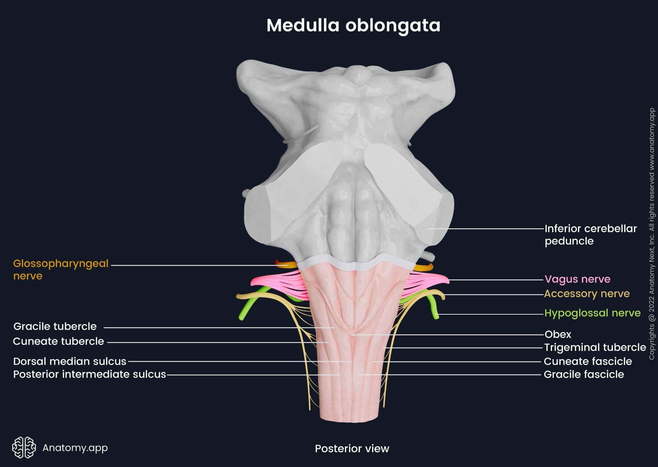
Along the midline of the dorsal surface, the dorsal median sulcus descends. Lateral to the dorsal median sulcus is the gracile fascicle and a bit more laterally - the cuneate fascicle. Superiorly, the fascicles form two prominences: the gracile tubercle medially and the cuneate tubercle more laterally. The two mentioned tubercles contain their respective nuclei - the gracile nucleus and cuneate nucleus. Inferiorly, the posterior intermediate sulcus separates the cuneate and gracile fascicles. Lateral to the cuneate tubercle is another prominence called the trigeminal tubercle.
The internal anatomy of the medulla oblongata is very complex as it contains a multitude of nuclei (clusters of neuron cell bodies) and tracts (consisting of nerve fibers) originating from, crossing it and passing through it. To better understand these structures and their relations, the medulla oblongata is typically examined in three cross-sections: the level of pyramidal decussation, the level of medial lemniscus decussation, and at the level of the olives.
Pons
The pons is a part of the metencephalon and forms the middle portion of the brainstem. It is located beneath the midbrain and above the medulla oblongata in the posterior cranial fossa. Like the other structures of the brainstem, the pons contains a multitude of nuclei and is the site of passage and relay of numerous neural tracts. The pons regulates respiration, sleep, equilibrium and other vital functions of the body.
The pons is divided into two parts: the ventral (basilar) and dorsal (pontine tegmentum). The ventral surface is the anterior aspect of the basilar part of the pons. It appears convex and faces the clivus of the occipital bone. A central groove, called the basilar sulcus, runs along the anterior midline of the pons. Laterally, the pons narrows and transitions into the middle cerebellar peduncles - paired structures containing nerve tracts that connect the pons to the posteriorly lying cerebellum.
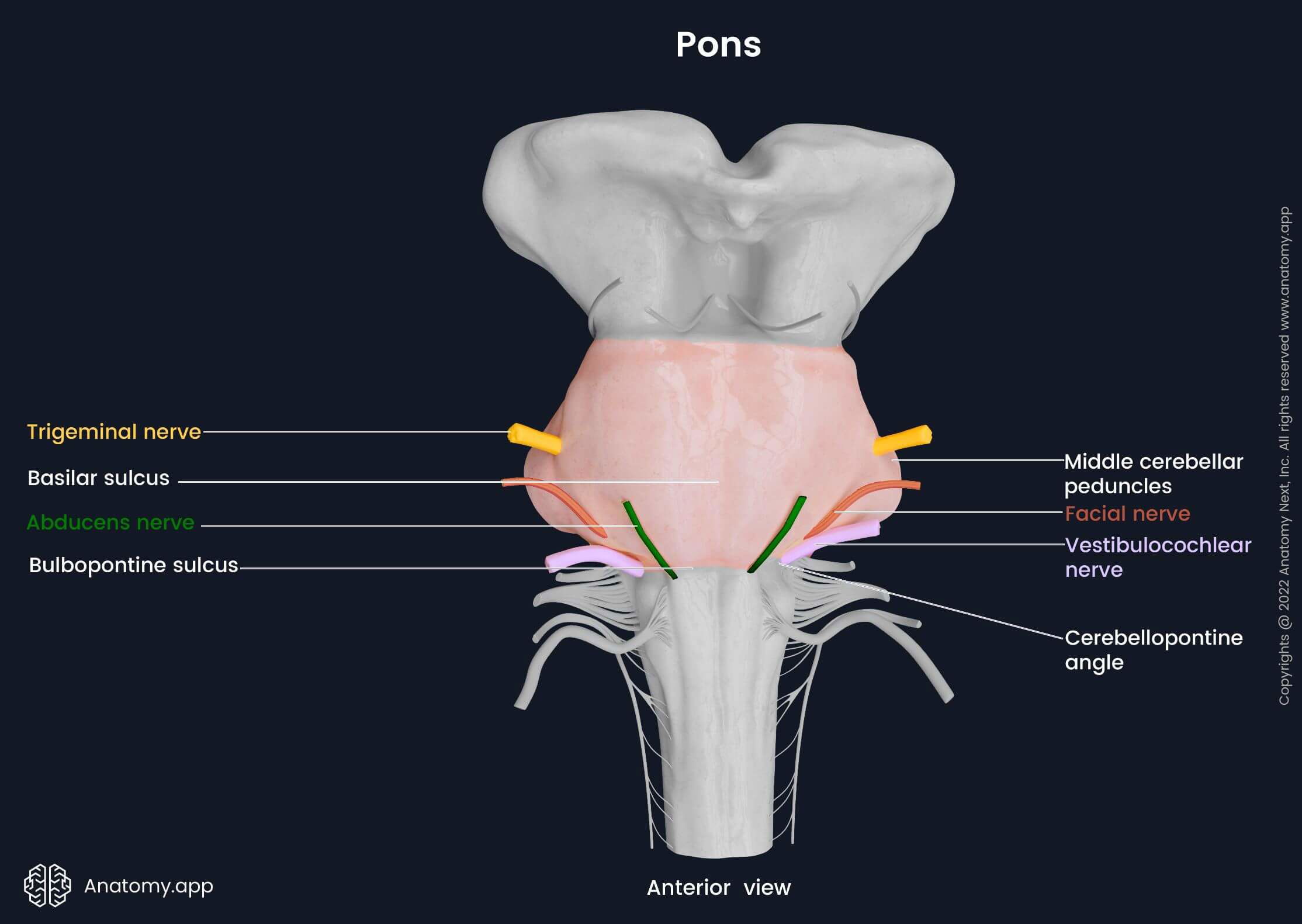
Four cranial nerves arise from the pons. The abducens nerve (CN VI) is the most medially positioned of all four. On each side, the nerve exits on the bulbopontine sulcus between the pyramid of the medulla oblongata and the pons. On the ventral surface of the pons at the border with the middle cerebellar peduncles, the trigeminal nerve (CN V) emerges. Lower, the facial (CN VII) and vestibulocochlear (CN VIII) nerves emerge from the cerebellopontine angle that is located on each side between the olive of the medulla oblongata and middle cerebellar peduncle.
The dorsal surface of the pons is covered by the cerebellum. It forms the upper part of the floor of the fourth ventricle. Moreover, the dorsal surface constitutes the upper half of the rhomboid fossa. Superiorly and laterally, it is limited by the superior cerebellar peduncles. Prominent peduncles, called the middle cerebellar peduncles, connect the pons to the cerebellum and contain pontocerebellar fibers. The superior medullary velum forms the upper border of the dorsal surface, and the striae medullares forms the inferior.
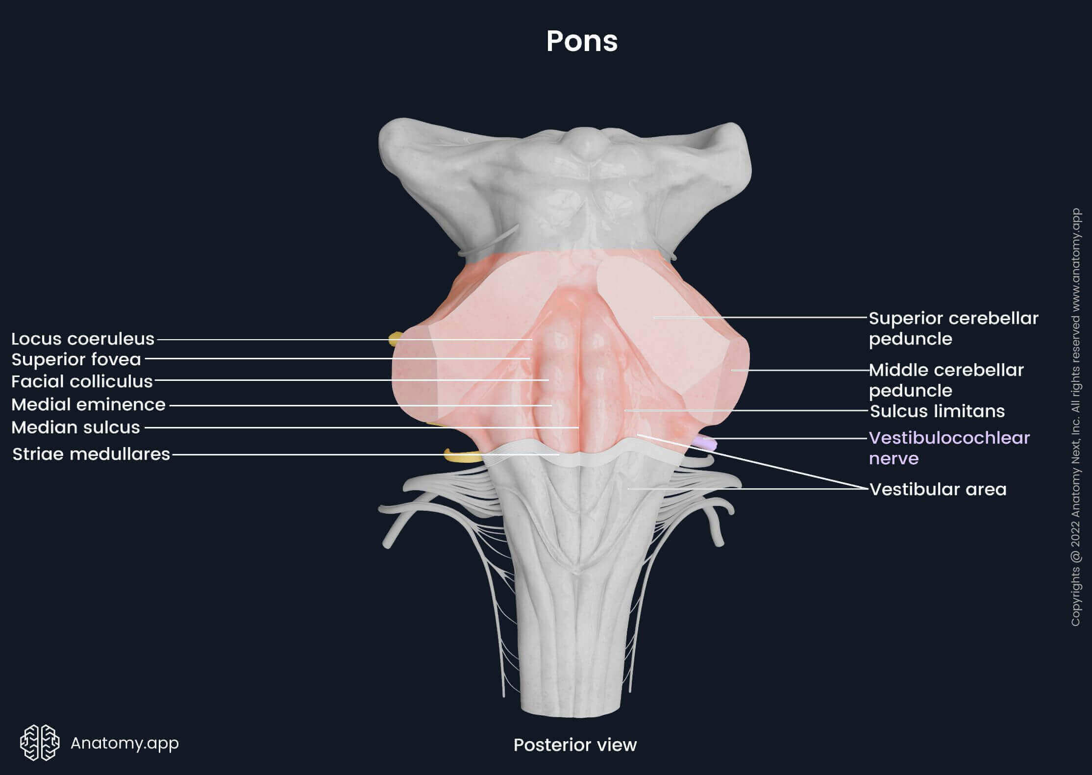
Vertically, the median sulcus runs along the midline of the dorsal pons. It separates a pair of structures called the medial eminences. Lateral to both medial eminences is the sulcus limitans, which widens at the level of the facial colliculus into a flattened depression - the superior fovea. Above it is the bilaterally located locus coeruleus, which contains the nucleus coeruleus.
The corticospinal tracts, corticobulbar tracts, and spinothalamic tracts pass through the pons. The anterior part contains the pontine nuclei. The pontine nuclei combine and form the cerebellar peduncles. The dorsal part of the pons includes structures of the reticular formation such as the medial longitudinal fasciculus, facial nucleus, medial and lateral lemniscus and the raphe nucleus, to name a few.
Midbrain
The midbrain, also known as the mesencephalon, is the uppermost part of the brainstem. The midbrain is located beneath the thalamus and above the pons in the posterior cranial fossa. The midbrain is involved in a wide array of vital functions, including regulating eye movement, movement planning, auditory processes, motivation, excitation, temperature, and others.
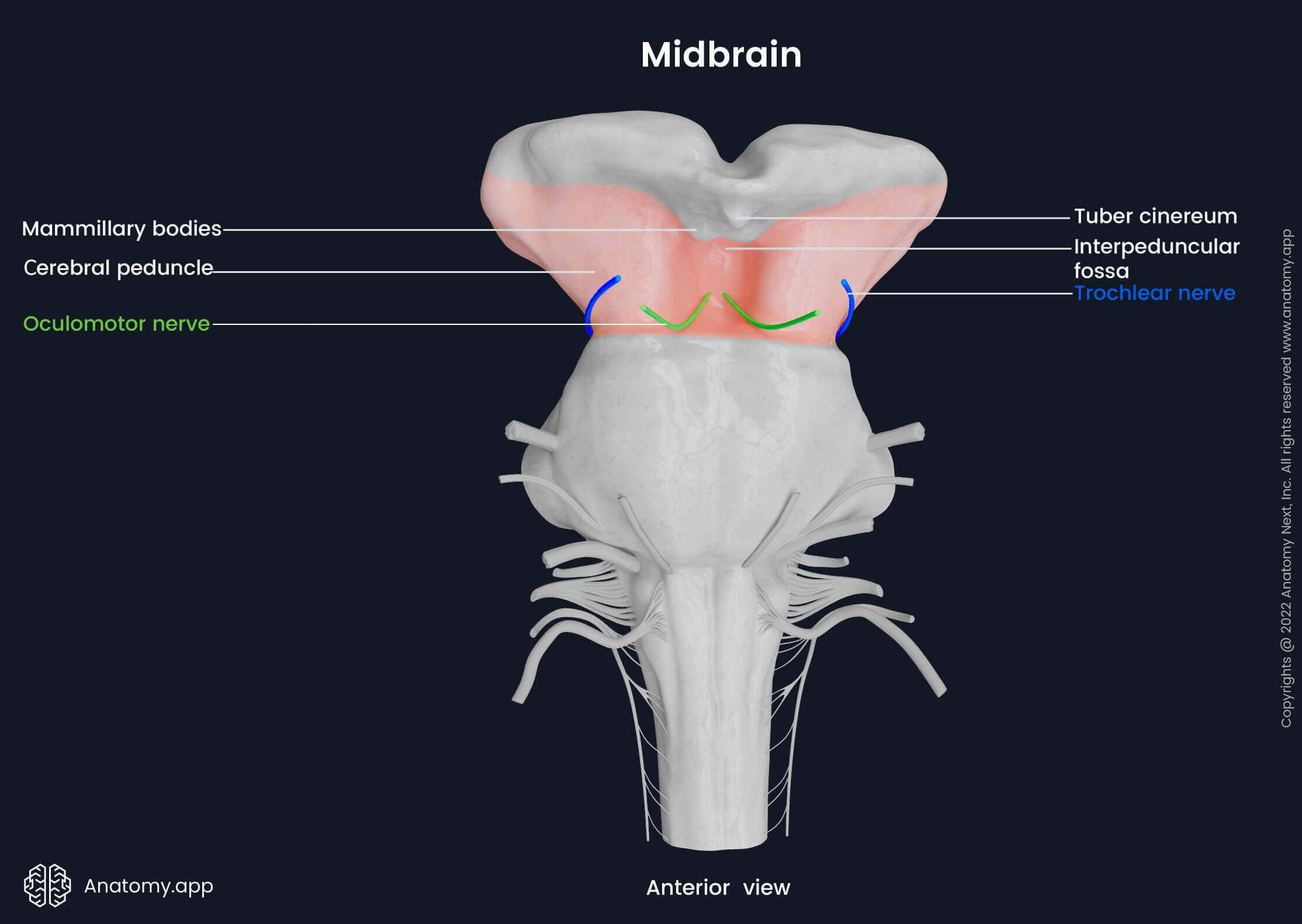
Externally, the midbrain has a ventral surface and a dorsal surface. On the ventral (anterior) surface of the midbrain, paired cerebral peduncles are situated. Both peduncles are separated by the interpeduncular fossa. Above the ventral surface of the midbrain mammillary bodies and the tuber cinereum can be seen, which are structures of the diencephalon. The oculomotor nerve (CN III) arises from the ventral surface of the midbrain.
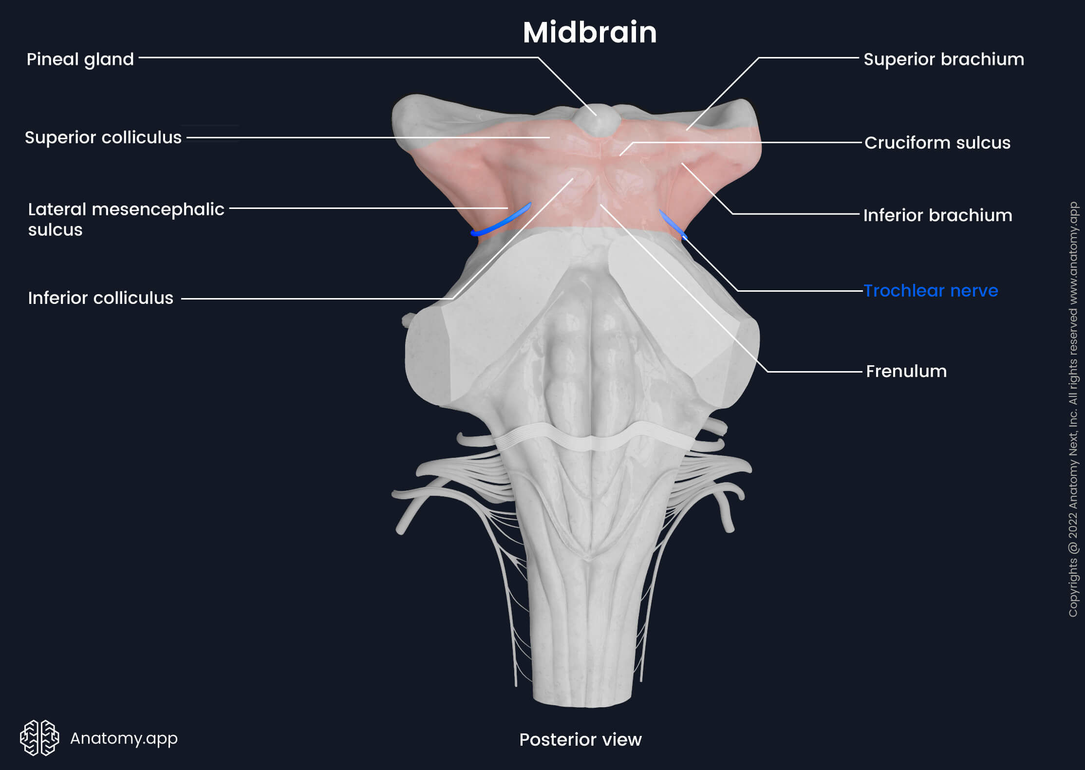
On the dorsal (posterior) surface of the midbrain four round formations known as the colliculi are seen. There are two superior and two inferior colliculi. Lateral from each colliculus, extends an eminence called a brachium (pl. brachia) containing white matter. There are two superior brachia and two inferior brachia. The colliculi are separated from each other by the cruciform sulcus, which extends caudally to the frenulum. The frenulum is a part of the medullary velum, separating both inferior colliculi from each other.
On the lateral surfaces of the midbrain, between the cerebral peduncles and the dorsal surface, is another sulcus – the lateral mesencephalic sulcus. On the dorsal surface of the midbrain emerge the right and left trochlear nerves (CN IV). After appearing, both nerves continue forward and laterally along the border between the midbrain and pons.
The midbrain is separated into the anterior and posterior parts by the cerebral aqueduct of Sylvius. The internal anatomy of the midbrain can be better understood when looked at from sagittal and transverse planes. In the sagittal cross-section, the midbrain is divided into crus cerebri, tegmentum and tectum. The crus cerebri forms the ventral part, the tegmentum the middle, and the tectum is the dorsal part of the midbrain. Crus cerebri is the anterior part of the cerebral peduncles, which contain motor tracts.
The substantia nigra separates the crus cerebri from the tegmentum. The red nuclei are located in the tegmentum. Other important nuclei of the midbrain are the superior and inferior colliculi, the nuclei of the trochlear and oculomotor nerves, medial geniculate nucleus, medial longitudinal fasciculus and others.
Diencephalon
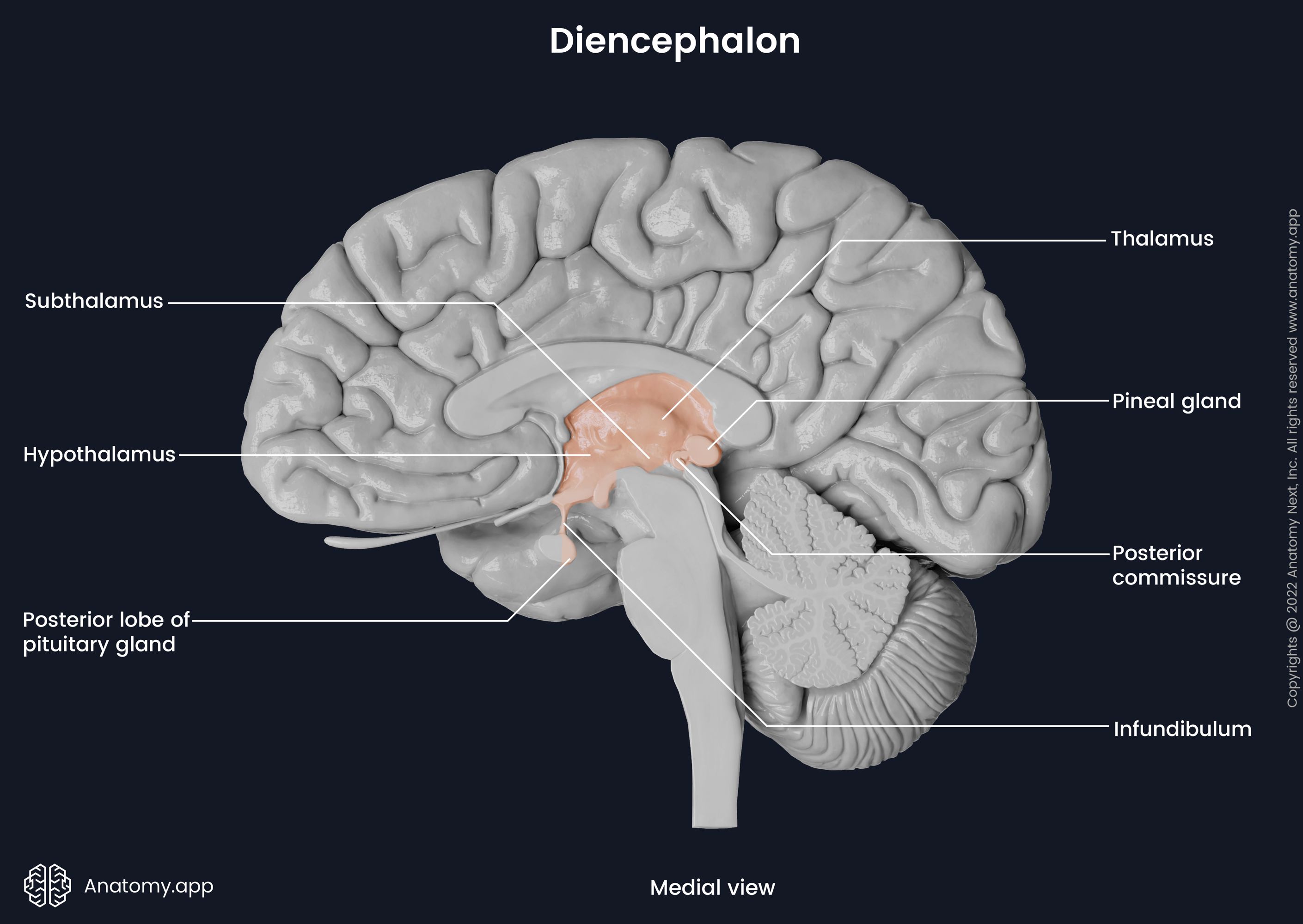
Hidden from view by the brain's cerebral hemispheres lies the diencephalon. It is a subcortical structure and consists of the thalamus, hypothalamus, epithalamus, subthalamus, and metathalamus. It lies above the midbrain and is considered to be the most rostral part of the brainstem. The diencephalon acts as a relay center for motor and sensory information, communicates with the other subcortical systems, regulates hormonal production, and more.
The thalamus consists of grey matter and is the largest structure of the diencephalon. It is formed of two symmetric oval masses connected by a bridge called the interthalamic adhesion. The thalamus has an anterior and a posterior pole. The thalamus houses five groups of nuclei: anterior, lateral, medial, reticular, and intralaminar nuclei. Four surfaces of the thalamus can be distinguished: the superior, inferior, medial, and lateral surface.
The thalamus is located beneath the corpus callosum. Its superior surface is associated with the stria terminalis and is covered by a thin sheet of white matter called stratum zonale. The medial surface of the thalamus forms the lateral wall of the third ventricle. The lateral surface separates the thalamus from the basal ganglia, and it is also covered by a thin white tissue layer called the external medullary lamina. The inferior surface is associated with the hypothalamus.
The hypothalamus is located anterior to and below the thalamus and forms the ventral part of the diencephalon. It is divided into three zones: medial, lateral, and periventricular. The medial zone forms part of the third ventricle's lateral wall. The hypothalamus houses four groups of nuclei: the preoptic, supraoptic, infundibular, and mammillary nuclei.
The mammillary nuclei are found in the mammillary bodies that are small rounded structures in the posterior aspect of the hypothalamus. Inferiorly, the hypothalamus connects to the pituitary gland by the pituitary stalk, also known as the infundibulum. Note that only the posterior part of the pituitary gland is part of the diencephalon.
The thalamus processes, integrates and relays all sensory information going to the higher regions of the brain, while the hypothalamus is critical for homeostasis, the maintenance of the body's internal environment. The hypothalamus regulates endocrine functions that further affect the whole body via the hypothalamic-pituitary axis.
The epithalamus is the posterior part of the diencephalon. The epithalamus has five parts: stria medullaris, posterior commissure, habenular nuclei, pineal body, and paraventricular nuclei. The epithalamus connects the limbic system to the other parts of the brain. It controls the body's circadian rhythms through the pineal body, which is a small piriform endocrine gland. Additionally, it regulates motor pathways, emotions and the sleep cycle.
The subthalamus is located below the dorsal part of the thalamus. It contains the subthalamic, reticular and perigeniculate nucleus. The subthalamic nucleus is functionally considered part of the basal ganglia. The metathalamus is formed by the bilaterally located medial and lateral geniculate bodies found on the caudal surface of the diencephalon. The geniculate bodies are important relay stations for the auditory and optic pathways.
Cerebellum
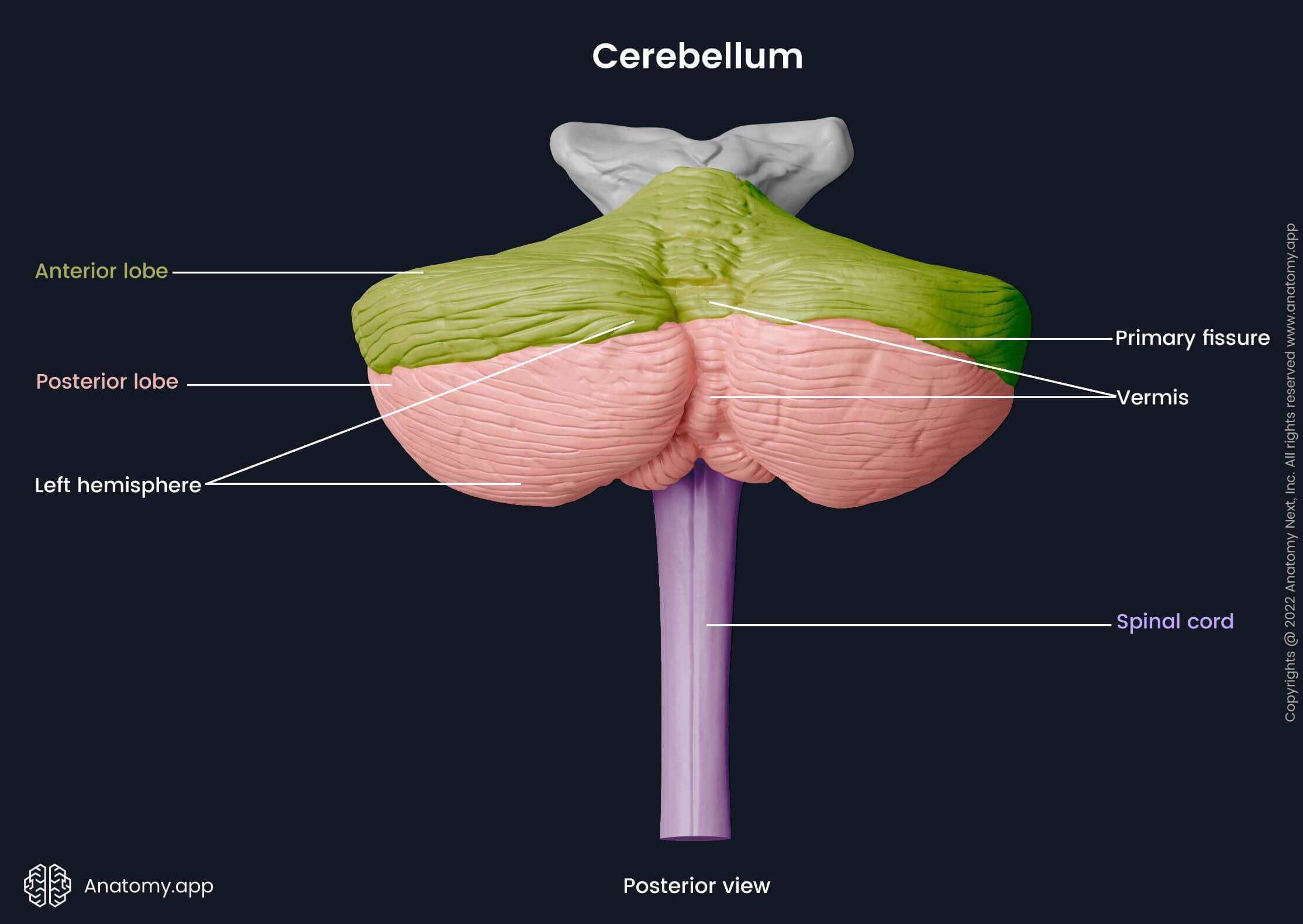
The cerebellum is located in the posterior cranial fossa dorsally to the pons and medulla oblongata. Together with the pons, it forms the metencephalon. Like the cerebrum, the cerebellum also consists of two hemispheres. Both hemispheres are joined together by the vermis. The cerebellum is divided into three lobes, and further subdivisions called lobules. The cerebellum is mainly responsible for motor functions, coordination, balance, and proprioception.
The structures connecting the cerebellum to the anteriorly located brainstem are three pairs of cerebellar peduncles, with each pair of peduncles connected to different parts of the brainstem. The superior cerebellar peduncles connect the cerebellum to the midbrain, the middle cerebellar peduncles to the pons, and the inferior cerebellar peduncles to the medulla oblongata.
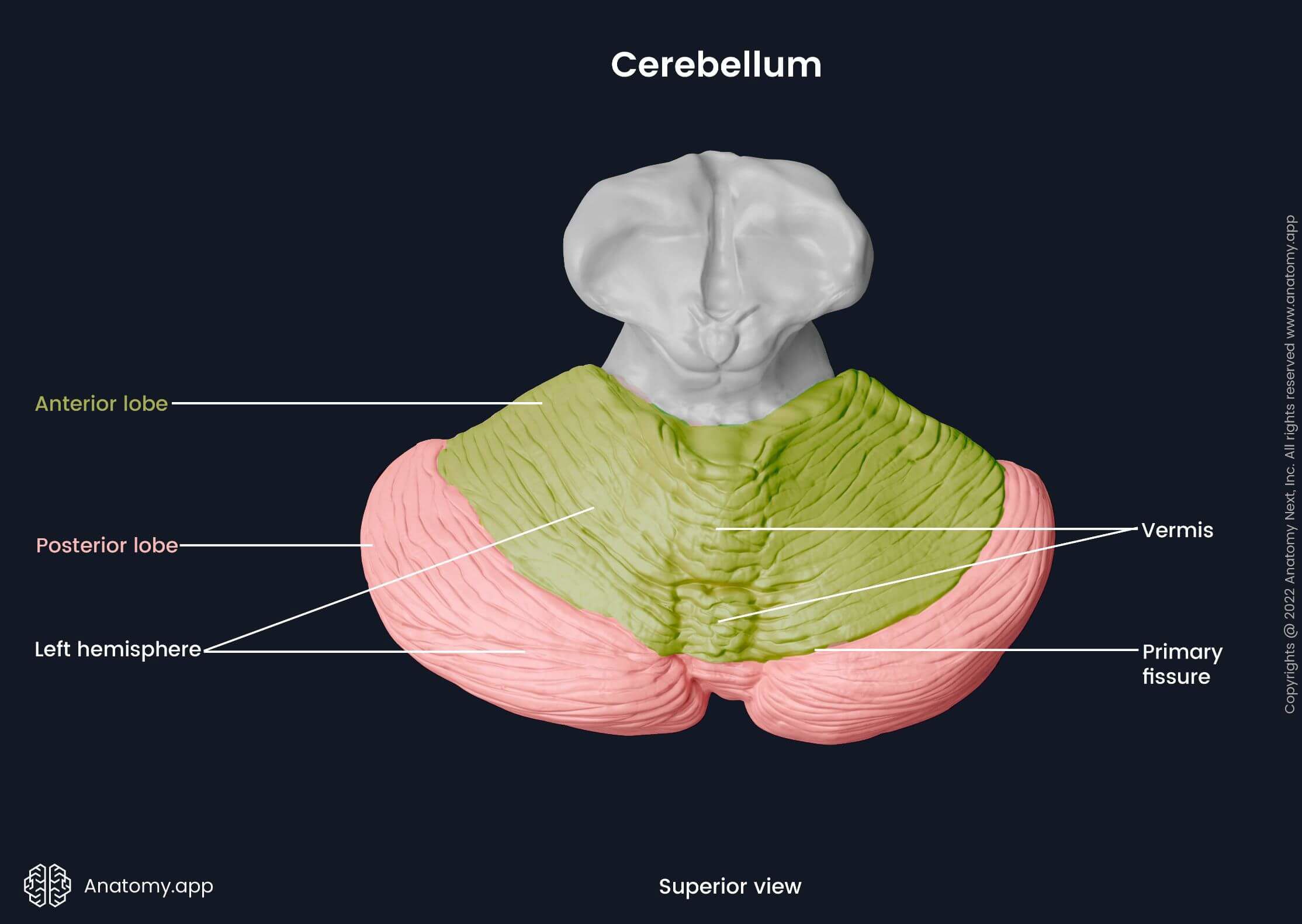
Each hemisphere of the cerebellum is divided into three lobes: the anterior, posterior and flocculonodular lobes. Between the two hemispheres at the midline is the vermis, connecting both hemispheres. Inferiorly, where the vermis meets the posterior lobe, two prominences are present - the cerebellar tonsils. Anteriorly, the superior and inferior medullary velum of the cerebellum and the cerebellar peduncles form the roof of the fourth ventricle.
The cerebellum has two surfaces: the superior (tentorial) and the inferior (occipital) surface. Grooves and small elevations called folia can be seen on the surfaces of the cerebellum. The more minor grooves are called sulci, and the bigger ones are called fissures. There are five fissures seen on the cerebellar surfaces: the primary fissure, postlunate fissure, horizontal fissure, posterolateral fissure and the retrotonsillar fissure.
The primary fissure travels horizontally across the superior surface and separates the anterior and posterior lobes of the cerebellum. The postlunate fissure lies parallel and inferior to the primary fissure. Further down is the horizontal fissure that travels horizontally around the posterior lobe. The posterolateral fissure is located anteriorly between the flocculonodular lobe and posterior lobe. Inferiorly, the retrotonsillar fissure runs along the cerebellar tonsils.
The cerebellar cortex is made up of gray matter forming tight folds known as folia. The previously described fissures are formed between these folds. The inner core of the cerebellum is composed of white matter where four bilaterally paired cerebellar nuclei are found: the fastigial, globose, emboliform, and dentate nuclei. Efferent fibers originate from these nuclei. The white matter "branches" between the grey matter of the cortex, forming the arbor vitae (from Latin, meaning "tree of life").
Cerebrum
The most superior and largest part of the brain is called the cerebrum. The cerebrum is also known as the telencephalon, and it is made up of two symmetrical cerebral hemispheres. It is located in the cranial cavity. The outer part of the cerebrum is the cortex. The cortex is formed of gray matter, which is mainly composed of cell bodies. The inner part of the cerebrum is formed by subcortical structures and white matter, which is primarily comprised of myelinated axons.
Each hemisphere has three surfaces and three margins, and is divided into lobes. The three surfaces are the medial, superolateral, and inferior surface. The medial surface is at the surface facing medially the opposing hemisphere's medial surface. The superolateral surface is the laterally facing convex part of the hemisphere. The inferior surface is a horizontal surface found at the bottom of the hemisphere. The margins are found at the meeting points of these surfaces.
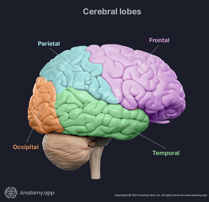
The three margins are: superior, inferomedial, inferolateral. The superior margin runs along the edge where the superolateral and medial surfaces meet. At the confluence of the medial and inferior surface, the inferomedial margin is formed. The inferolateral margin is seen on the border between the superolateral and inferior surfaces. Each cerebral hemisphere can be further divided into six cerebral lobes:
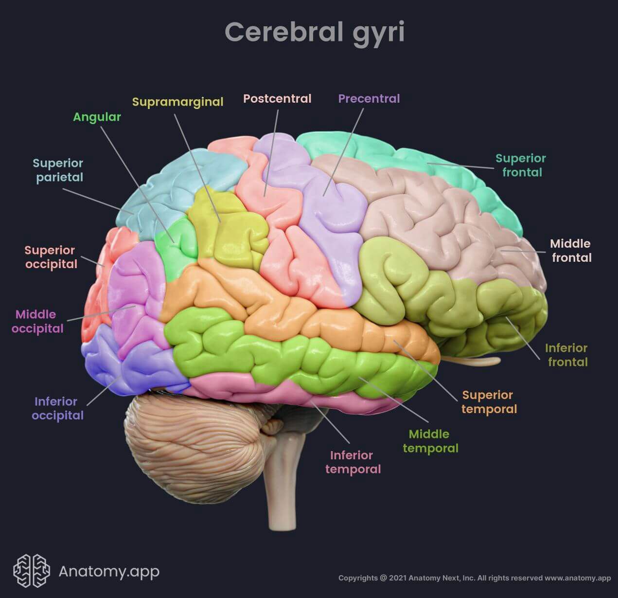
As mentioned, the cerebral cortex is a layer of gray matter forming the outer part of the cerebral hemispheres. On the cerebral cortex, there are folds known as cerebral gyri (sing. gyrus) and grooves called cerebral sulci (sing. sulcus). The deeper grooves are known as fissures. These cerebral folds and grooves increase the surface area of the cerebral cortex, thus increasing the amount of information that can be processed.
Functionally, the cerebral cortex is divided into the motor, sensory, and association areas. Each lobe contains numerous gyri separated by sulci responsible for selective functions ranging from memory to speech and innumerable others. Sensory areas receive sensory input, motor areas are involved in voluntary movement, and association areas provide complex functions such as thinking, learning, writing and speaking, among others. The major cerebral gyri are as follows:
- Frontal lobe:
- Precentral gyrus (anterior paracentral gyrus on the medial surface)
- Superior frontal gyrus
- Middle frontal gyrus
- Inferior frontal gyrus
- Parietal lobe:
- Postcentral gyrus (posterior paracentral gyrus on the medial surface)
- Superior parietal lobule (precuneus on the medial surface)
- Inferior parietal lobule, formed by two gyri:
- Temporal lobe:
- Occipital lobe:
- Superior occipital gyrus (cuneus on the medial surface)
- Middle occipital gyrus
- Inferior occipital gyrus
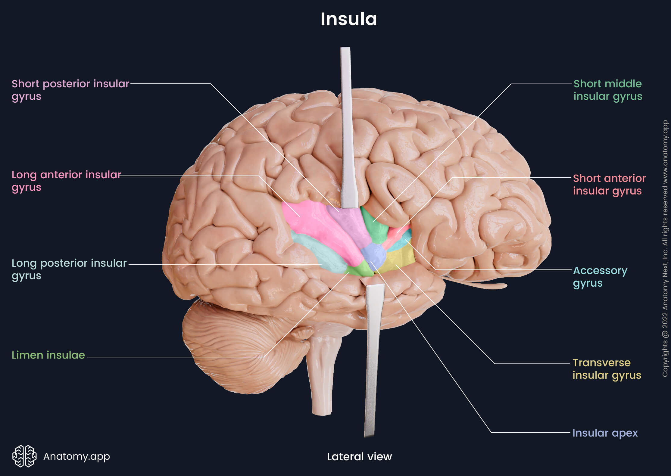
- Insula:
- Transverse insular gyrus
- Accessory gyrus
- Short anterior insular gyrus
- Short middle insular gyrus
- Short posterior insular gyrus
- Long anterior insular gyrus
- Long posterior insular gyrus
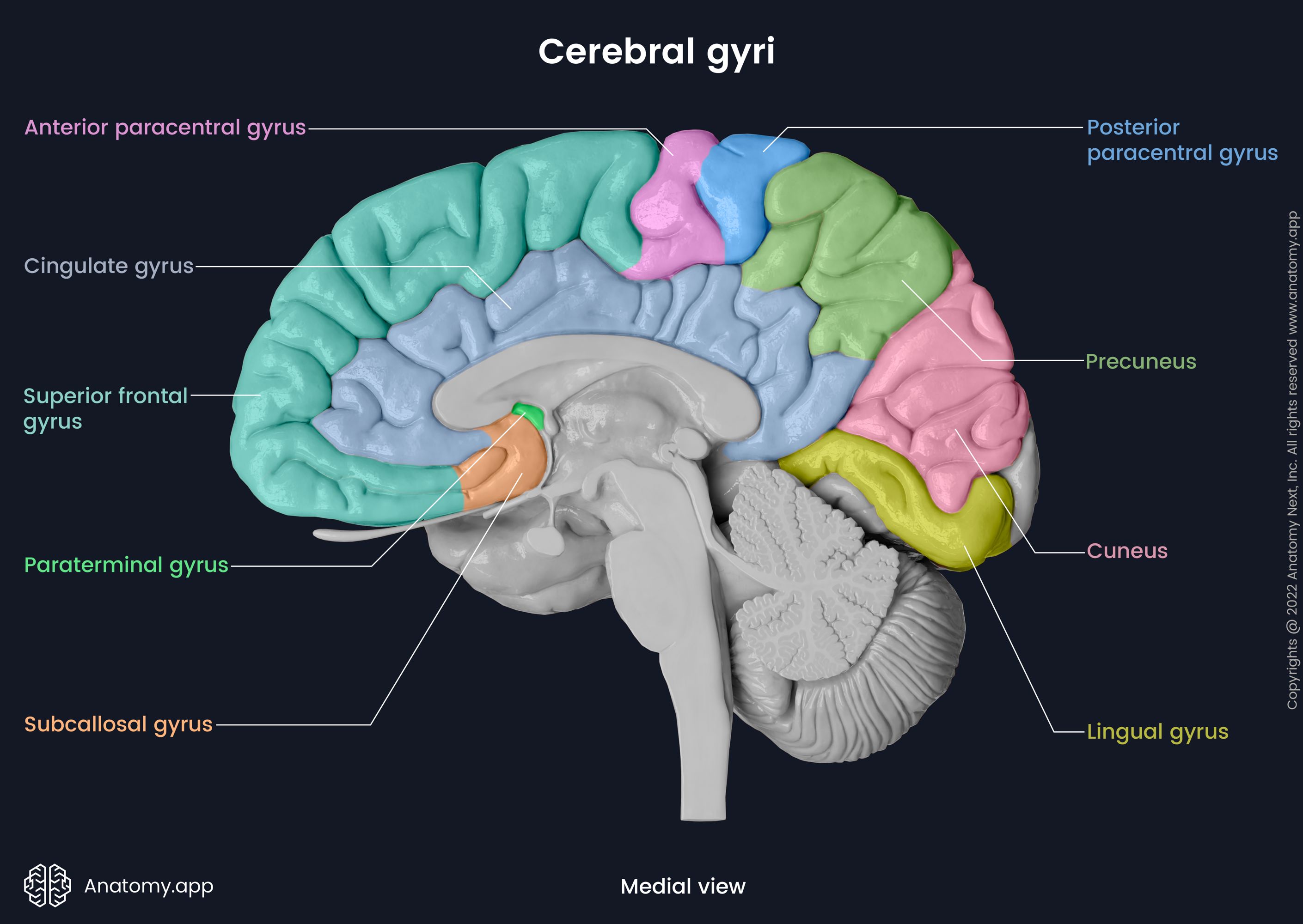
Some of the previously mentioned cerebral gyri extend on the inferior and medial surfaces of the cerebrum. On the medial surface they are known as follows:
- Superior frontal gyrus (superior frontal gyrus of the frontal lobe)
- Anterior paracentral gyrus (precentral gyrus of the frontal lobe)
- Posterior paracentral gyrus (postcentral gyrus of the parietal lobe)
- Precuneus (superior parietal lobule of the parietal lobe)
- Cuneus (superior occipital gyrus of the occipital lobe)
Besides the mentioned gyri, the medial surface of the cerebrum also contains the following gyri:
- Cingulate gyrus
- Paraterminal gyrus
- Subcallosal gyrus
- Lingual gyrus
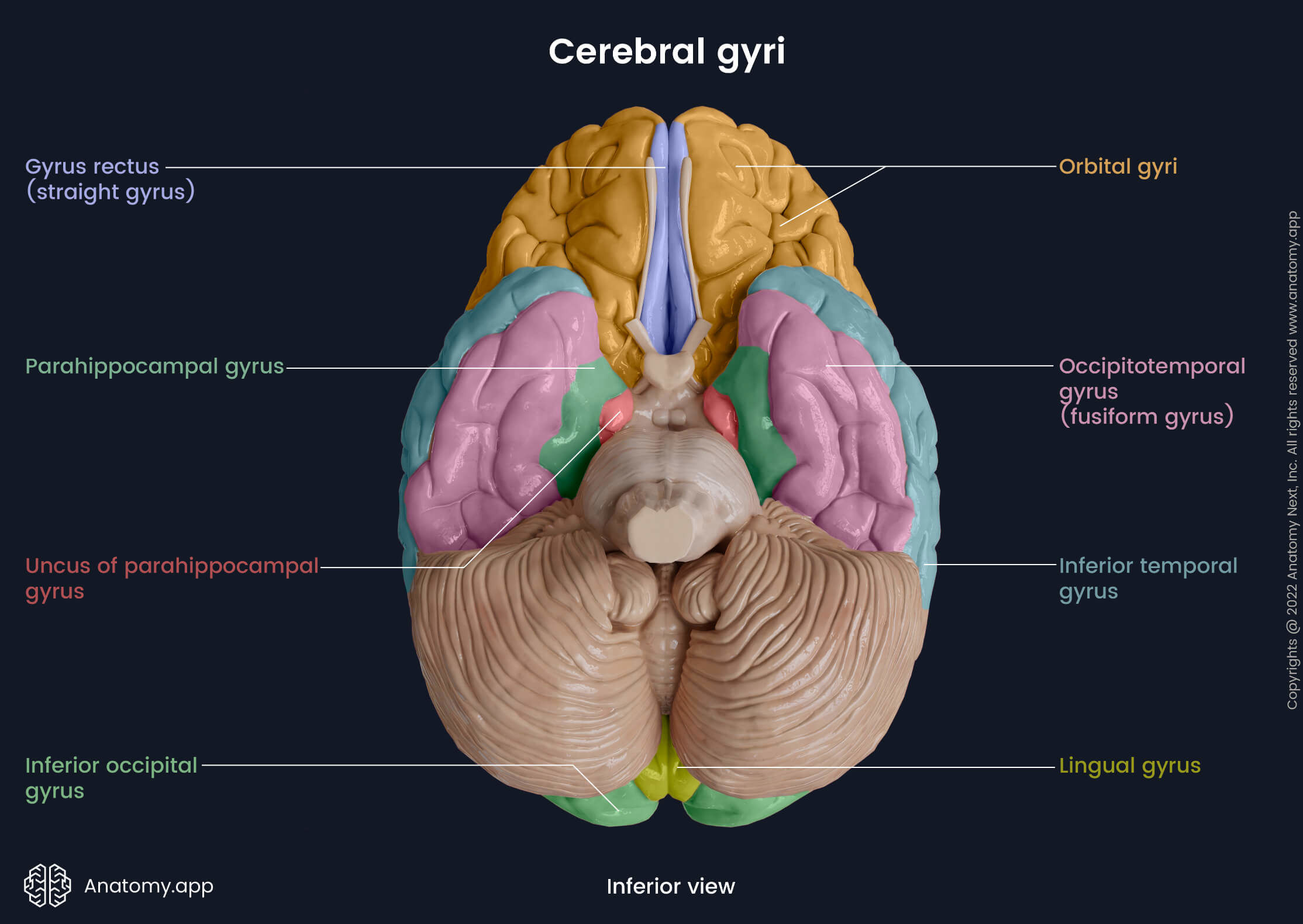
The inferior surface of the cerebrum presents the following gyri:
- Straight gyrus (gyrus rectus)
- Orbital gyri
- Inferior temporal gyrus
- Occipitotemporal (fusiform) gyrus
- Parahippocampal gyrus
- Lingual gyrus
- Inferior occipital gyrus
Both cerebral hemispheres are separated at the top by the longitudinal fissure and inferiorly are bound together by the corpus callosum. Besides the longitudinal fissure, each hemisphere contains three major cerebral sulci (between the cerebral lobes):
- Central sulcus - located on the superolateral surface and separates the frontal and parietal lobes;
- Lateral sulcus - lies at the bottom of the central sulcus and separates the parietal and temporal lobes;
- Parieto-occipital sulcus - situated on the medial surface between the parietal and occipital lobes.
Nevertheless, each cerebral lobe presents its own sulci. For example, the frontal lobe has the superior frontal and inferior frontal sulci amongst others. And the major sulci of the temporal lobe are the superior temporal and inferior temporal sulci.
The frontal lobes are located in the anterior cranial fossa. These are the biggest lobes of the brain and are most prone to traumatic injury. Anteriorly, the prefrontal cortex is located. It is responsible for complex cognitive processes such as thinking, planning, problem-solving, and social behavior. Anterior and parallel to the central sulcus, the primary motor cortex is located in the precentral gyrus. The motor cortex is responsible for initiating voluntary movement.
The superior and inferior frontal sulci divide the anterior part of the frontal lobe into the superior, middle and inferior frontal gyri. The middle frontal gyrus contains the somatomotor association area, which coordinates and forms complex movements. Below the inferior frontal sulcus, the inferior frontal gyrus is situated. The Broca's area is located in the dominant hemisphere's inferior frontal gyrus. For most people the left hemisphere is the dominant one. Broca's area is responsible for speech production.
The occipital lobes are situated in the posterior cranial fossa and make up the rear part of the cerebrum. They are divided into the superior, medial and inferior occipital gyri. The primary visual cortex is located at the posterior pole, and the visual association area is anterior to it. The primary visual cortex detects simple visual stimuli. The visual association area processes visual information. Moreover, it is also responsible for the formation of visual memory.
Superiorly, between the frontal and occipital lobe lies the parietal lobe. Right behind the central sulcus is the postcentral gyrus. It houses the primary somatosensory cortex, which receives multisensory information from the whole body. The data is further processed in the behind lying somatosensory association area. Inferiorly, the supramarginal gyrus contains Wernick's area that is responsible for speech and language comprehension.
Beneath the parietal lobe is the temporal lobe. It is located in the lateral part of the middle cranial fossa. On the temporal lobe, the superior and inferior temporal sulci are seen. They run parallel to the lateral sulcus. These sulci form three gyri on the temporal lobe's surface: the superior, middle and inferior temporal gyrus. The superior temporal gyrus houses the primary auditory cortex and the auditory association area. They detect and process auditory stimuli and sound characteristics such as tone and loudness.
The insula is located deep within the lateral sulcus and is covered by the frontal, parietal and temporal lobes. The insula is divided into two lobules - anterior and posterior - by the oblique central sulcus. At the bottom rear corner of the anterior lobule is the insular apex from which gyri and sulci radiate out. On the posterior lobule are the postcentral insular sulcus and two gyri. The insula is involved in controlling autonomic functions.
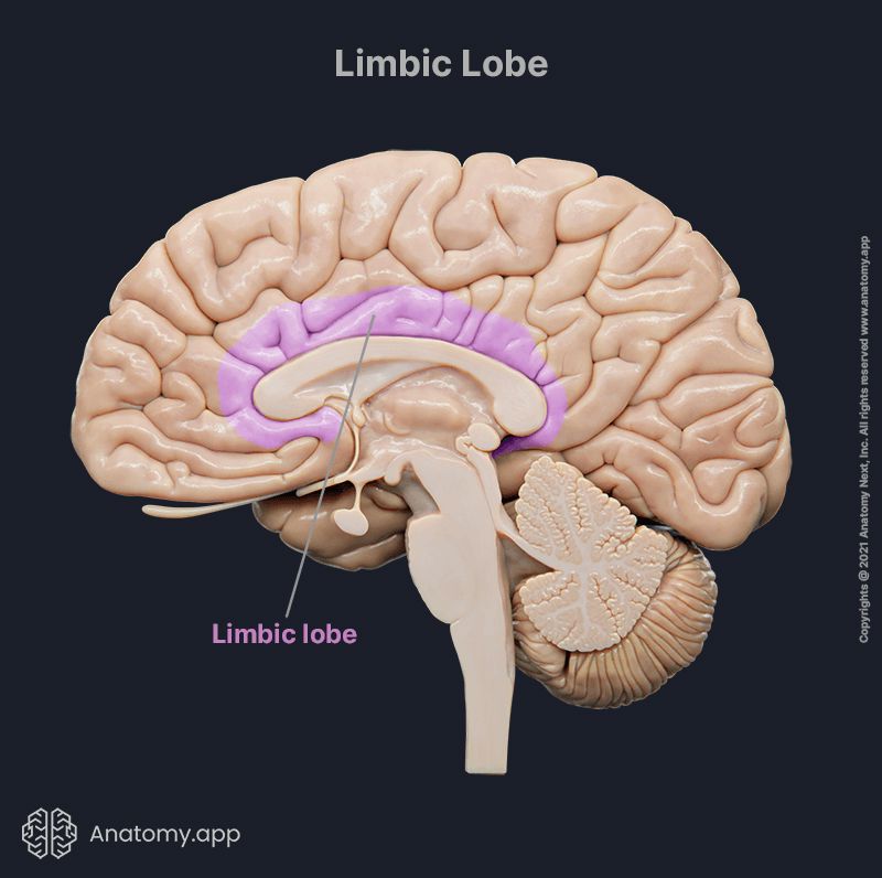
The limbic lobe is found deep in the cerebrum above the corpus callosum. In some literature, it is described as part of the limbic system and not a separate lobe. The limbic lobe is located in the medial aspect of each cerebral hemisphere. It is arc-shaped and contains parts of the frontal, parietal and temporal lobes. The limbic lobe modulates emotions, learning, memory, and visceral, hormonal and automatic functions.
Subcortical structures
Subcortical structures are neural formations in the brain located deeper below the cerebral cortex, not visible from the outside of the brain. They are part of the cerebrum and diencephalon. Subcortical structures include the previously described diencephalon, the limbic structures, basal ganglia, pituitary and pineal glands. They partake in the regulation of emotion, memory, pleasure, control, hormonal production and more.
The pituitary gland sits in the sella turcica in the sphenoid bone, and it connects to the hypothalamus. The pituitary gland has an anterior and posterior lobe. The gland secretes hormones that further affect other organ systems. The anterior lobe secretes the growth hormone, prolactin, follicle-stimulating hormone, luteinizing hormone, thyroid-stimulating hormone, and adrenocorticotropic hormone. The posterior lobe secretes oxytocin and vasopressin.
The limbic structures together are known as the limbic system. It is the main regulatory center for emotions and social behavior. The structures are responsible for modulating reproductive behavior, motivation, stress, motherly instincts, memory, satiety and hunger. To execute and regulate all the previously mentioned mechanisms, the limbic system communicates closely with the basal ganglia, diencephalon, and other brain parts.
The limbic system is composed of both cortical and subcortical structures. The cortical structures are the neocortex, orbital frontal cortex and insular cortex, the hippocampus, and the cingulate, subcallosal and parahippocampal gyri. The subcortical structures of the limbic system are also known as the limbic lobe. These include the olfactory bulb, hypothalamus, septal nuclei, amygdala, and the anterior and dorsomedial thalamic nuclei.
The basal ganglia are a group of subcortical nuclei found deep in the white matter of the brain. They are located within the telencephalon, midbrain, and diencephalon and closely communicate with the motor cortex and cerebellum. The basal ganglia are part of the extrapyramidal motor system, and all the nuclei are primarily responsible for motor function. There are five pairs of nuclei: the subthalamic nucleus, globus pallidus, caudate nucleus, substantia nigra, and the putamen.
Meninges of brain
Three membranous layers known as meninges envelop the brain and spinal cord. The outermost layer is the dura mater, the middle layer is the arachnoid mater, and the pia mater is the deepest membrane. The meninges protect the brain and spinal cord. They are divided into the spinal and cranial meninges, which are continuous with each other. Between the meninges are spaces filled with cerebrospinal fluid (CSF). These spaces are called the epidural, subdural, and subarachnoid spaces.
The epidural space surrounds the spinal cord between the dura mater and vertebral wall. It is filled with fat cells and small blood vessels. The subdural space can appear between the dura mater and arachnoid mater post-mortem or in case of trauma or as a result of other pathological processes. The subarachnoid space is the area between the arachnoid and pia mater. It is filled with cerebrospinal fluid, connective tissue trabeculae and houses the cranial arteries and veins.
The pia mater is composed of thin fibrous tissue. Blood vessels pass through it and supply the brain. The pia mater closely covers and follows the involutions of the brain. Above, the pia mater is separated from the arachnoid mater by the subarachnoid space. The arachnoid mater is the middle meningeal layer, and it is a thin spider web-like membrane. Inferiorly, small projections called arachnoid trabeculae pass through the subarachnoid space and connect it to the pia mater.
The dura mater is a superficial layer of the meninges closest to the skull. It is the hardest of all meninges. It is composed of an outer periosteal and inner meningeal layer. The dura mater contains dural sinuses and forms continuations of its meningeal layer called dural partitions that project into the cranial cavity. The dural partitions are the falx cerebri, tentorium cerebelli, falx cerebelli and diaphragma sellae.
Dural partitions
The falx cerebri descends from the dura mater and runs along the midline of the frontal and parietal bones. Anteriorly, it is attached to the ethmoid bone's crista galli and the frontal crest of the frontal bone. It advances along the coronal plane between the two cerebral hemispheres. Posteriorly, it attaches to and merges with the tentorium cerebelli. The inferior border runs along the midline of the longitudinal fissure.
The tentorium cerebelli is located in the posterior cranial fossa and goes along the transverse plane. It separates the cerebellum from the posterior parts of the cerebral hemispheres. Posteriorly, the tentorium cerebelli attaches to the occipital bone. Laterally, it is connected to the petrous portion of the temporal bone. In the middle, it forms an oval opening through which the midbrain passes. And anteriorly, the tentorium cerebelli attaches to the clinoid process of the sphenoid bone.
The falx cerebelli is a small dura mater projection located in the posterior cranial fossa. It runs between the two cerebellar hemispheres. Posteriorly, the falx cerebelli attaches to the internal occipital crest of the occipital bone. Superiorly, the falx cerebelli connects to the tentorium cerebelli. The projection is situated along the sagittal axis, and its anterior border is free.
The diaphragma sellae is a small dural projection that covers the hypophyseal fossa. The hypophyseal fossa is found in the sella turcica, a midline depression in the sphenoid bone. At the center of the diaphragm, there is an opening. The infundibulum that connects the hypothalamus to the pituitary gland passes through this opening accompanied by blood vessels.
Dural sinuses
The dural sinuses are venous sinuses found on the inside of the cranial cavity. They are situated between the outer periosteal and inner meningeal layer of the cranial dura mater. Some sinuses are found in the projections of the dural partitions. The dural sinuses empty mainly into the internal jugular vein. Epithelial cells line the inside of the venous sinuses, and they receive blood from the cerebral veins and CSF from the subarachnoid space.
The falx cerebri houses the superior and inferior sagittal sinus. The straight, transverse and superior petrosal sinuses are located in the tentorium cerebelli. The falx cerebelli is home to the occipital sinus. On both lateral sides of the diaphragma sellae, the cavernous sinuses pass it by. The diaphragma sellae itself contains the anterior and posterior intercavernous sinus.
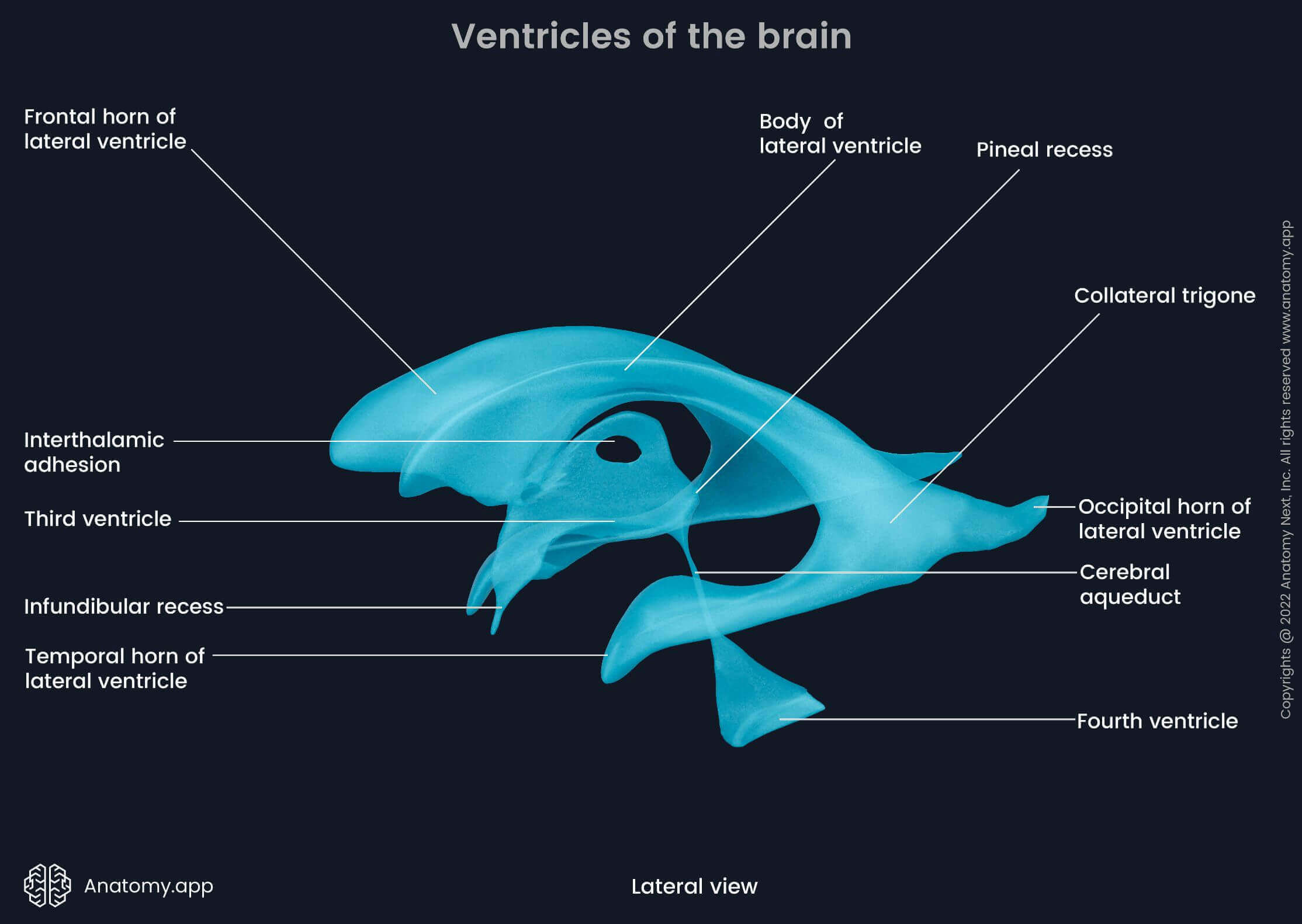
Ventricles of brain
The ventricles are an interlinked network of cavities in the brain. They are filled with cerebrospinal fluid. The cerebrospinal fluid supplies the brain and the spinal cord with glucose and other nutrients and removes waste products. Moreover, the CSF protects the brain and spinal cord through shock-absorption. Together there are four ventricles of the brain: two lateral ventricles, the third, and the fourth ventricle.
The CSF is produced by the choroid plexus. Extension of the pia mater's vascular layer called tela choroidea forms the choroid plexus. The tela choroidea folds to increase its surface area and is covered by the ependyma and choroid epithelium. Its capillaries are fenestrated and have high permeability. There is a choroid plexus located in each ventricle.
The two bilaterally located lateral ventricles can be further divided into the central part and three horns - the frontal, temporal and occipital horns. They are the most prominent ventricles and are situated in both cerebral hemispheres. The right lateral ventricle is located in the right hemisphere, and the left - in the left hemisphere. Both lateral ventricles connect to the third ventricle via the interventricular foramen of Monro at the frontal horn.
The third ventricle is located largely in between the two thalami in the middle of the cerebrum. It can be divided into the following parts (recesses): the supraoptic, suprapineal, pineal and infundibular recesses. The choroid plexus enters the third ventricle anteriorly through the interventricular foramen of Monro. Posteriorly and inferiorly, the third ventricle transitions into the fourth ventricle through the cerebral aqueduct of Sylvius.
The fourth ventricle is located in the brainstem. The rhomboid fossa forms its floor. And its roof is constructed by the superior and inferior medullary vela of the cerebellum. Inferiorly, the fourth ventricle flows into the cerebral canal through the obex. Additionally, connect the fourth ventricle to the cisterns. The cisterns are widenings of the subarachnoid space. They include the interpeduncular, chiasmatic, prepontine and corpus callosum cisterns.
The lateral ventricles produce most of the cerebrospinal fluid (CSF). The CSF flows further into the third ventricle through the interventricular foramen of Monro, where there is additional production of the fluid. The CSF continues into the fourth ventricle through the aqueduct of Sylvius. From there, the fluid continues into the spinal canal through the obex and into the cisterns through the foramina of Luschka and foramen of Magendie. The cerebrospinal fluid is then uptaken by the arachnoid granulations into the dural venous sinuses and joins the systemic circulation from the cisterns.
Brain disorders
The most common brain diseases and neurological disorders are autoimmune diseases, neurodegenerative diseases, infections, seizures, trauma and tumors. These categories can overlap and combine in many ways. The exact prevalence and incidence of brain diseases differ according to the population studied, location and other specific factors.
Autoimmune diseases that affect the brain are, for example, multiple sclerosis and vasculitis. Multiple sclerosis may involve the brain and spinal cord. It is an autoimmune disease in which the immune system attacks myelin sheaths covering axons of neurons. This deteriorates the nerves and decreases the body's ability to communicate via the nerves effectively. The clinical picture depends on which nerves are affected.
Neurodegenerative diseases are degenerative diseases of the brain and spinal cord that result in progressive loss of motor functions and decline in cognitive functioning (dementia). Their prevalence is increasing, and this is mainly attributable to aging populations and many other factors. The cause of neurodegenerative diseases is a complex interplay between genetics and other external factors such as tumors, trauma, stroke, infections, the use of toxic substances, including alcohol, and more.
Alzheimer's disease and Parkinson's disease are two of the most common neurodegenerative disorders affecting the brain. Alzheimer's disease causes shrinkage of the brain and atrophy of brain cells, often due to abnormal buildup of proteins in and around the brain cells leading to a progressive and continuous decline in cognitive functions, including memory, thinking, reasoning, and changes in social behavior.
Parkinson's disease is a degenerative disease that affects the ability to control movements. It has a slow onset and progresses over time. In the later stages of Parkinson's disease, people have problems controlling movements, including completing tasks such as walking and talking. The main symptoms of this disease include impaired balance, shaking, stiffness, disturbed motor coordination.
Common inflammatory diseases that affect the brain are meningitis and encephalitis. Meningitis is an inflammation of the meninges and can be caused by viral or bacterial agents and other microorganisms. Other common causes include injuries, cancer and certain drugs. Meningitis presents with symptoms such as stiff neck, severe headaches, nausea, and more. In the case of encephalitis, inflammation affects the functional tissue of the brain. It is most commonly caused by an infection or an autoimmune process. Encephalitis usually manifests with similar symptoms to meningitis. However, it may also present with seizures.
References:
- Crossman, A. R., & Neary, D. (2019). Neuroanatomy: an Illustrated Colour Text (6th ed.). Elsevier.
- Vanderah, T. W., & Gould, D. J. (2020). Nolte's The Human Brain (8th ed.). Elsevier.
- Goetz, C. (2007). Textbook of Clinical Neurology (3rd edition). Saunders.
- WHO. (2006). Neurological disorders : public health challenges. https://www.who.int/mental_health/neurology/neurological_disorders_report_web.pdf
Anatomy.app
Contact information
- For questions regarding business inquiries. Please contact:
- info@anatomy.app