- Anatomical terminology
- Skeletal system
- Joints
- Muscles
- Heart
- Blood vessels
- Lymphatic system
- Nervous system
- Respiratory system
- Digestive system
- Urinary system
- Female reproductive system
- Male reproductive system
- Endocrine glands
- Eye
- Ear
Lymphatic system
The lymphatic system (Latin: systema lymphoideum), also known as the lymphoid system, is a complex system that consists of a network of lymphatic vessels (including capillaries, tracts, and ducts) and various lymphoid organs. Through the lymphatic system flows yellowish lymphatic fluid called lymph, and lymphoid organs produce and store lymphatic cells (lymphocytes). Additionally, lymph is filtered through various lymphoid tissue collections called lymph nodes.
The components of the lymphatic system are found throughout all body parts and tissues. The lymphatic system is considered a part of the circulatory system, as well as a part of the immune system. It has a crucial role in immune defense and is important for maintaining fluid balance and absorbing fatty acids in the gastrointestinal tract. Overall, it collects excessive extracellular fluid from the tissue and provides it drainage back into the bloodstream, where it gets eliminated.
The network and branching pattern of lymphatic vessels are very similar to the vascular system, and lymphatics go along with the blood vessels. They are found in every organ and part of the human body except for the epidermis, cartilages, bone marrow and some structures of the eye. Overall, tissues that lack blood vessels also do not contain any lymphatics. The most significant amount of lymphatic vessels are found within the liver and intestines.
Components of lymphatic system
As mentioned above, the lymphatic system consists of numerous lymphatic vessels and various lymphoid organs.
Lymphatic vessels
The lymphatic system arises from a network of lymphatic capillaries that collect excessive extracellular fluid from the tissue. Lymphatic capillaries are the smallest lymphatic vessels. They appear as thin-walled vessels arising blindly within the extracellular space of various tissues.
The lymphatic capillaries of the small intestine are called lacteals. They are found within the villi, and they contribute to the absorption of dietary fats. The lymphatic capillaries further form large capillary networks called lymphatic plexuses and merge to form even larger lymphatic vessels.
The lymphatic vessels can be subdivided into superficial and deep vessels. The superficial vessels are found within the subcutaneous layer of the skin, while the deep vessels are located more deeply and drain internal organs. The superficial vessels eventually will drain into the deep lymphatic vessels.
There is one more difference between the superficial and deep lymphatic vessels. The first group is usually accompanied by veins and venous plexuses, while the deep vessels go alongside the arteries.
Like the veins, the collecting and larger lymphatic vessels have one-way valves preventing lymph from flowing backward. The lymphatic capillaries do not contain valves. Overall, lymph through the lymphatic vessels is moved by the pressure that is put from the arteries on the walls of the lymphatic vessels, pulsation of the arteries (smooth muscle contractions), contractions of the skeletal muscles, and body and respiratory movements.
The superficial and deep lymphatic vessels pass through various lymph nodes that serve as lymph filters. Lymphatic vessels that convey unfiltered lymph from the body tissues to the lymph nodes are known as the afferent lymphatics, while vessels that leave the lymph nodes and convey filtered lymphatic fluid from lymph nodes to subsequent nodes or into the venous system are the efferent lymphatic vessels.
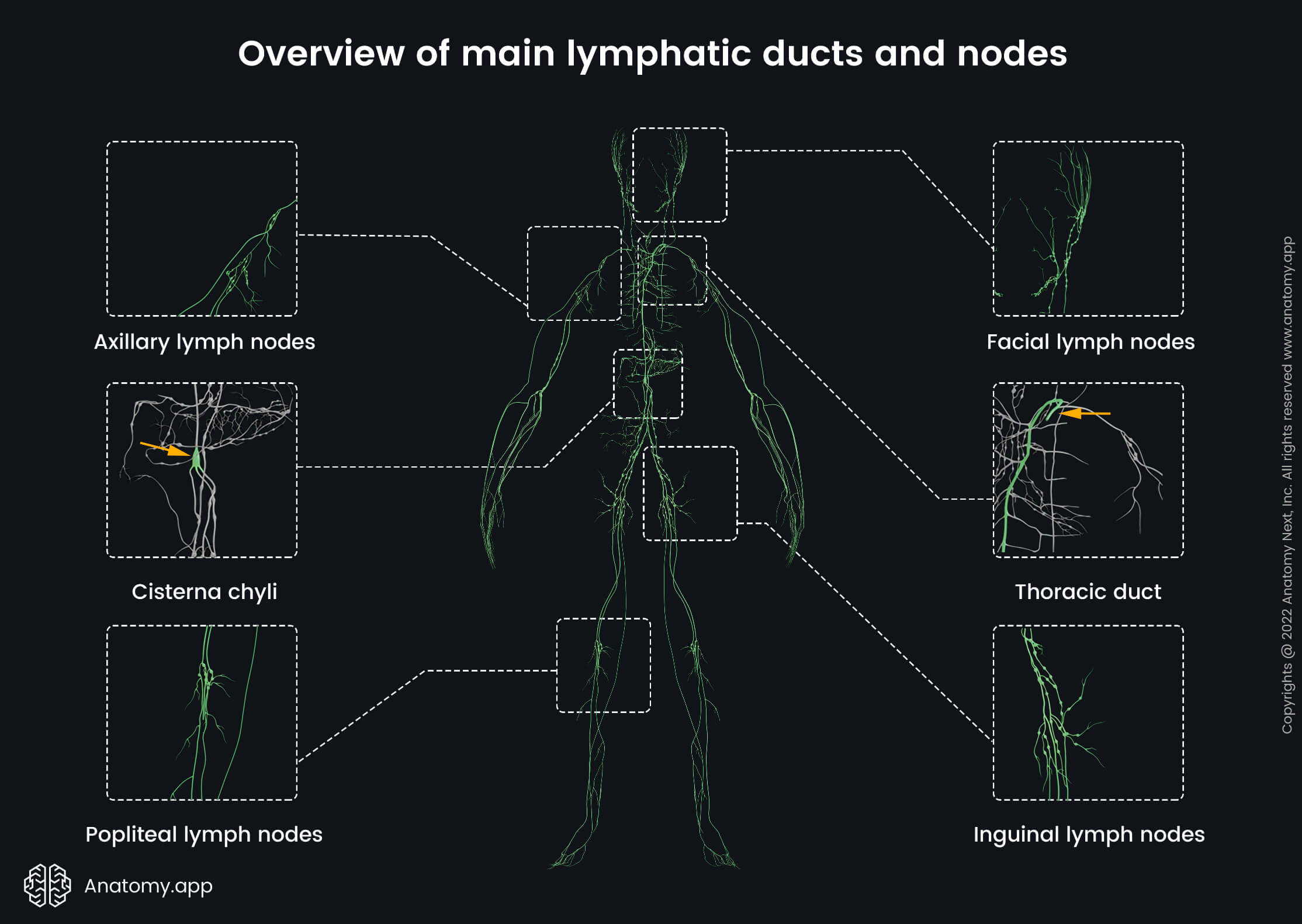
The efferent lymphatic vessels further merge and form larger vessels called lymphatic trunks. Each person has four paired and one unpaired lymphatic trunks:
- Right and left jugular trunks (2) - located in the neck; drain the cervical lymph nodes, which receive lymph from the head and neck;
- Right and left subclavian trunks (2) - found in the upper part of the chest below the clavicle; drain lymph from the axillary lymph nodes that receive lymph from the upper limb;
- Right and left bronchomediastinal trunks (2) - go within the thoracic cavity; drain lymph from the organs of the thorax, including the heart, lungs, and trachea;
- Right and left lumbar trunks (2) - drain lymph from the lower limbs and pelvis;
- Intestinal trunk (1) - the only unpaired trunk that receives lymph from the intestines.
The mentioned trunks are named after the region they drain the lymph from. The intestinal trunk collects lymph from most of the organs of the gastrointestinal tract. It empties in the cisterna chyli, which is a dilated lymphatic sac. The cisterna chyli is formed at the first and second lumbar vertebrae ( L1 - L2) level by the confluence of the intestinal and both lumbar lymphatic trunks.
And finally, lymphatic trunks unite to form lymphatic ducts. The cisterna chyli is the origin site for the largest lymphatic vessel in the human body, called the thoracic duct. Besides the thoracic duct, there is one more lymphatic duct formed by the merging of lymphatic trunks, and it is known as the right lymphatic duct.
Right lymphatic duct
The right lymphatic duct is one of two main ducts in the human body. As the name suggests, it is found on the right side of the body and drains most of the right upper side, including the right side of the head and neck, the right upper extremity and the right upper trunk.
The origin and termination sites are variable. However, the right lymphatic duct usually arises from the confluence of the right bronchomediastinal, jugular and subclavian trunks. It is found in the neck anterior to the anterior scalene muscle.
Overall, the right lymphatic duct is a short trunk. From its origin, the right lymphatic duct extends only for 0.4 - 0.8 inches (1 - 2 cm) and then flows into the venous system at the junction site of the right internal jugular vein and the right subclavian vein, or it flows into the right brachiocephalic vein.
Thoracic duct
The thoracic duct, or the left lymphatic duct, is the largest lymphatic vessel in the human body. It drains most of the body except the parts that are drained by the right lymphatic duct. Opposite the right lymphatic duct, the thoracic duct is a relatively long vessel, and it is 14.2 - 17.8 inches (36 - 45 centimeters) long. Its diameter is about 0.08 - 0.24 inches (2 - 6 mm).
Like other vessels of the lymphatic system, the thoracic duct is a highly variable vessel. It arises in the abdomen from the superior aspect of the cisterna chyli at the level of the twelfth thoracic vertebra (T12) and ends at the root of the neck.
The thoracic duct leaves the abdominal cavity via the aortic hiatus. Then it ascends in the thoracic cavity going anterior and to the right side of the spine between the thoracic aorta and azygos vein.
Upon entering the inferior aspect of the superior mediastinum, it typically crosses to the left side of the spine. It happens at the level of the fourth to fifth thoracic vertebrae (T4/T5). Further, the thoracic duct ascends vertically through the superior mediastinum.
Eventually, the thoracic duct drains into the left internal jugular vein, the left subclavian vein, or in the angle formed by the junction of the left subclavian vein and the left internal jugular vein.
Lymphoid organs
Besides the various lymphatic vessels going through the human body, the lymphatic system is also composed of various lymphoid organs that are involved in the production of lymphocytes. All lymphoid organs can be subdivided into primary and secondary organs.
Primary lymphoid organs
The primary lymphoid organs are the primary sites where the lymphocytes are produced and formed. These include the bone marrow and the thymus. The bone marrow is the primary organ for the lymphocyte production. The thymus is positioned behind the sternum anterior to the pericardium.
There are two main types of lymphocytes - B and T lymphocytes. B lymphocytes fully develop in the bone marrow, while T lymphocytes originate from the stem cells in the bone marrow but mature and further develop in the thymus. The primary lymphoid organs provide antigen-independent development of lymphocytes.
Secondary lymphoid organs
The secondary lymphoid organs are the sites where lymphocytes provide their functions. These include the spleen, tonsils, lymph nodes, vermiform appendix and lymphoid tissue of the mucosae (MALT).
After the lymphocytes finish the maturation process in the primary lymphoid organs, they travel to the secondary organs, where they meet the antigens for the first time and undergo the final stage of maturation before they fight against specific pathogens. The secondary lymphoid organs provide antigen-dependent activation.
The spleen is the largest organ of the lymphatic system. It appears somewhat purplish and is around the size of a fist. It is positioned in the left upper abdominal quadrant. Besides its immune functions, it also serves as a blood filter and is a site where senescent and damaged red blood cells are degraded and recycled.
The tonsils are lymphatic tissue aggregates that serve as filters to prevent antigens from entering the body. Overall, each person has unpaired pharyngeal and lingual tonsils and paired palatine and tubal tonsils. Some mucous membranes also contain lymphatic tissue aggregates that provide the same function as the tonsils.
The lymph nodes are small lymphatic tissue collections that are found along the course of the lymphatic vessels. They serve to monitor the content of lymph. Lymph nodes work as filters, and they drain excess extracellular fluid and leaked plasma proteins, as well as eradicate pathogens. They get swelled during infections due to the buildup of lymph, antigens, immune cells, debris and other particles.
Lymph
Lymph or lymphatic fluid is a yellowish, watery, transparent biological fluid that flows through lymphatic vessels. As blood travels via arterioles, most components of blood plasma get through the walls of the arterioles into the interstitial space due to an increase in the hydrostatic pressure.
Most of the fluid is later reabsorbed back into the bloodstream via venules. However, up to 10 - 15% of the fluid stays in the tissue. It is called interstitial fluid, and as it accumulates, it further gets absorbed along with other substances into the lymphatic capillaries, where it forms lymph.
Lymph is similar in composition to blood plasma, and the majority of it consists of water. Also, it contains carbohydrates (mainly glucose), proteins, lipids, ions and some cells (mainly lymphocytes). The composition of lymph can vary depending on the region of the body.
As mentioned previously, the lymphatic vessels within the small intestine are called lacteals. Lymph in the gastrointestinal tract is called chyle. Additionally, it contains fatty acids, cholesterol, glycerol, and fat products, and that is why chyle appears as a milky white fluid.
Functions of lymphatic system
The lymphatic system serves several crucial functions for maintaining homeostasis. Its primary function is maintaining the fluid balance within the body, as it balances the volume of interstitial fluid and drains it into the venous circulation. If the fluid does not get drained, it can accumulate within the tissue and cause localized tissue swelling called lymphedema.
Together with the excess fluid, lymph also transports molecules that are too large to diffuse through the capillary walls, including the excess protein and lipid molecules. This also explains why the small intestine has a vast network of lymphatic vessels. Its lymphatics play a part in the absorption of fat-soluble vitamins and fats. They are absorbed via lacteals and further carried to the venous circulation.
The lymphatic system also participates in immune surveillance, fighting against various foreign antigens. If an antigen is detected, the immune cells produce antibodies and start an immune response to destroy and eliminate the foreign particle, preventing the body from damage.
Despite previous information, some latest studies also show that lymphatic vessels are found within the meninges, and they are involved in the cerebrospinal fluid outflow from the central nervous system.
Anatomy.app
Contact information
- For questions regarding business inquiries. Please contact:
- info@anatomy.app