- Anatomical terminology
- Skeletal system
- Skeleton of trunk
-
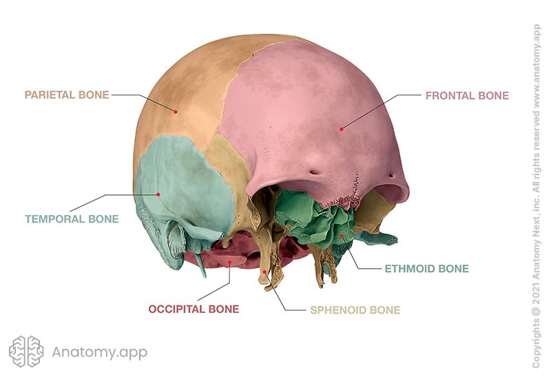
Skull
- Neurocranium
- Viscerocranium
- Auditory ossicles
- Sutures of skull
- Topography of skull
- Skeleton of upper limb
- Skeleton of lower limb
- Joints
- Classification of joints
- Joints of skull
- Joints of spine
- Joints of lower limb
- Muscles
- Heart
- Blood vessels
- Lymphatic system
- Nervous system
- Respiratory system
- Digestive system
- Urinary system
- Female reproductive system
- Male reproductive system
- Endocrine glands
- Eye
- Ear
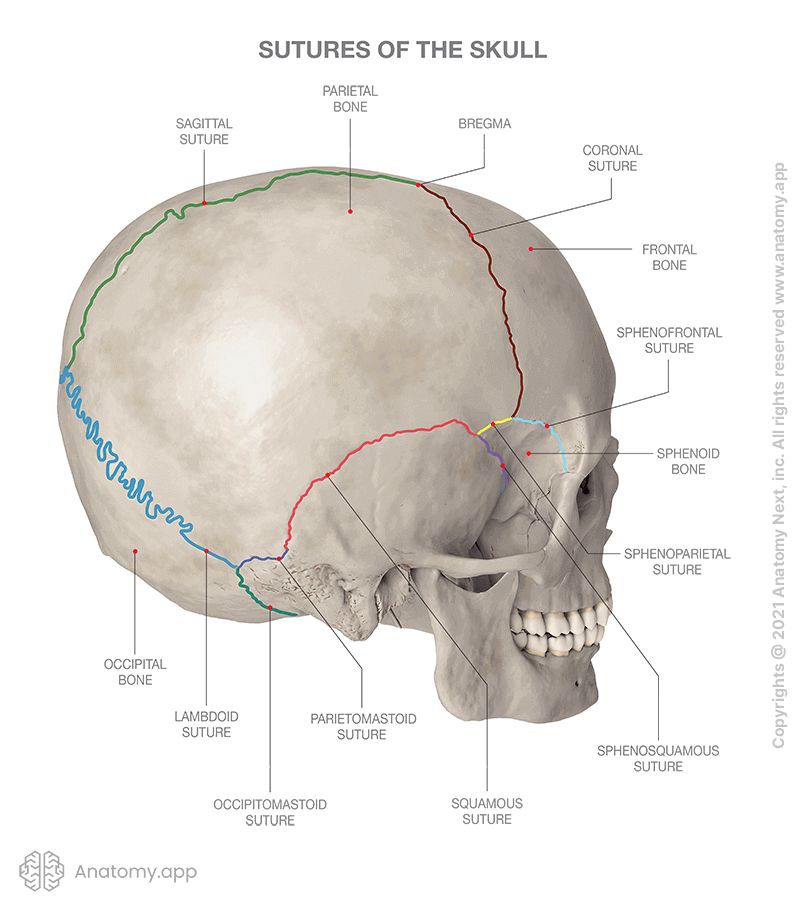
Sutures of skull
Sutures of the skull, also known as cranial sutures, are fibrous joints with a fracture-like appearance found between the bones of the skull. Sutures are formed during embryonic development. They are sites for bone expansion, ensuring craniofacial growth during the embryonic, postnatal and later growth periods. The cranial sutures ossify at different rates, but most sutures have ossified by the age of 20.
Sutures of an adult skull are categorized as synarthroses. Synarthrosis is a type of joint that, under normal circumstances, is immobile. It also doesn't have a capsule or joint cavity. Sutures are connected by intervening fibrous connective tissue composed mainly of collagen. They form immobile articulations between the bones of the skull.
Depending on the age and skeletal growth stage, the different sutures are seen at various stages of fusion and ossification. The ossification phase can be used for estimating the age of death. Where the sutures intersect, bony landmarks like elevations or depressions can be observed. Cranial landmarks, such as the bregma and lambda, can be used as craniometric reference points in radiology and anthropological measurements.
Many sutures join together the bones of the skull. They connect and form junctures with each other. Sutures can be further subdivided according to their location into sutures of the neurocranium, sutures of the viscerocranium, and the sutures between the neuro- and viscerocranium. The latter are seen on the border between cranial bones belonging to the neurocranium and those belonging to the viscerocranium.
Development of sutures
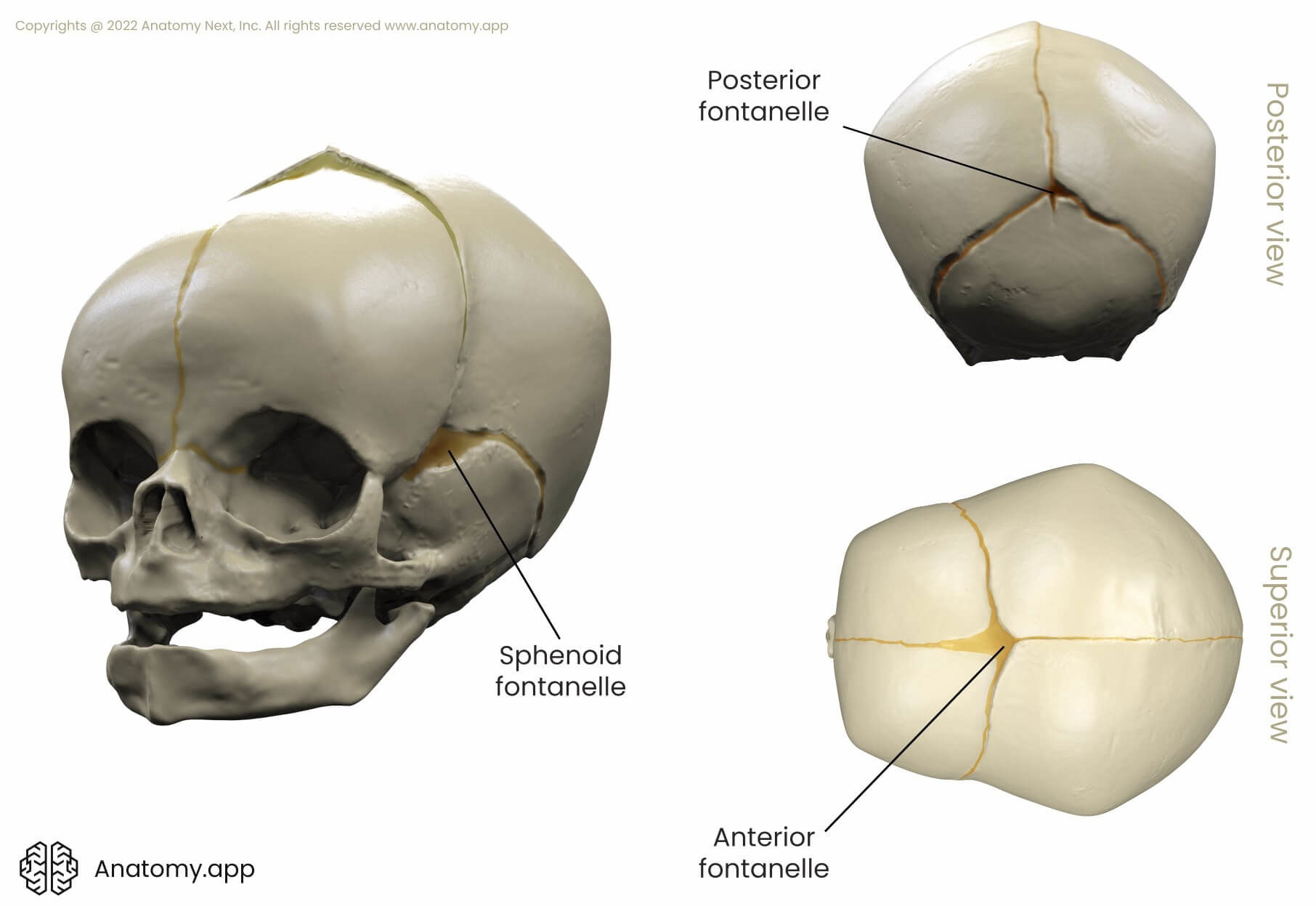
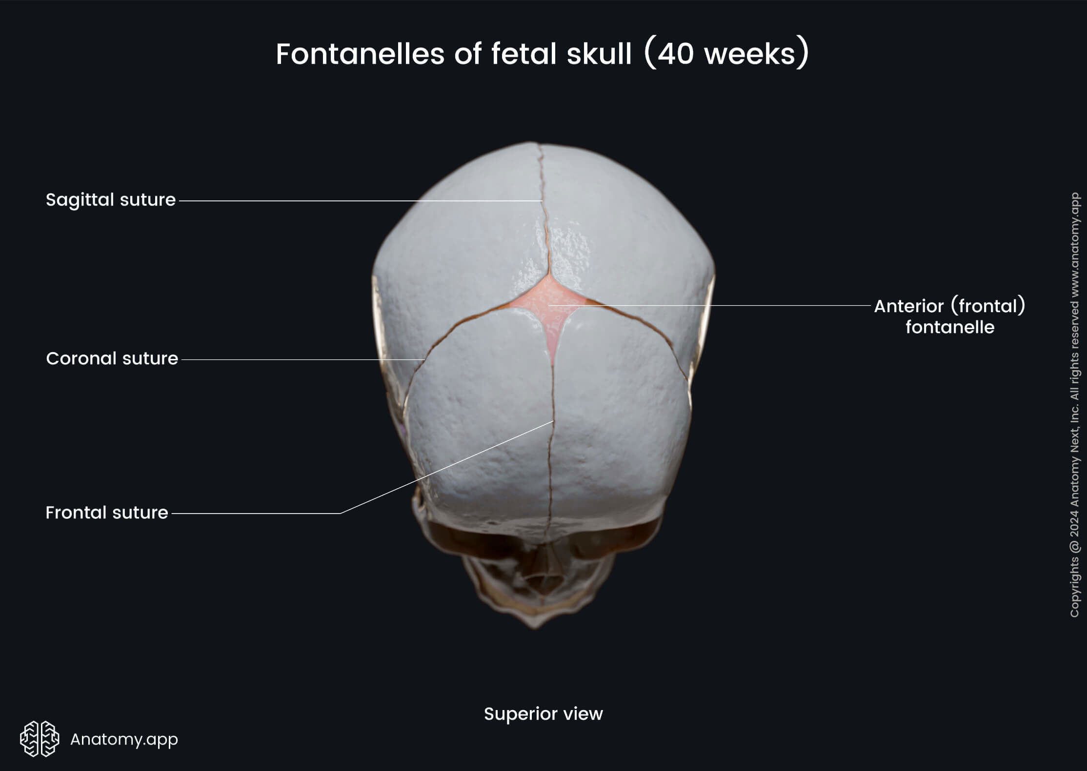
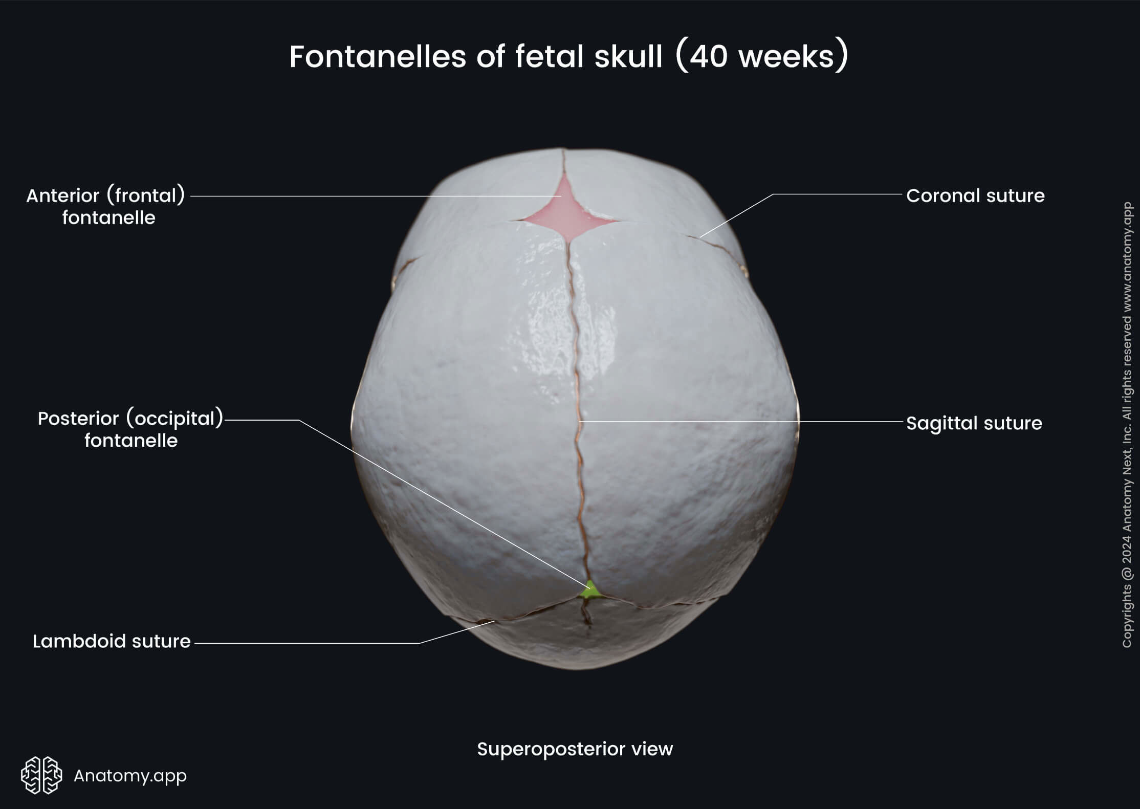
The rate at which sutures fuse and ossify is physiologically relevant. During childbirth, the fibrous joints provide a malleable quality to the child's head and allow the bones to move. In neonates, the sutures are incompletely fused, leaving membranous gaps called fontanelles. Fontanelles are also often called soft-spots. There are six fontanelles: frontal, occipital, mastoid (2), and sphenoid fontanelles (2).
Additionally to the sutures, small naturally occurring irregular bones called wormian bones, also called sutural bones, can be seen along or within the cranial sutures. They often appear as single bones, commonly in the lambdoid suture. They mainly arise from separate ossification centers at the major and minor fontanelles. Wormian bones can be mistaken for fractures on radiological imaging.
During embryological development and childhood, sutures function as intramembranous bone growth sites and only fuse and ossify later in life. New bone is produced at the sutural edges as a result of scrupulously coordinated external stimuli. Delayed or restricted bone growth results in suture agenesis and wide-open fontanelles. Moreover, excessive bone growth can also result in deformities of the skull.
Fontanelles
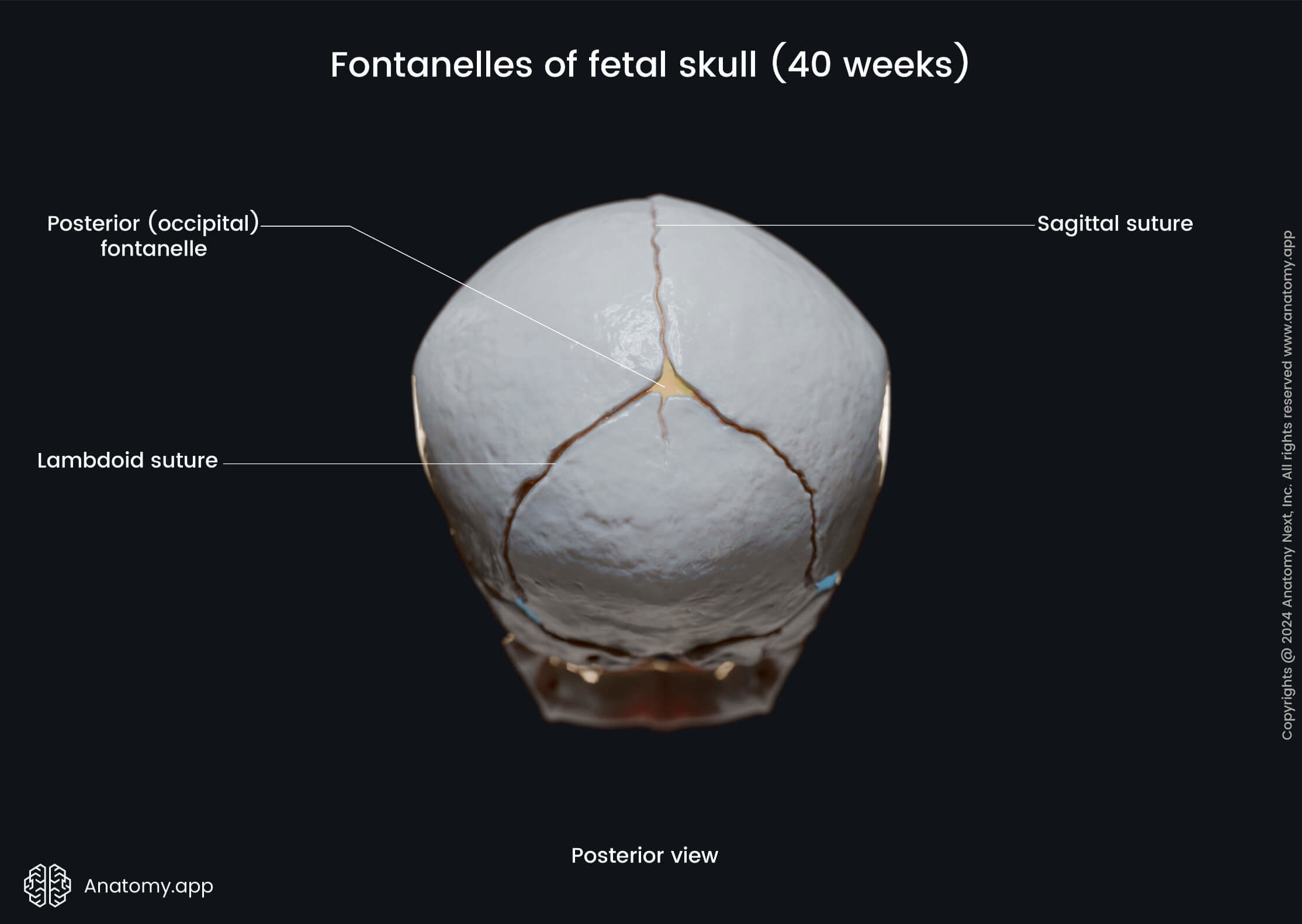
The two major fontanelles are the frontal and occipital fontanelles. The frontal fontanelle, also known as the anterior fontanelle, is found at the junction of the coronal and sagittal sutures. The frontal fontanelle closes between 12 and 18 months of age. The occipital fontanelle, also known as posterior fontanelle, located at the junction between the sagittal and lambdoid suture, typically closes around the first and second months of age.
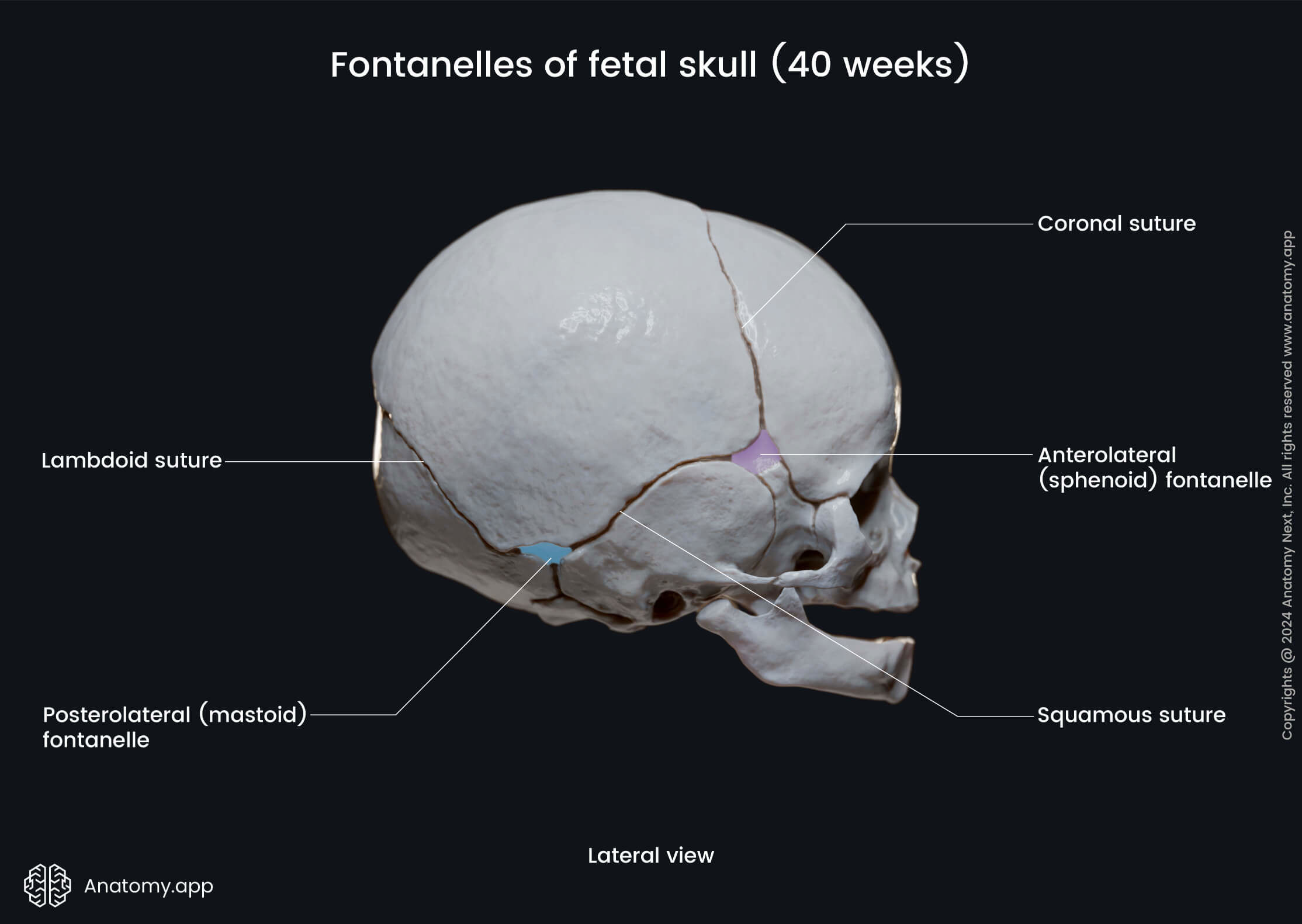
The minor fontanelles are paired. They are smaller and situated on the sides of the skull. These are the sphenoid and mastoid fontanelles. The sphenoid fontanelle is located between the sphenoid, temporal, frontal, and parietal bones. The mastoid fontanelle is situated between the temporal, occipital, and parietal bones.
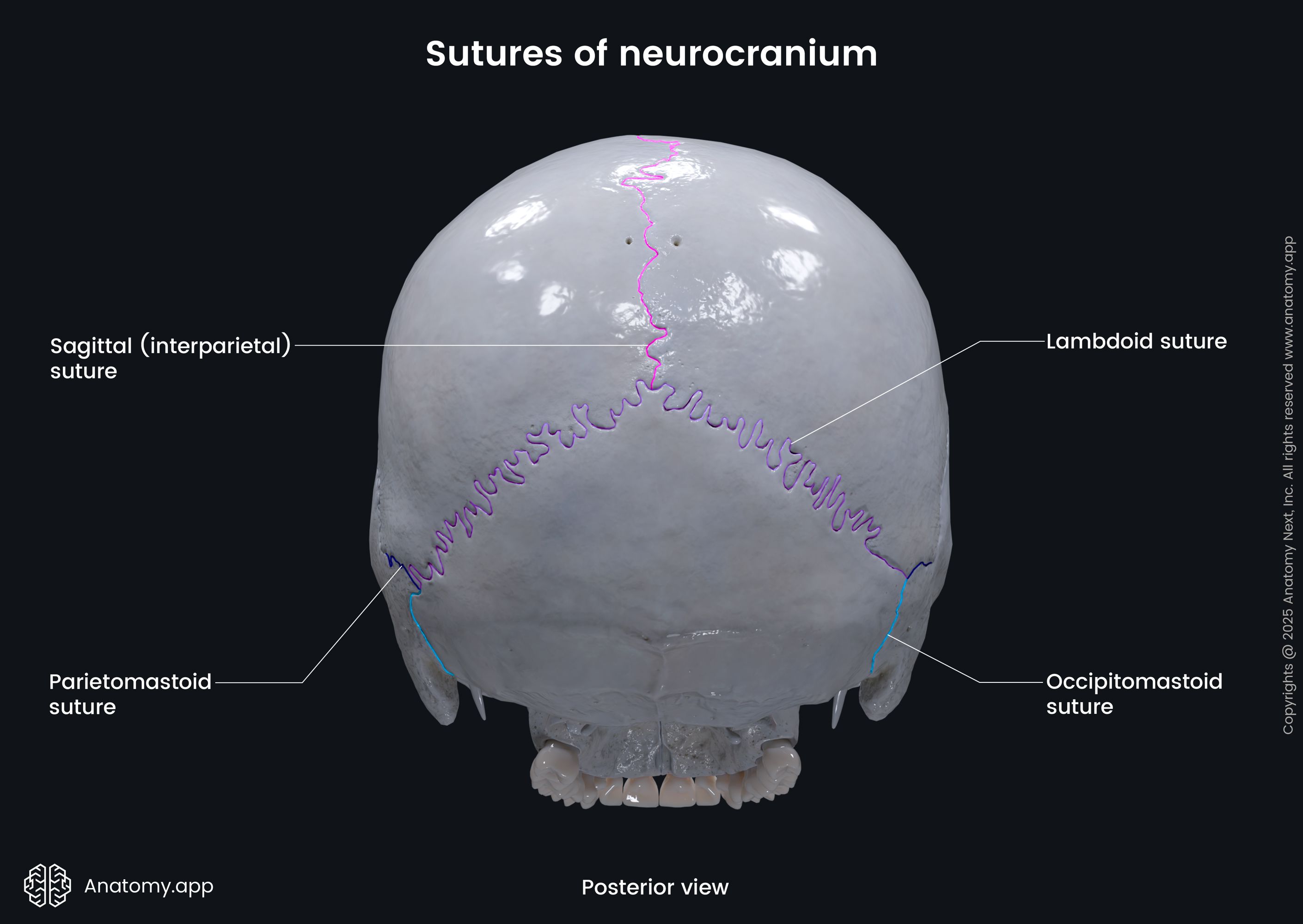
Sutures of neurocranium
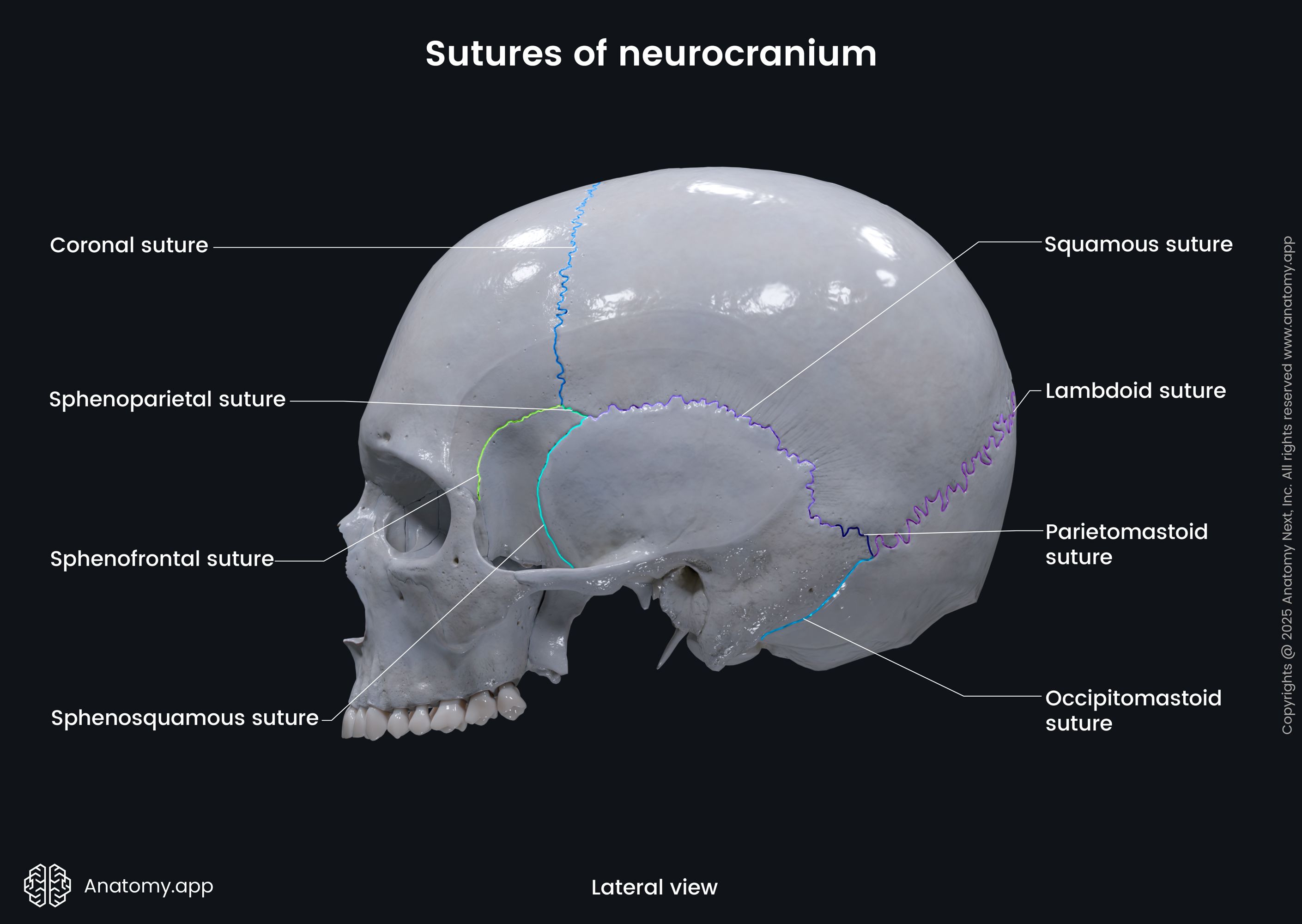
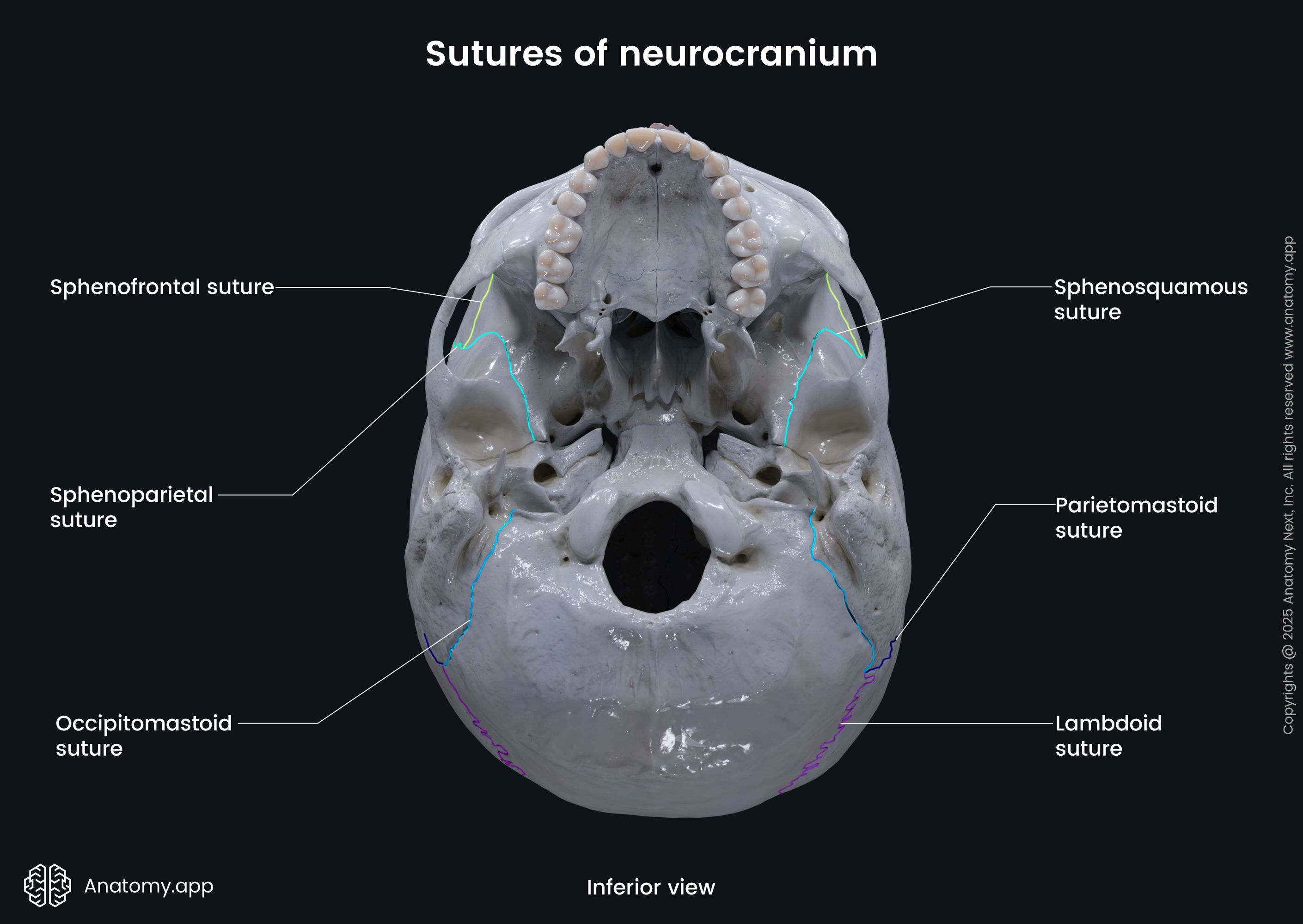
Sutures of the neurocranium are found on the superior, posterior, lateral, and inferior parts of the skull. Some sutures are seen running along the midline. However, most are paired and located on both sides of the skull. Neurocranial sutures are:
- Coronal suture (2)
- Frontoethmoidal suture (2)
- Lambdoid suture
- Metopic suture
- Occipitomastoid suture(2)
- Petrosquamous suture (2)
- Petroclival suture (2)
- Sagittal (interparietal) suture
- Sphenofrontal suture (2)
- Sphenoparietal suture (2)
- Sphenopetrosal suture (2)
- Sphenosquamous suture (2)
- Sphenoethmoidal suture
- Squamous suture (2)
- Parietomastoid suture (2)
- Parietotemporal suture (2)
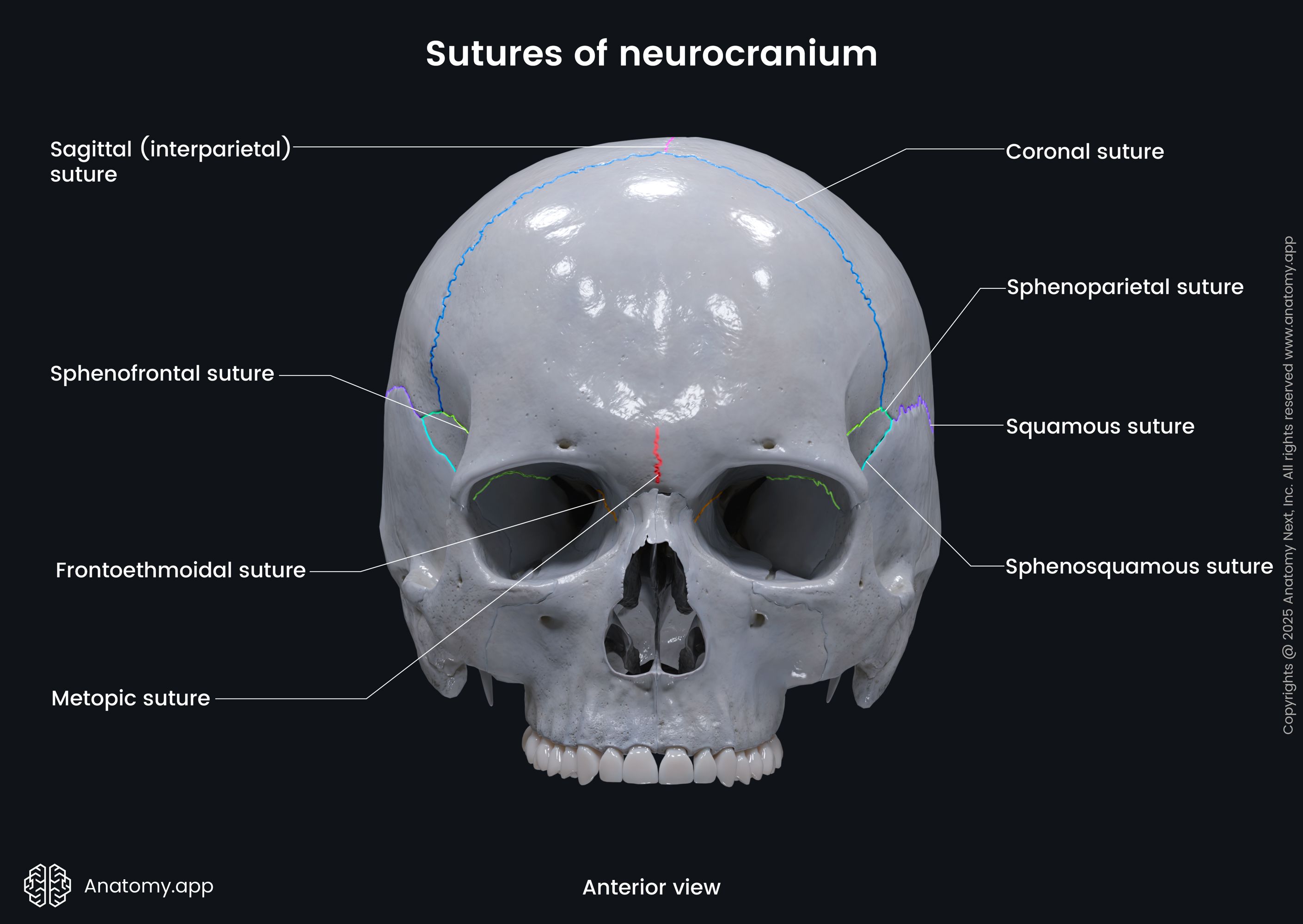
The coronal suture (Read more!) is seen on both the lateral and superior sides of the skull. It is formed at the junction between the parietal bones and frontal bone. It runs horizontally across the superior part of the skull. In the middle, it intersects with the opposite coronal suture and the metopic and sagittal sutures. The coronal suture usually fuses around age 2 and ossifies at around 24 years of age.
The frontoethmoidal suture is a small and short suture located in the anterior cranial fossa. It is a bilateral suture that forms part of the medial wall of the orbits and locates inferior to the anterior and posterior ethmoidal foramina. The frontoethmoidal suture is the articulation between the orbital plate of the frontal bone and the orbital plate of the ethmoid bone.
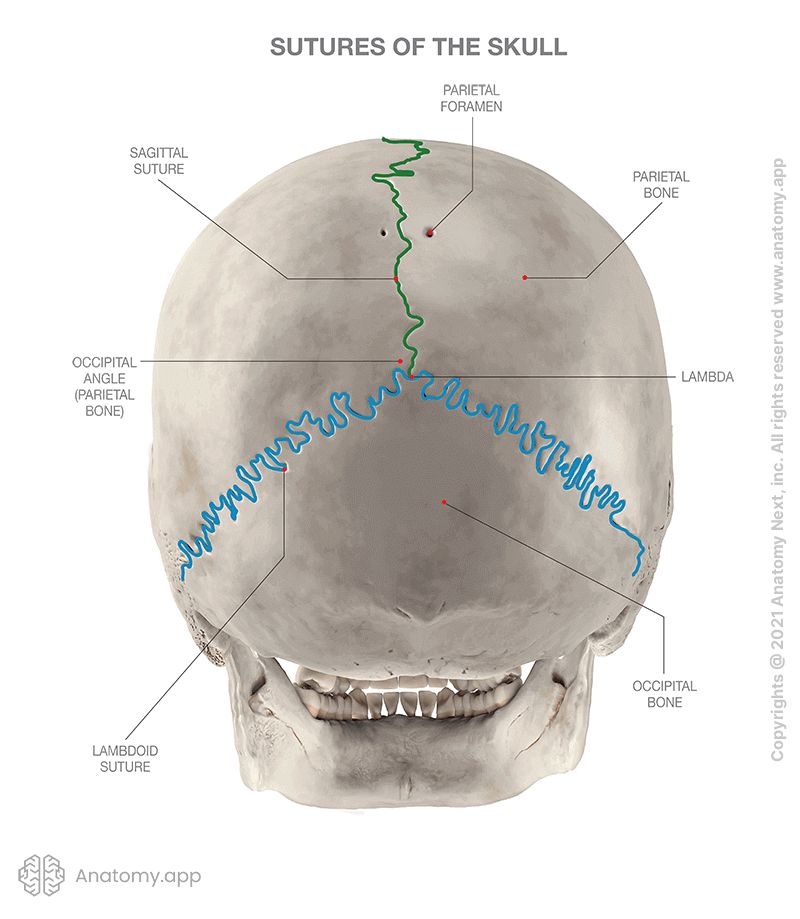
The lambdoid suture (Read more!) is an inverted U-shaped suture seen on the posterior cranium. It is formed at the articulation between the superior border of the occipital bone and the posterior edges of both parietal bones. In the middle of the posterior cranium, the lambdoid suture meets the sagittal suture forming a landmark called lambda. It is shaped like the Greek letter "λ," after which it is named. The lambdoid suture usually ossifies around 26 years of age.
The metopic suture is present in newborns. It is seen on the superior aspect of the skull. The metopic suture divides the frontal bone along the midline of the calvaria. Together with the coronal and sagittal sutures, it contributes to the formation of the frontal fontanelle. The metopic suture fuses between the ages of 3 to 7 months. In most people, the metopic suture has ossified by age 7.
The occipitomastoid suture (Read more!) is formed by the articulation of the squamous part of the occipital bone and the temporal bone's mastoid part. It looks like an oblique continuation of the lambdoid suture. The occipitomastoid, parietomastoid, and lambdoid sutures intersect on the posterolateral sides of the skull and form a landmark known as the asterion. The occipitomastoid suture ossifies around 16 years of age.
The petrosquamous suture, also called petrosquamous fissure, is located between the medial petrous and the lateral squamous parts of the temporal bones. The petroclival suture, also called the petro-occipital or petroclival fissure, is the articulation between the lateral surface of the occipital bone's basilar part and the apex of the temporal bone's petrous part.
The sagittal (interparietal) suture (Read more!) runs across the midline of the calvaria. It is formed by the two parietal bones articulating with each other at their superior margins. The sagittal suture is seen on the superoposterior part of the skull. Posteriorly, it meets the lambdoid suture and contributes to the formation of the lambda. Ossification of the sagittal suture usually happens around 22 years of age but can also occur much later.
The sphenofrontal suture (Read more!) is seen in the roof of the orbits. It forms at the articulation between the greater wings of the sphenoid bone and the frontal bone. The sphenofrontal suture ossifies around 15 years of age. Laterally to the sphenofrontal suture and intersecting with it is the sphenoparietal suture (Read more!). It is a short suture located between the greater wings of the sphenoid bone and the sphenoid angle of the parietal bone. Both sutures contribute to the formation of a landmark known as the pterion.
The sphenopetrosal suture can be seen in the middle cranial fossa behind the foramen ovale, and it contributes to the formation of the foramen lacerum. It is also often called the sphenopetrosal fissure or synchondrosis. The sphenopetrosal suture is the articulation between the greater wing of the sphenoid bone and the temporal bone's petrous part.
The sphenosquamous suture (or sphenosquamosal suture) (Read more!) is found on both lateral sides of the skull. It is a vertical suture between the anterior aspect of the temporal bone's squamous part and the greater wing of the sphenoid bone. Superiorly, it contributes to the formation of the H-shaped pterion. It usually ossifies at around age 10. The sphenoethmoidal suture is located in the anterior cranial fossa. It is formed by the sphenoid bone and the cribriform and perpendicular plates of the ethmoid.
The squamous suture, also called squamosal suture, is a bilaterally found suture seen on the lateral sides of the skull. It is formed by the lateral margin of the parietal bone and the squamous part of the temporal bone. Anteriorly, the squamous suture contributes to the formation of the pterion. During growth, the squamous suture ensures vertical growth of the neurocranium. The squamous suture typically ossifies later in life until 60 years of age.
Closer to the mastoid angle of the parietal bone, the squamous suture transforms into and continues as the parietomastoid suture (Read more!). At the most posterior parietal bones articulation, between the parietal and temporal bones, is the parietotemporal suture. Both parietomastoid and parietotemporal sutures are direct posterior continuations of the squamous suture, and their exact borders are not always clearly defined.
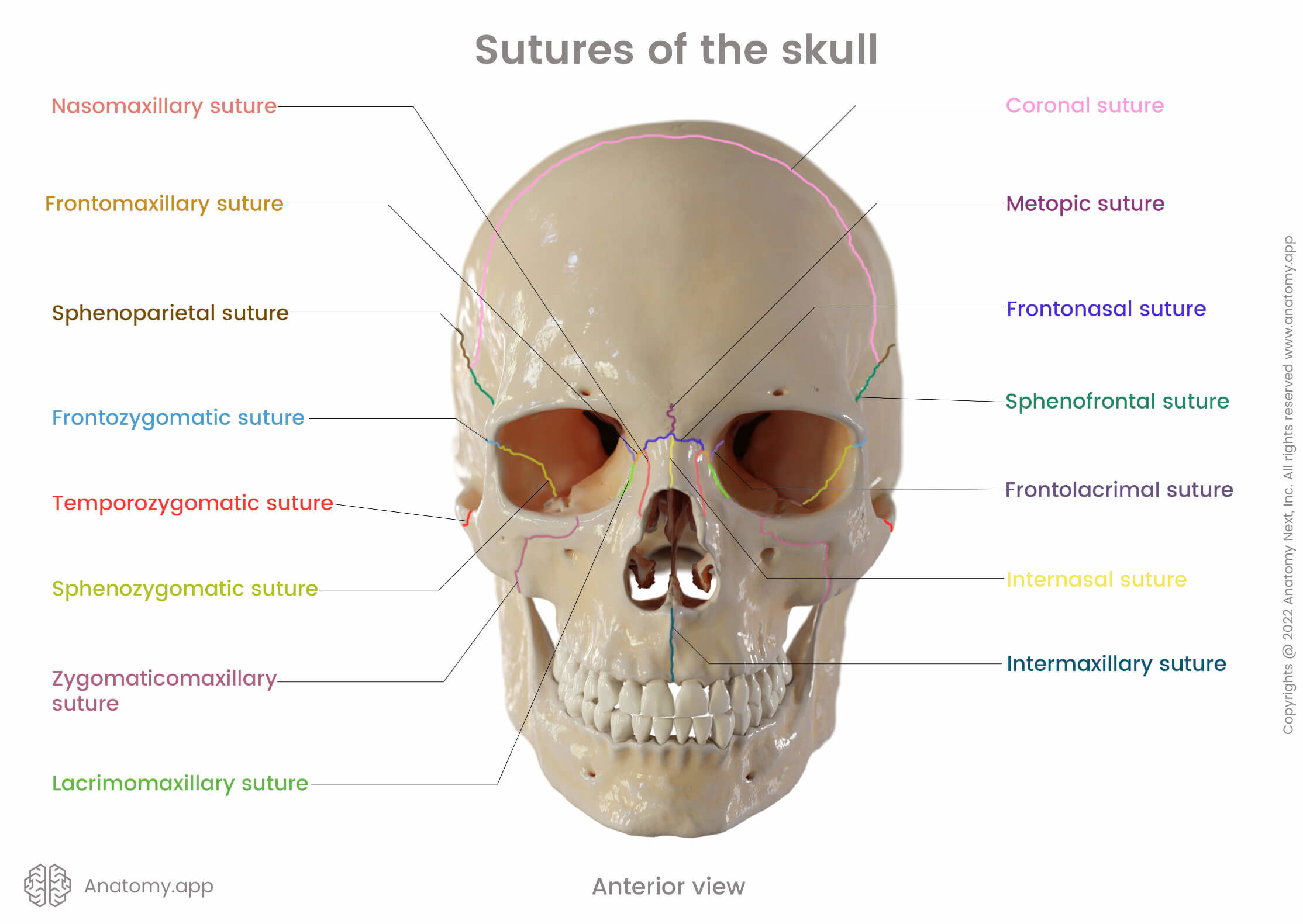
Sutures of viscerocranium
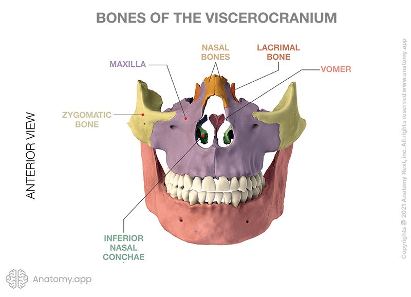
Facial sutures are seen on the anterior part of the skull. For the most part, they are formed between bones of the viscerocranium. The sutures of the viscerocranium are:
- Intermaxillary suture
- Median palatine suture
- Lacrimomaxillary suture (2)
- Nasomaxillary suture (2)
- Zygomaticomaxillary suture (2)
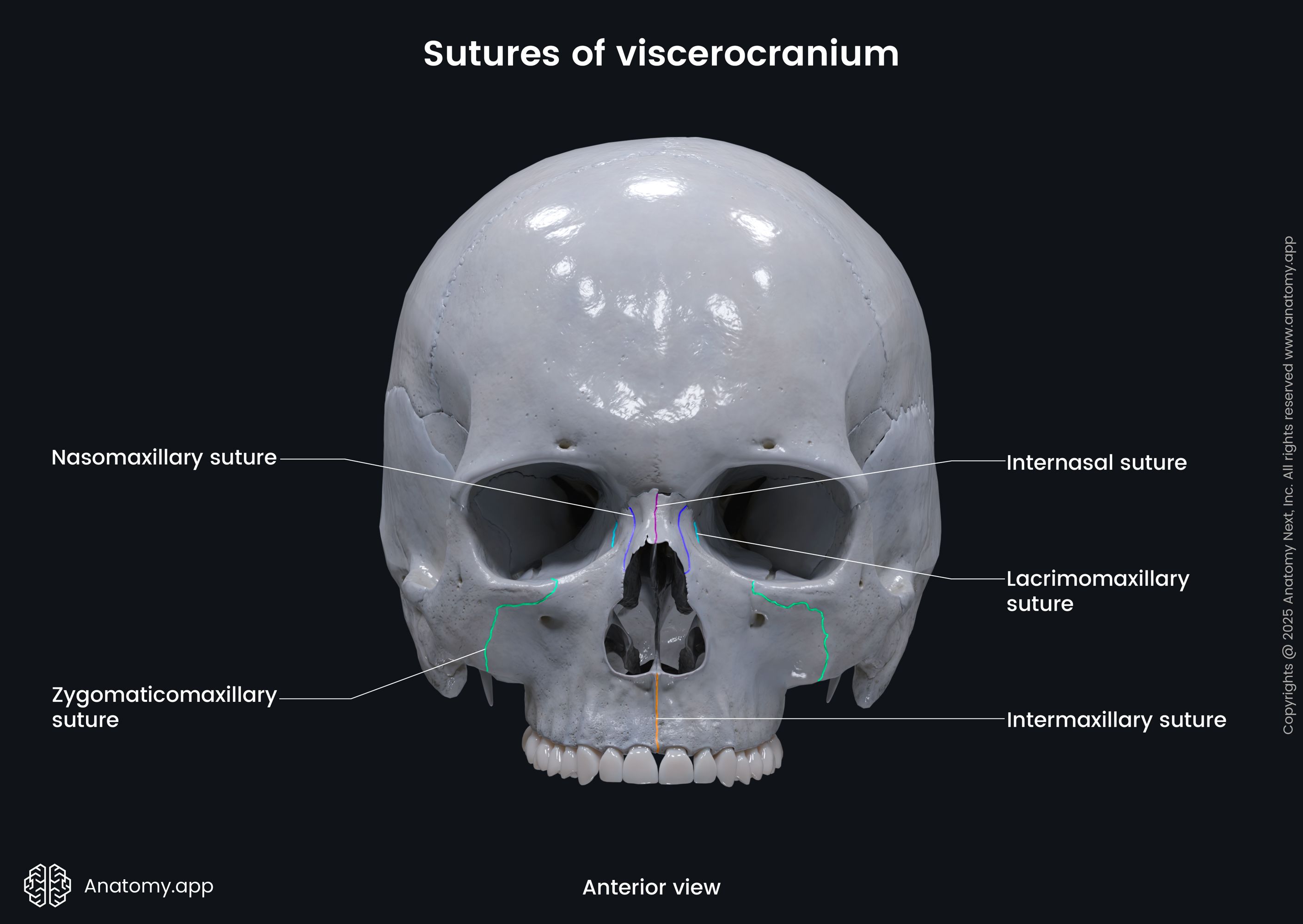
The intermaxillary suture (Read more!) is a single suture seen running along the sagittal plane, right below the nose. It is formed by the fusion of the two separate maxillary bones creating the maxilla. Internally, it continues as the median palatine suture between the two palatine processes of the maxillary bones. It serves as the growth site of the maxilla.
The lacrimomaxillary suture (Read more!) is a bilateral suture located on the inferior part of the anteromedial wall of each orbit. It is created by the articulation between the anterior border of the lacrimal bone and the frontal process of the maxilla. The nearby nasomaxillary suture (Read more!) is formed by the later sides of the nasal bones and the frontal process of the maxillary bones.
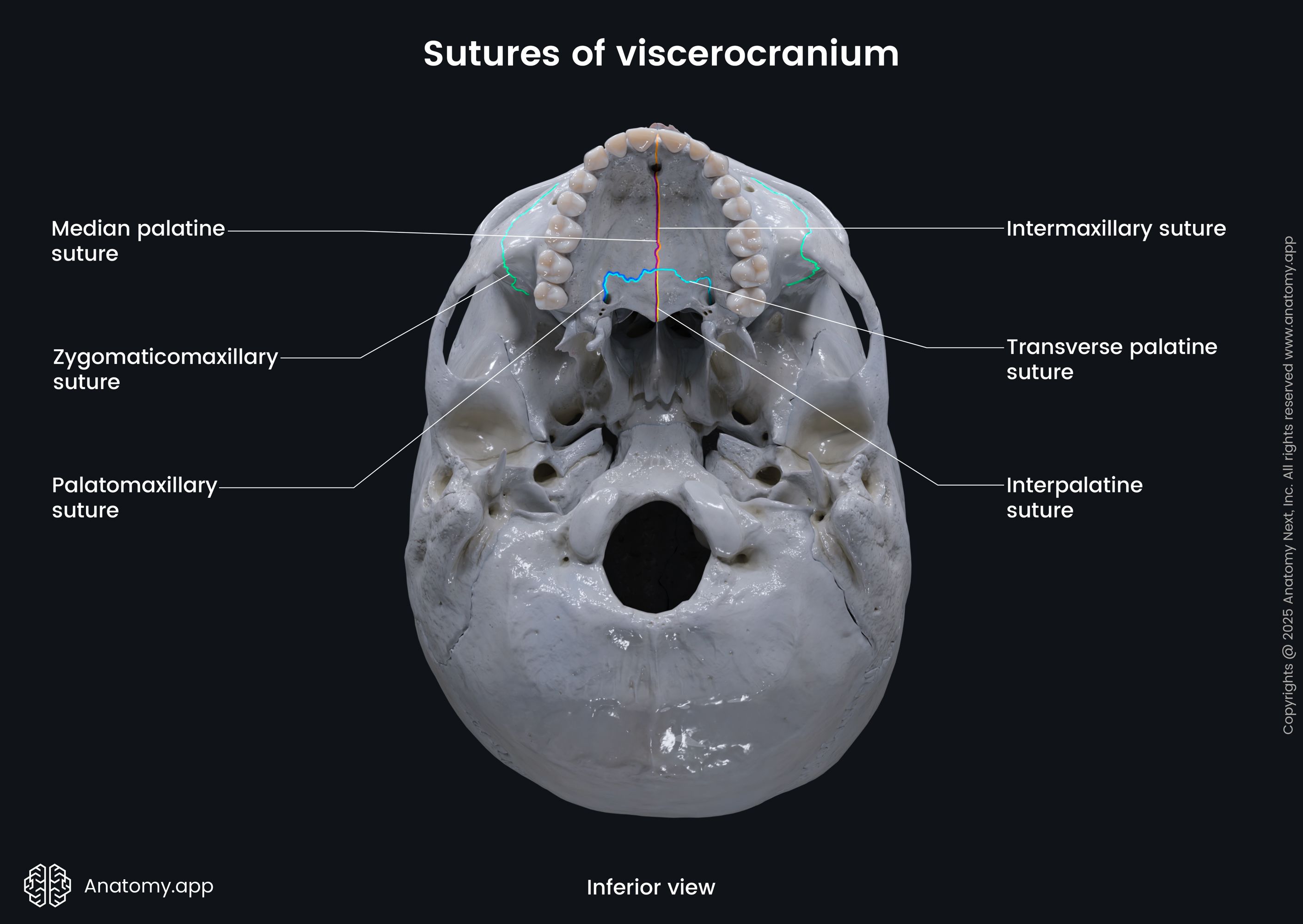
The more laterally situated zygomaticomaxillary suture (Read more!) is a paired cranial suture seen on both sides of the viscerocranium. It is formed between the maxillary process of the zygomatic bone and the zygomatic process of the maxillary bone.
Sutures between neuro- and viscerocranium
Sutures between the neuro- and viscerocranium bones are sutures that are seen on the border between cranial bones belonging to the neurocranium, and cranial bones belonging to the viscerocranium. These include:
- Frontolacrimal suture (2)
- Frontonasal suture (2)
- Frontomaxillary suture (2)
- Frontozygomatic suture (2)
- Sphenozygomatic suture (2)
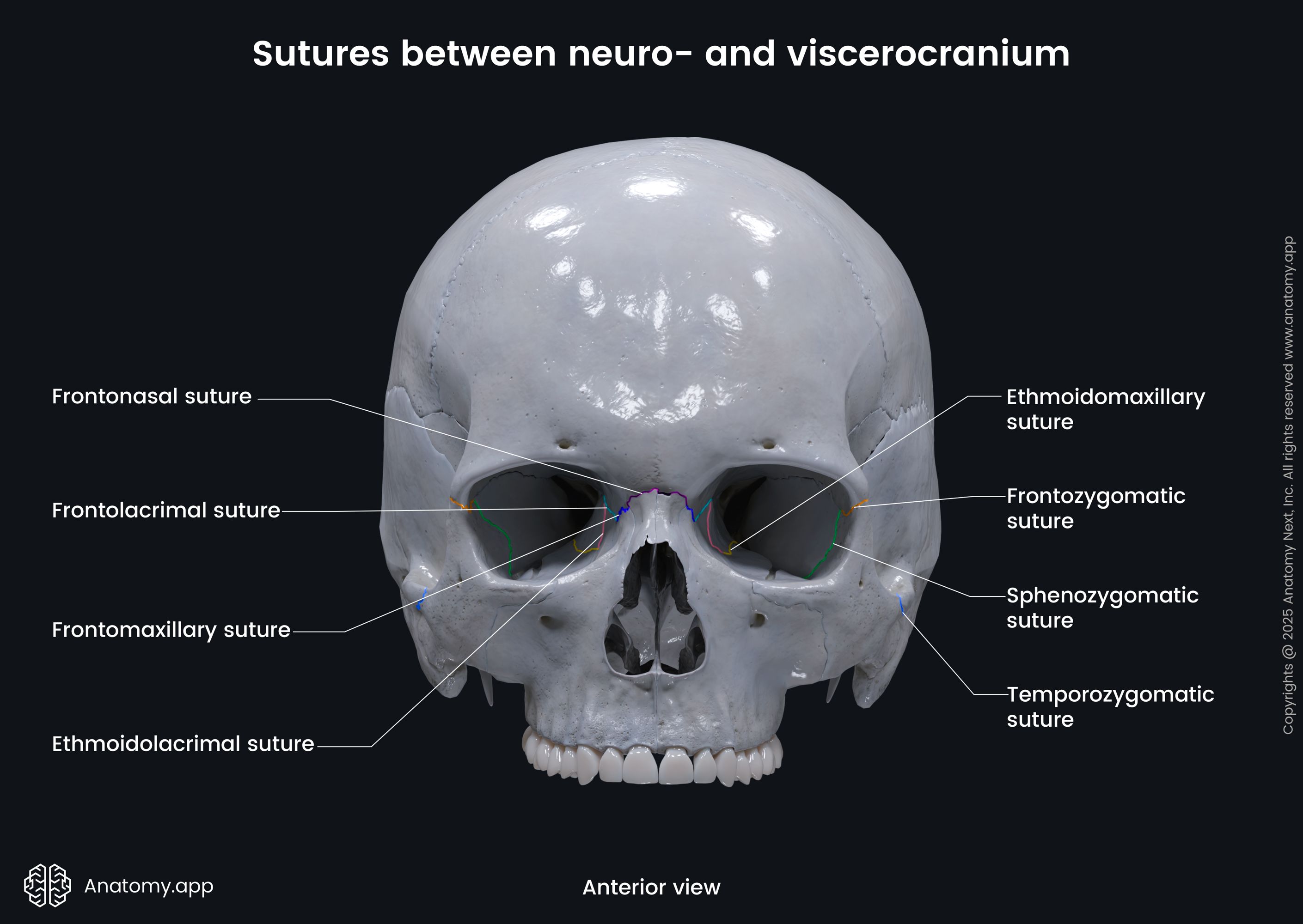
The frontolacrimal suture (Read more!) is a tiny suture formed between the inferior border of the frontal bone's orbital part and the superior border of the small and delicate lacrimal bones. The frontonasal suture (Read more!) is found between the nasal part of the frontal bone and the superior border of the nasal bones. The frontonasal sutures fuse medially with each other.
The frontomaxillary suture (Read more!) is a short suture situated on both sides of the nasal bones between the frontonasal suture and frontolacrimal suture. It is found where the nasal part of the frontal bone articulates with the superior border of the maxilla's frontal process. The combination of the six sutures - two frontolacrimal, two frontomaxillary and two frontonasal sutures - looks like one continuous suture.
The frontozygomatic suture (Read more!) is formed by the articulation between the frontal bone's zygomatic process and the frontal process of the zygomatic bone. It's a relatively short cranial suture seen on both lateral sides of the anterior part of the skull. In most people, the frontozygomatic suture ossifies around the age of 80. In others - even later or never.
The sphenozygomatic suture (Read more!) is another bilateral suture of the skull. It connects the greater wing of the sphenoid bone with the zygomatic bone and its medial part participates in forming the lateral wall of the orbit. The lateral part of the suture participates in forming the anterior cranial fossa.
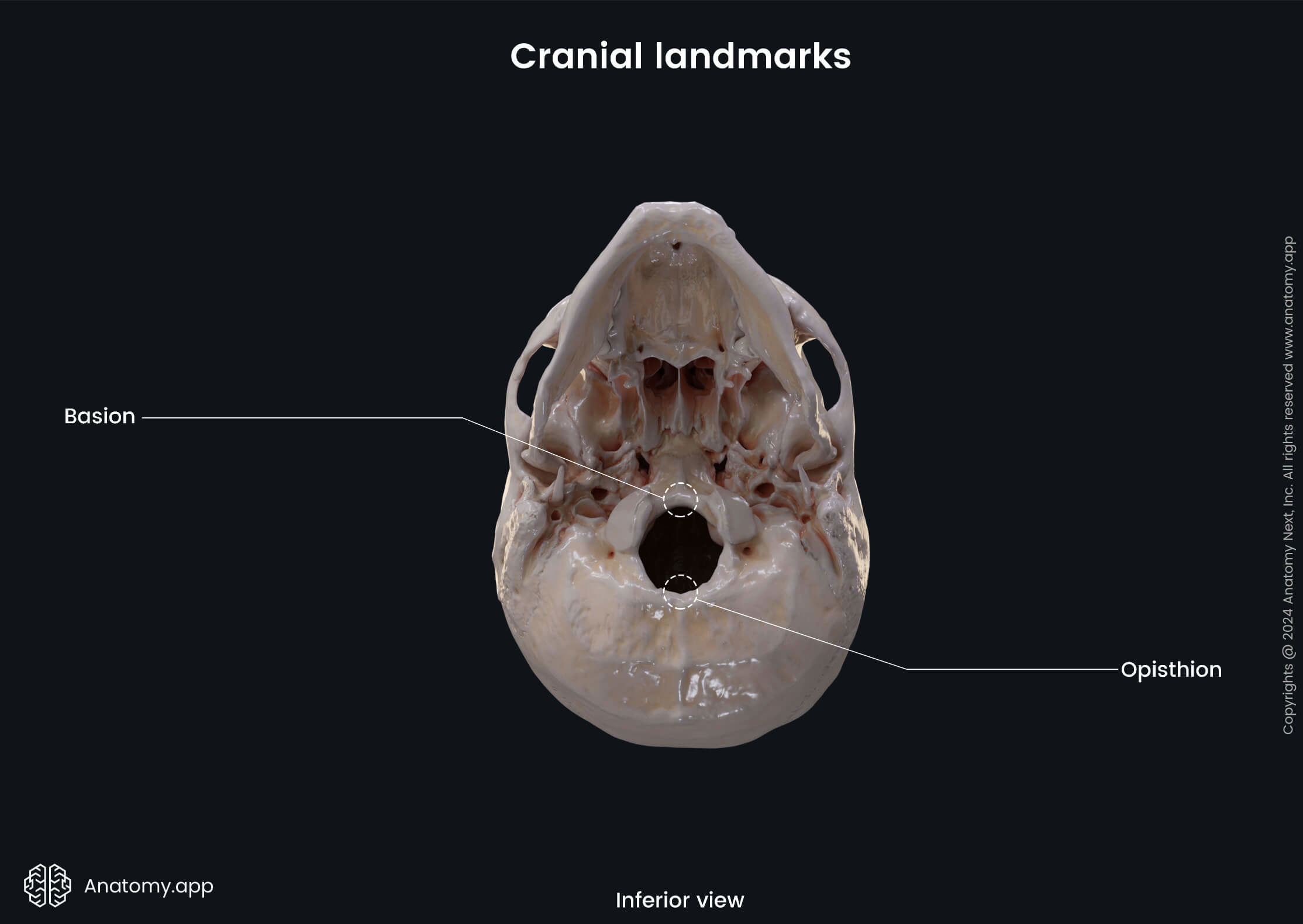
Cranial landmarks
Cranial landmarks are anatomically important features, and some are used as craniometric points of reference in radiology and anthropological measurements. They can be bony formations, elevations or depressions of the cranial bones. Some of these landmarks are formed where sutures intersect, once started to fuse and ossify, or previously contained fontanelles. The most important landmarks associated with sutures are:
- Bregma
- Lambda
- Asterion (2)
- Nasion
- Obelion
- Pterion (2)
- Vertex
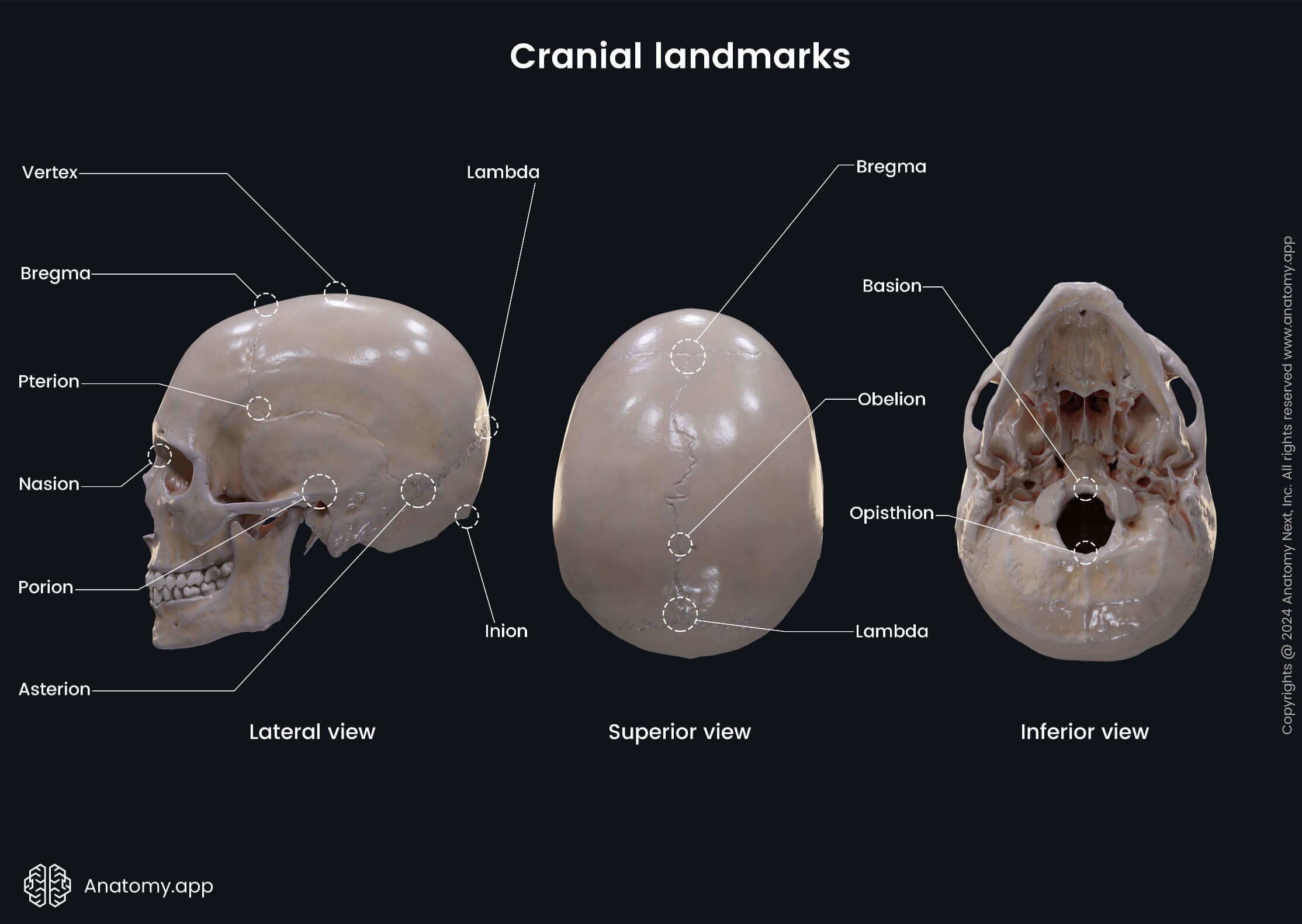
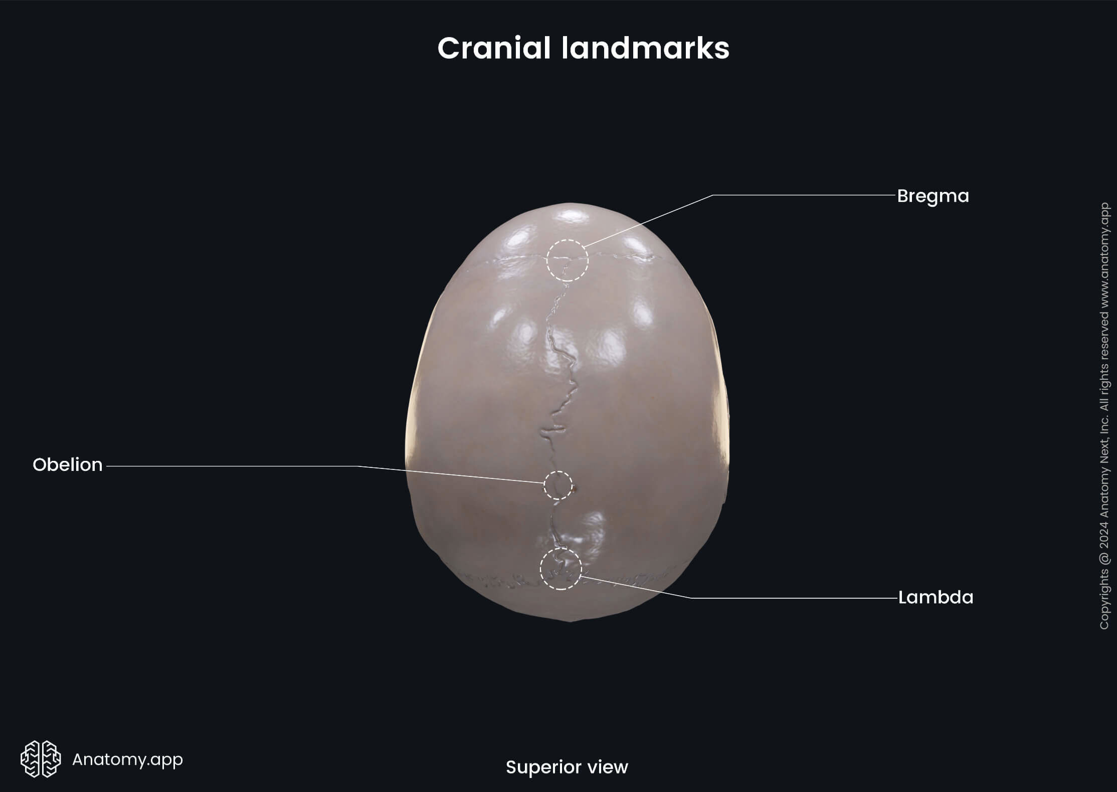
The bregma and lambda are two major suture-associated landmarks of the neurocranium. These landmarks are used as reference points in anthropological measurements, stereotactic surgical procedures, and radiology.
The bregma is formed on the anterior part of the calvaria. It is the intersection of the coronal and sagittal sutures. The bregma is the site of the frontal fontanelle in neonates and young children, which usually fuses around the age of 2.
The lambda, as previously mentioned, is formed at the convergence between the sagittal and lambdoid sutures. Moreover, it is the site where once the occipital fontanelle was located, which usually closes around 2 months of age.
The obelion is formed at the intersection of the sagittal suture and an imaginary line that connects the two parietal foramina. The obelion represents the site at which the fusion and ossification of the sagittal suture has started.
The asterion is seen on the posteroinferior part of the skull at the junction of the parietomastoid, occipitomastoid and lambdoid sutures. Additionally, it is the site of the previously located mastoid fontanelle.
The nasion is a midline bony depression between the two orbits. It corresponds to the continuous suture formed by the two frontolacrimal, two frontomaxillary and two frontonasal sutures between the frontal bone and the lacrimal, maxillary and nasal bones.
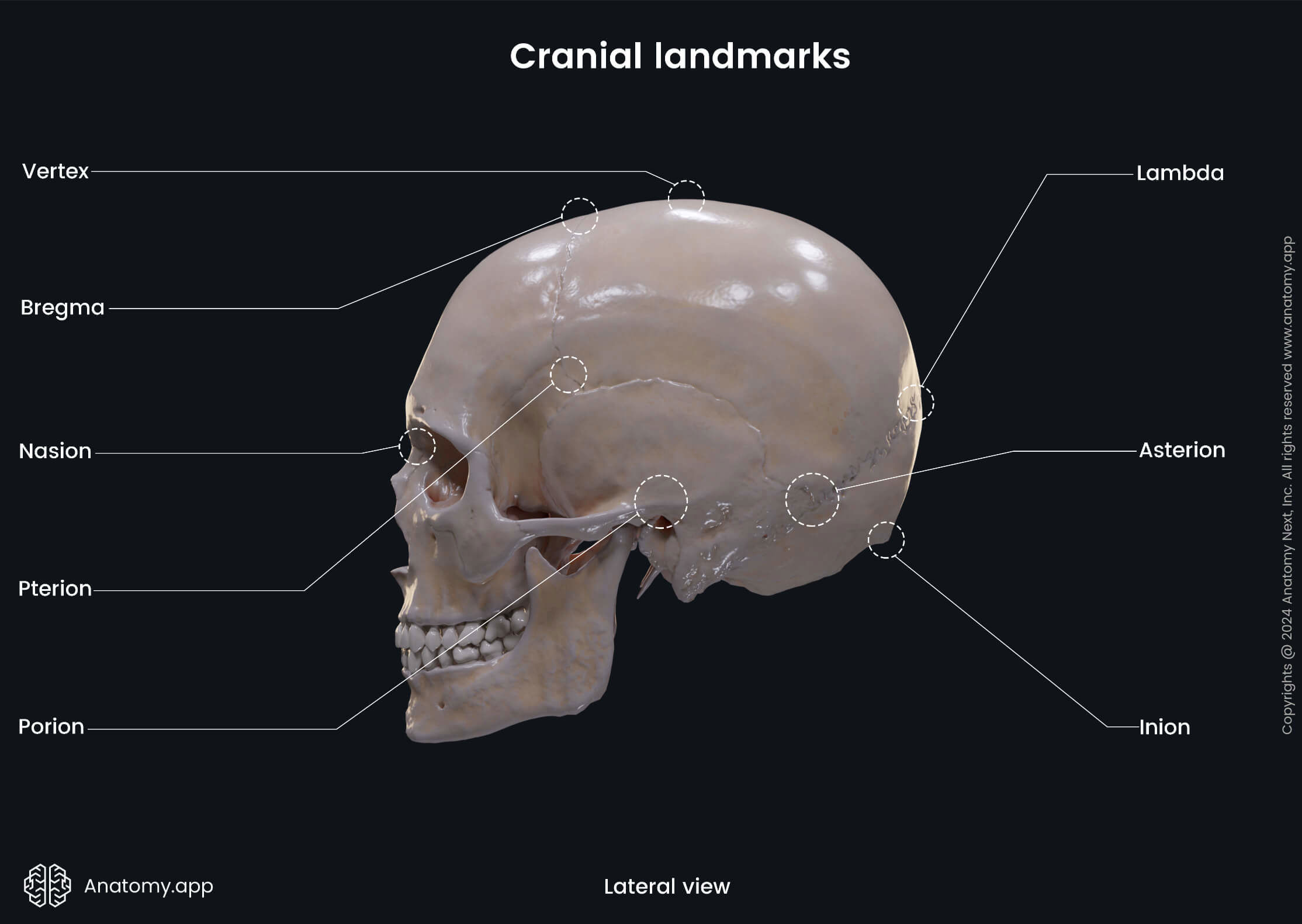
The pterion is a bilateral landmark seen on the lateral sides of the skull. It is an H-shaped point of junction between four bones: the sphenoid, temporal, frontal and parietal bone. It is formed by the sphenofrontal, coronal, sphenoparietal, sphenosquamous, and squamous sutures.
The pterion is clinically relevant due to the many junctions. It is a weak spot of the skull and prone to fractures. Directly underneath it is the middle meningeal artery, a large branch of the maxillary artery. Trauma to the pterion can rupture the underlying artery and cause an epidural hematoma.
The vertex is the highest (most superior) bony landmark of the skull. It is a single midline landmark found on the top of the calvaria between the bregma and lambda, almost at the midpoint of the sagittal suture. The vertex of the human skull is formed at the juncture of four bones: frontal bone, parietal bones (2) and occipital bone.
References:
- Angelieri, F., Franchi, L., Cevidanes, L. H. S., Hino, C. T., Nguyen, T., & McNamara, J. A. (2017). Zygomaticomaxillary suture maturation: A predictor of maxillary protraction? Part I — A classification method. NCBI. https://www.ncbi.nlm.nih.gov/pmc/articles/PMC5503123/
- Cranial Suture - an overview | ScienceDirect Topics. (2017). ScienceDirect. https://www.sciencedirect.com/topics/medicine-and-dentistry/cranial-suture
- Crossman, A. R., & Neary, D. (2019). Neuroanatomy: An Illustrated Colour Text (6th ed.). Elsevier.
- Fonseca, R. J. (2017). Oral and Maxillofacial Surgery (Third Edition). Elsevier.
- J. Lipsett, B., Reddy, V., & Steanson, K. (2021). Anatomy, Head and Neck, Fontanelles. NCBI. https://www.ncbi.nlm.nih.gov/books/NBK542197/
- Katsianou, M. A., Adamopoulos, C., Vastardis, H., & Basdraa, E. K. (2016). Signaling mechanisms implicated in cranial sutures pathophysiology: Craniosynostosis. NCBI. https://www.ncbi.nlm.nih.gov/pmc/articles/PMC5144105/
- Mamaa, M., Edsrc, R., Fhea, R. S., & Hutchings, R. T. (2016). Skull, skull bone articulations and teeth. In McMinn's Color Atlas of Head and Neck Anatomy (5th ed., pp. 1–85). Elsevier.
- Norton, N. S. P. D. (2016). Osteology. In Netter's Head and Neck Anatomy for Dentistry (Netter Basic Science) (3rd ed., pp. 25–64). Elsevier.
- Opperman, L. A. (2000). Cranial sutures as intramembranous bone growth sites. PubMed. https://pubmed.ncbi.nlm.nih.gov/11084647/
- Turgut, M., Tubbs, S. R., Turgut, A. T., & Dumont, A. S. (2021). The Sutures of the Skull: Anatomy, Embryology, Imaging, and Surgery (1st ed. 2021 ed.). Springer.
- Young Sim, S. M. D., Han Yoon, S. M. D., & Yong Kim, S. M. D. P. D. (2012). Quantitative Analysis of Developmental Process of Cranial Suture in Korean Infants. NCBI. https://www.ncbi.nlm.nih.gov/pmc/articles/PMC3291703/
Anatomy.app
Contact information
- For questions regarding business inquiries. Please contact:
- info@anatomy.app