- Anatomical terminology
- Skeletal system
- Joints
- Muscles
- Heart
- Blood vessels
- Lymphatic system
- Nervous system
- Respiratory system
- Digestive system
- Urinary system
- Female reproductive system
- Male reproductive system
- Endocrine glands
- Eye
- Ear
Ear

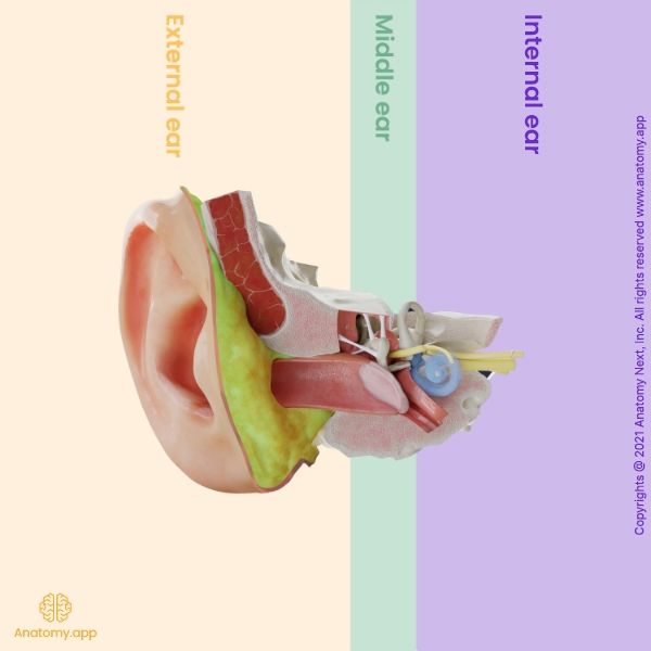
The human ear (Latin: auris) is a paired sensory organ responsible for the sense of hearing and balance. This complex anatomical structure is also referred to as the vestibulocochlear apparatus.
Most of the structures of the vestibulocochlear apparatus are located inside the temporal bone of the skull. The human ear consists of three parts:
- External (outer) ear
- Middle ear
- Internal (inner) ear
The first two parts conduct sound vibrations to the internal ear, which contains the receptors of hearing and balance.
External ear
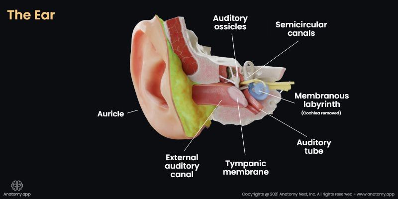
The external ear (or outer ear) is the outer part of the vestibulocochlear apparatus that consists of the auricle and the external auditory meatus. The structures of the external ear participate in conducting and focusing sound vibrations to the eardrum and further to the middle ear and the inner ear.
Middle ear
The middle ear includes the tympanic cavity and the auditory tube that connects the tympanic cavity with the pharynx. At the deep end of the external acoustic meatus, separating the external ear from the tympanic cavity (of the middle ear) lies the tympanic membrane (eardrum). The middle ear is a complex system of openings and canals. Most of this system is located in the temporal bone. The tympanic cavity houses three tiny bones, the auditory ossicles, which transfer the vibrations of the tympanic membrane into the waves in the fluid and membranes of the inner ear.

Internal ear
The internal ear (or inner ear) is located within the petrous part of the temporal bone. It is formed by a bony labyrinth and a membranous labyrinth. The bony labyrinth of the inner ear consists of the vestibule, three semicircular canals, and the cochlea. The membranous labyrinth is located within the bony labyrinth, and it includes two sacs (utricle and saccule), three semicircular ducts, and the cochlear duct. The space inside the membranous labyrinth is filled with the endolymphatic fluid, while outside the membranous labyrinth space is filled with perilymph.
The vestibulocochlear apparatus contains two types of receptors located in the inner ear: the organ of Corti for receiving the sound stimulus - located in the cochlear duct, and the receptors of the vestibular apparatus for appreciation of the impact of gravitation (static balance) - located in the utricle and saccule, and acceleration (kinetic balance) - located in the semicircular ducts.
Transmission of sound
The transmission of the sound or the auditory pathway starts with the auricle of the external ear that turns sound waves into a unidirectional wave. The wave is directed into the external acoustic meatus. Through the external acoustic meatus, the wave reaches the tympanic membrane causing it to vibrate. The amount of the vibration depends on the sound - the louder the sound, the bigger vibration it causes.
The tympanic membrane articulates with the handle of the malleus, while the malleus also articulates with the incus. The incus articulates with the stapes through its lenticular process. The stapes causes the oval window to move in and out with sounds. The round window moves out when the oval window moves in. Without the stapes, the transmission of the sound waves into the inner ear would not be possible.
The sound waves are transmitted up the scala vestibuli to the apex of the cochlear duct. Then it continues down the spiral cochlear organ in the scala tympani. During the movement of sound waves within perilymph in the scala vestibuli and scala tympani, the vibrations cause the basilar membrane to move.
On the basilar membrane is also the organ of Corti that is responsible for changing the vibrations into electrochemical signals. The organ of Corti contains the hair cells that have stereocilia. The basilar membrane vibration causes the hair cells to bend and potassium channels to open.
The opening of the channels causes the potassium influx and leads to a local current and action potential that is traveling through the cochlear nerve from the vestibulocochlear nerve. The vestibulocochlear nerve sends this information to the cochlear nuclei in the brainstem.
The cochlear nerve gives the information to the ventral and dorsal cochlear nuclei. The information from these nuclei is sent to the superior olivary complex, the contralateral superior olivary complex. The second neurons are sent to the inferior colliculus. Most of the connections eventually end in the auditory cortex.
The superior olivary complex is responsible for detecting the time difference between the sound reaches the ear, so it helps to localize where the sound is coming from. The complex consists of the lateral superior olive and the medial superior olive. The difference between them is that the lateral is responsible for understanding the difference in sound intensity, while the middle locates the angle where the sound is coming from.
The inferior colliculus also takes part in localizing where the sound comes from.
Physiology of balance
The balance is controlled by the semicircular ducts, the utricle, and the saccule. As a reminder:
- The semicircular ducts detect movements within their specific planes.
- The saccule recognizes movements of the head and accelerations in the vertical plane.
- The utricle detects movements of the head and accelerations in the horizontal plane.
Due to the movement of endolymph that fills the semicircular ducts, the utricle, and the saccule, the stereocilia on the hair cells generate an electrical impulse. Endolypmh's movement is caused by moving and tilting the head. The hair cells from each semicircular duct, the saccule, and the utricle send information to their according nerve.
From the anterior semicircular duct to the anterior ampullary nerve; from the posterior semicircular duct to the posterior ampullary nerve; from the lateral semicircular duct to the lateral ampullary nerve; from the saccule to the saccular nerve; from the utricle to the utricular nerve.
The anterior and lateral ampullary nerves, together with the utricular nerve, create the utriculoampullary nerve. The utriculoampullary nerve synapses with the posterior ampullary and the saccular nerve in the vestibular ganglion. The vestibular ganglion is the starting point for the vestibular nerve that, together with the cochlear nerve, forms the vestibulocochlear nerve.
The vestibular nerve ends in the vestibular nuclei. The axons from the vestibular nerve go in different directions: through the vestibulospinal tract to the motor nuclei of the spinal cord frontal horns; to the olivary nuclei; adds to medial longitudinal fasciculus; goes to the cerebellum; travels to the cortex. From the olivary nuclei, the axons travel to the cerebellum via the olivocerebellar tract and to the motor nuclei in the spinal cord frontal horns via the olivospinal tract.