- Anatomical terminology
- Skeletal system
- Joints
- Muscles
- Heart
- Blood vessels
- Lymphatic system
- Nervous system
- Respiratory system
- Digestive system
- Urinary system
- Female reproductive system
- Male reproductive system
- Endocrine glands
- Eye
- Ear
Urinary system
The urinary system is a multicomponent organ system mainly consisting of hollow and tube-like structures that are continuous with each other. It starts with the kidneys in the abdominal cavity and extends into the pelvic cavity, ending with the urethra and external urethral orifice. The kidneys are the main components of the urinary system.
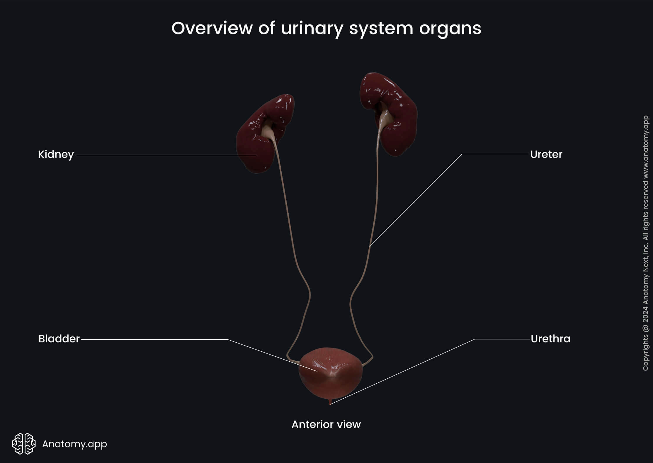
The urinary system includes six organs. Besides the paired kidneys, it is also composed of the paired ureters, single urinary bladder and unpaired urethra. Together, these organs are responsible for urine production, collection, transportation, storage, and excretion from the body into the outer environment.
The urinary system is also known as the excretory system because kidney-produced urine is excreted from the body in the outer environment during micturition, and it contains various metabolic products and toxic wastes. These wastes include such by-products as ammonia, urea and uric acid, creatinine, and drugs, as well as excess body water and electrolytes (salts) are removed from the body during urination.
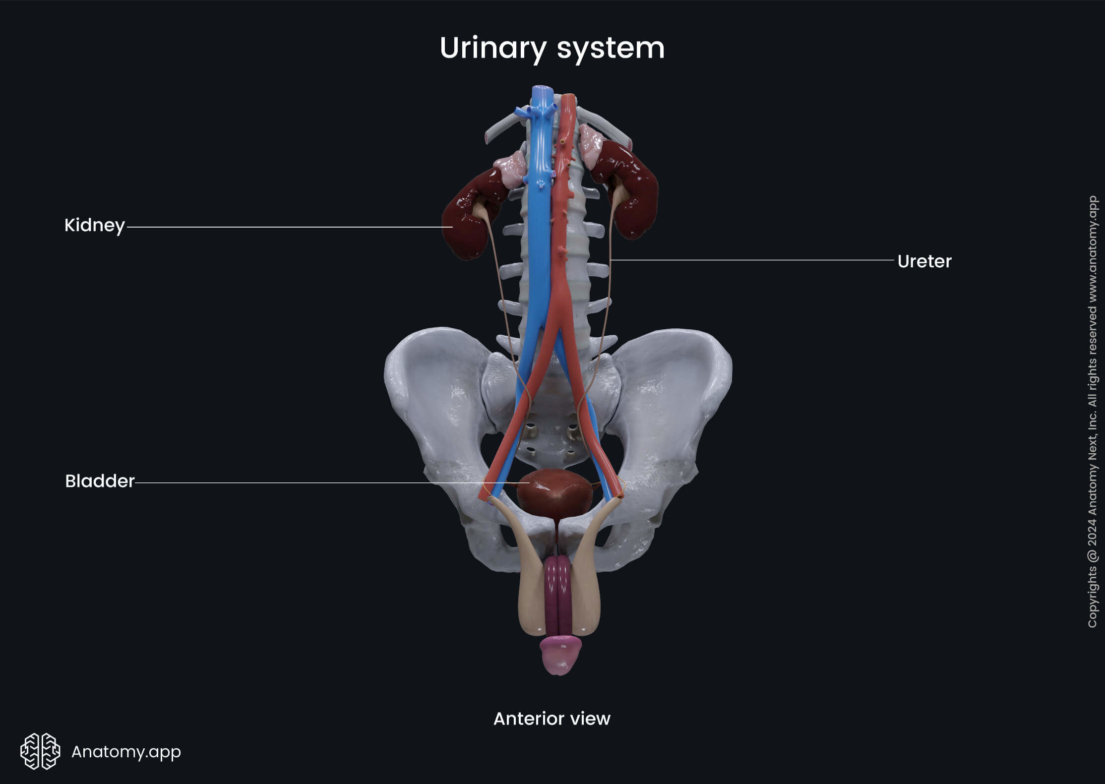
To sum up, the urinary system plays a vital role in maintaining homeostasis and appropriate levels of water and electrolytes in the blood.
Divisions of urinary system
The urinary system can be subdivided into two divisions - the upper urinary tract and the lower urinary tract. The kidneys and ureters belong to the organs forming the upper urinary tract while the urinary bladder and urethra form the lower urinary tract.
The primary functions of the upper urinary tract are the formation of urine and the production and transportation of urine from the kidneys to the urinary bladder. In contrast, the lower urinary tract is responsible for urine storage and controlled excretion from the urinary bladder through the urethra into the outer environment.
Kidneys
The kidneys are the main organs of the urinary system. They filter and modify the blood. The kidneys are paired structures that appear bean-shaped with medial concavity and lateral convexity. Within the medial concavity is the hilum - a deep vertical slit that serves as an entry and exit point for the renal blood vessels, ureter, nerves, and lymphatic vessels.
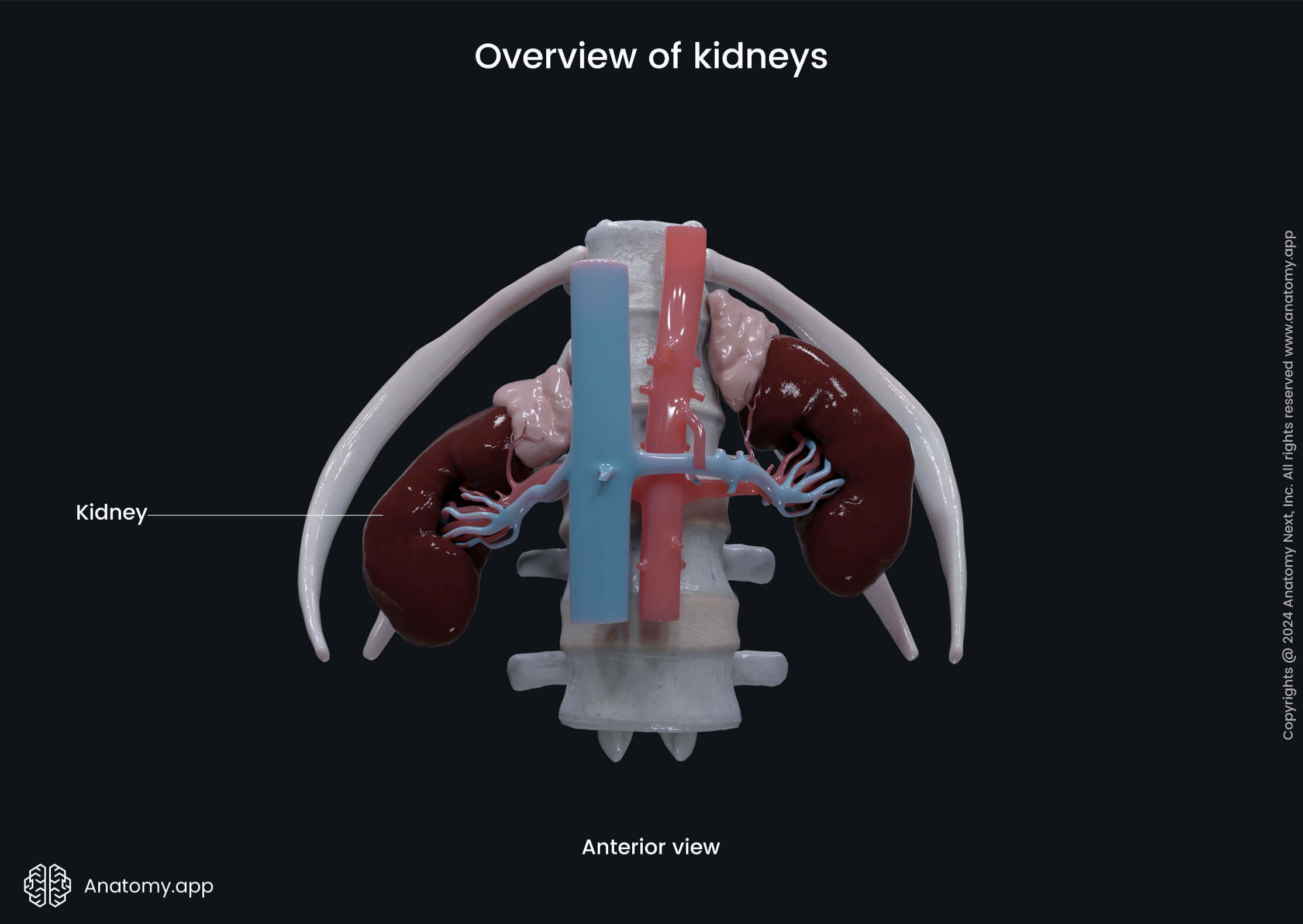
There is a particular order in which structures entering and leaving the kidney are usually positioned within the hilum. The most anterior structure is the renal vein, followed by the renal artery, behind which is the ureter (VAU). However, the placement of hilar structures frequently varies.
The kidneys are positioned just below the diaphragm on either side of the lumbar spine near the posterior wall of the abdomen. Therefore, they are protected by the lower rib cage. The kidneys are retroperitoneal organs.
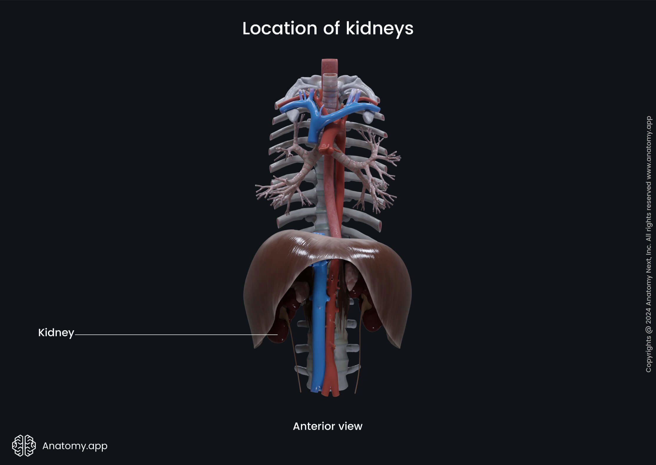
Both kidneys are similar in size and shape. However, the left kidney is longer and more slender than the right and lies closer to the midline. The right kidney lies a bit lower than the left due to its relation to the liver.
- The left kidney extends from the T11/T12 vertebra superiorly to the L2 vertebra inferiorly.
- The right kidney extends from approximately the T12/L1 vertebra superiorly to the L3 vertebra inferiorly.
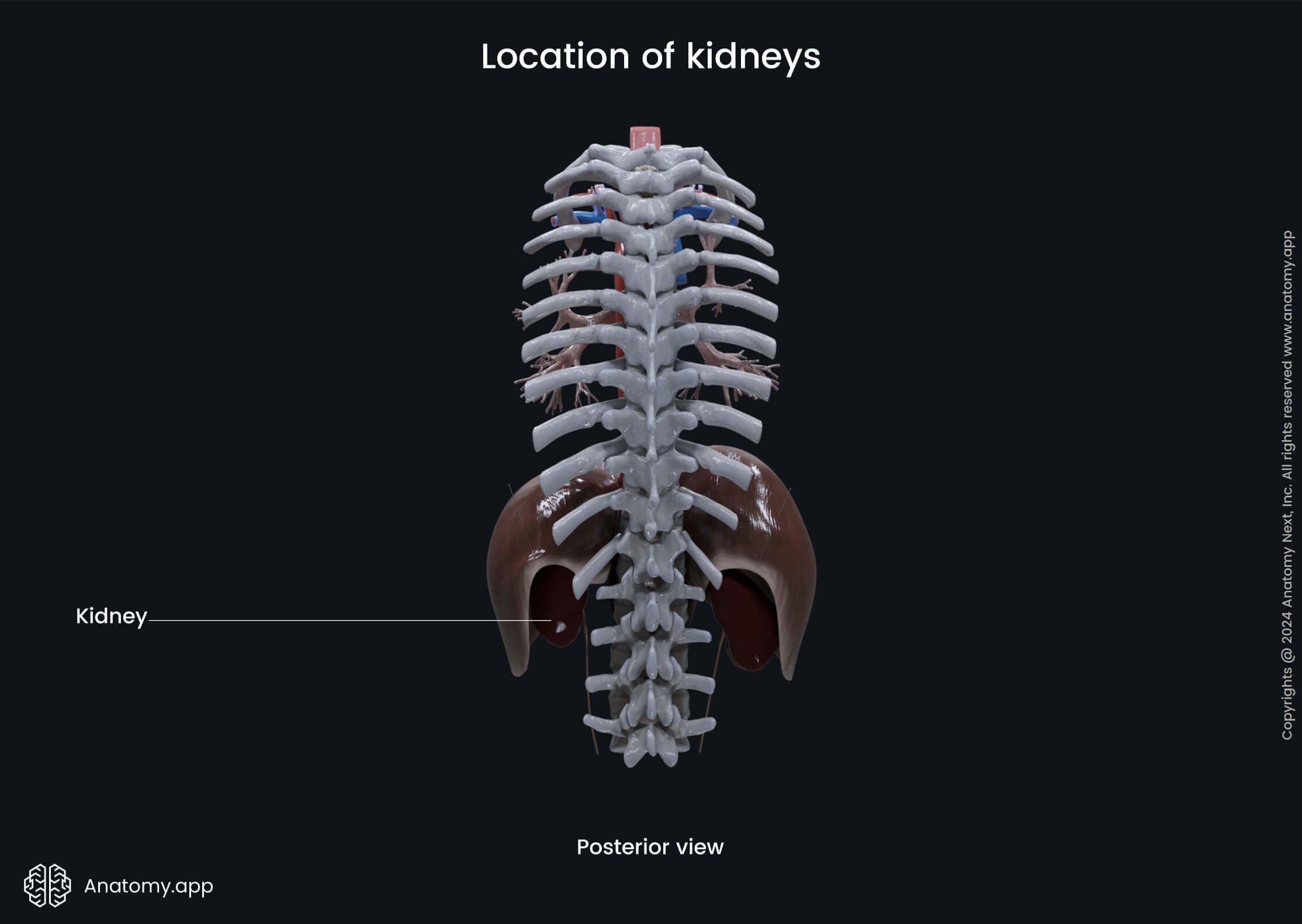
Internally, the parenchyma of each kidney consists of two parts - an outer renal cortex and an inner renal medulla. Both these regions accommodate the functional units of the kidneys - the nephrons. They are the filtering structures of the kidneys. Each adult has approximately 2 million nephrons (about 1 million in one kidney).
Functions of kidneys
The kidneys produce urine, and their main functions include maintaining appropriate levels of water and salts. They eliminate excess water, salts and metabolic by-products.
The kidneys regulate blood osmolarity, acid-base (pH) balance, blood volume and pressure and filter the blood of unnecessary substances. Besides that, they also take part in vitamin D metabolism by participating in the production of calcitriol.
In addition, these organs release erythropoietin when necessary, which further stimulates red blood cell production. Overall, the kidneys are essential organs regulating body homeostasis.
Ureters
Once the urine is formed in the kidneys, it travels down the ureters. The ureters are two thin muscular tubes that connect each kidney with the urinary bladder.
The ureters move urine using rhythmic muscle contractions known as peristalsis, which ensures a consistent urine flow from the kidneys to the bladder. Because of the peristalsis, the wall of the ureter is very muscular (it is composed of three muscle layers).
Like the kidneys, the ureters are also retroperitoneal structures. The ureters pass through both the abdominal and pelvic cavities. Within the abdominal cavity, they descend, lying on the psoas major muscle. The ureters usually enter the lesser (true) pelvis at the iliac bifurcation.
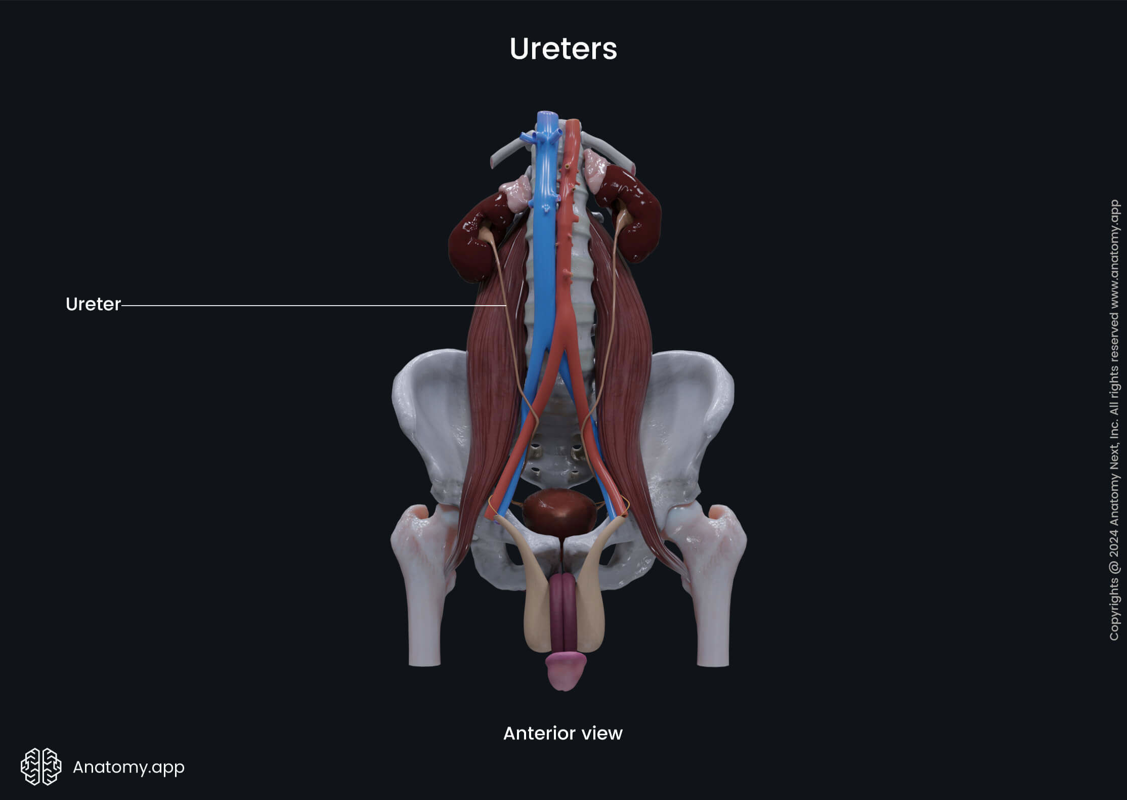
Each ureter crosses either the common iliac artery or the beginning of the external iliac artery at the level of the sacroiliac joint. They can also be found within the cleft formed by both iliac arteries.
Once the ureter enters the lesser (true) pelvis, it descends further, first traveling in the posterolateral direction along the anterior aspect of the greater sciatic notch. When it reaches the ischial spine, it turns in the anteromedial direction to reach the urinary bladder. The ureter enters the bladder via the ureteric orifice.
It is important to add that the ureter goes through the wall of the urinary bladder in an oblique manner. This creates an antireflux mechanism, preventing urine from flowing back into the ureter during detrusor muscle contractions.
Urinary bladder
The urinary bladder is a hollow, expandable organ with muscular walls that stores urine until it is ready to be excreted. In fact, it acts as both a temporary reservoir and a pump for urine.
The bladder is positioned in the lower abdomen directly behind the pubic symphysis. It lies within the lesser (true) pelvis, sitting on the pelvic floor. In males, posterior to it is the rectum, while in females, the vagina is positioned behind the bladder.
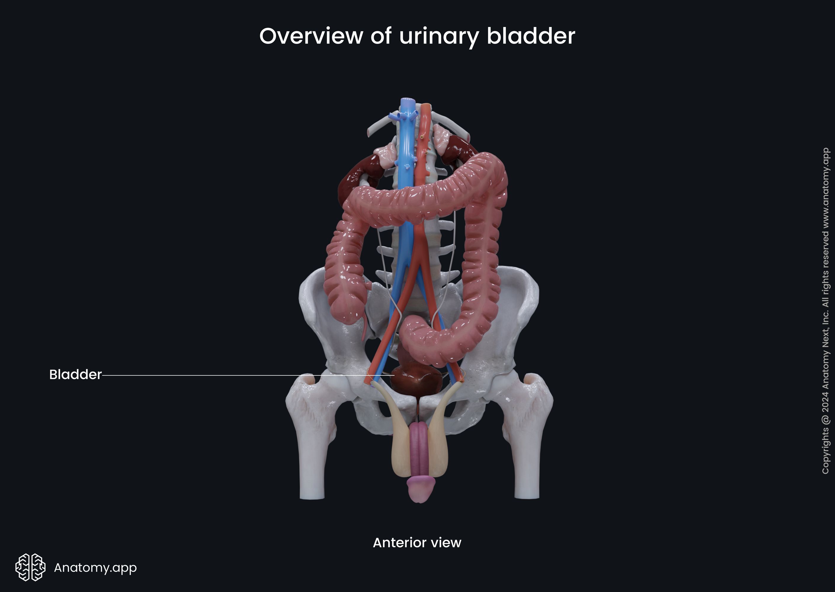
The position and appearance of the bladder vary depending on the amount of urine stored in it.
- An empty bladder appears flattened with thick walls and a slit-like cavity. When the urinary bladder is empty, its mucous membrane forms large folds that are known as the rugae.
- As the bladder fills with urine, it rises upward and becomes oval-like. When the bladder is full, it also extends into the abdominal cavity. The muscle fibers within its walls stretch, and rugae smooth out. Also, the walls of the bladder become thinner.
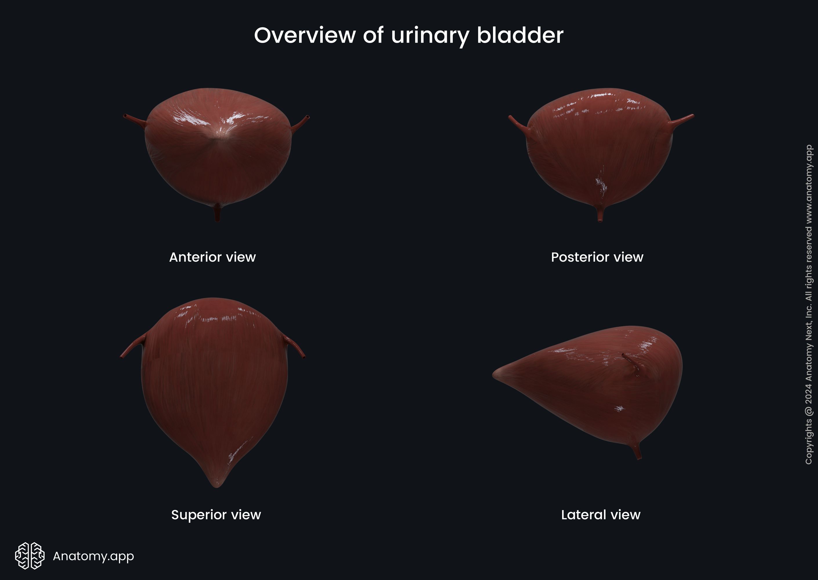
The muscular layer of the bladder wall forms the detrusor muscle. Its contractions allow the bladder to excrete urine in the urethra during urination. On the other hand, when the detrusor muscle is in a relaxed state, urine is held and stored in the bladder.
The detrusor muscle is composed of smooth muscle cells going in multiple directions and forming three sublayers - the inner and outer layers are longitudinal, while the middle layer is circular. The middle sublayer forms an involuntary internal urethral sphincter around the inner opening of the urethra.
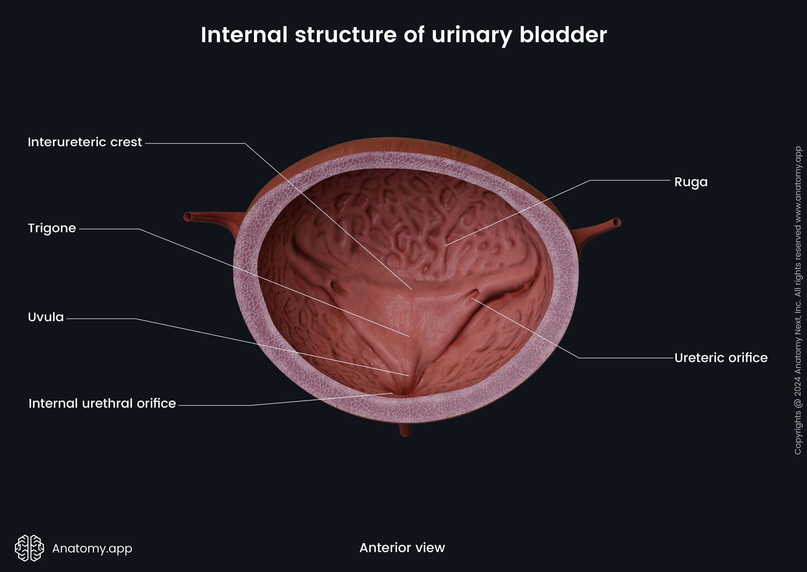
Additionally, the internal aspect of the urinary bladder features a smooth, inverted triangular-shaped area known as the trigone. Each corner of the trigone contains an opening. So, in total, the urinary bladder has three openings (two ureteric orifices and one internal urethral orifice).
Urethra
The urethra is the final passageway of the urinary system. It is a narrow tube-like structure that discharges urine out of the body. It conveys urine from the urinary bladder to the outer environment.
The urethra differs among females and males. Compared to the female urethra, the male urethra is much longer, and its course and structure are more complicated, as most of it goes through the penis. The course of the female urethra is straight, and it is embedded in the anterior wall of the vagina.
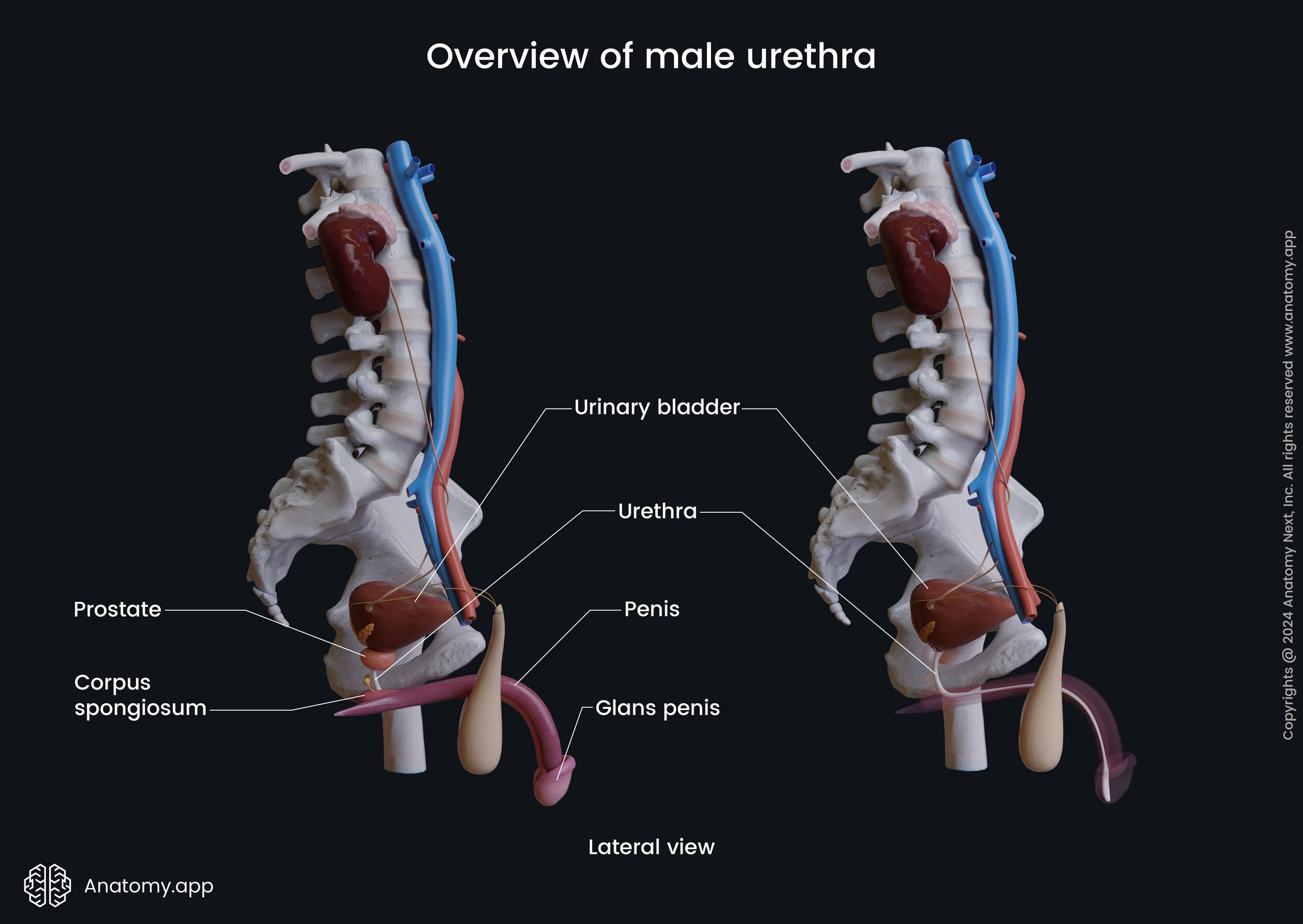
The female urethra is short - only about 1.57 inches (4 cm) long, while it is about 7.09 to 7.87 inches (18 - 20 cm) long in males. However, the length of the male urethra may vary from 6.3 to 8.66 inches (16 to 22 cm) or even more, depending on the length of the penis.
In contrast to the male urethra, the female urethra has only one function - it transports and discharges urine from the body. The male urethra is also an essential component of the male reproductive system - it not only transports urine but also conveys and discharges semen during ejaculation.
The male and female urethras have two sphincters - the involuntary internal urethral sphincter and the voluntary external urethral sphincter. Both sphincters help control the flow of urine. This mechanism ensures that urine is released at the right time, completing the process of waste elimination.
The urethra opens via the external urethral orifice. In females it is found on the vulva anterior to the vaginal orifice, while in males on the glans penis. The external opening of the male urethra presents as a vertical slit.
Functions of urinary system
The urinary system is responsible for a variety of functions and physiological processes. Primarily, this system filters the blood and creates urine. It is also accountable for osmoregulation (fluid and electrolyte balance) and blood volume and pressure regulation via the renin-angiotensin-aldosterone system. Besides that, the urinary system also maintains acid-base (pH) balance in the blood.
The excretory system also eliminates various waste products (such as metabolic and toxic waste (ammonia and urea)). Also, constant urine production and flow in the upper urinary tract and intermittent elimination from the lower urinary tract ensure that the urinary tract is cleansed and the body is getting rid of the microbes that might have entered it.
Finally, the urinary system participates in hormone production, calcium reabsorption and stimulation of red blood cell production with the help of erythropoietin. Also, the excretory system converts vitamin D to its active form of calcitriol.