Nasal cavity
The nasal cavity is an irregular, bilateral air-filled space located above the roof of the oral cavity. The nasal cavity forms the internal part of the nose. It is an initial part of the respiratory tract, and it lodges the organ of olfaction.
The bony frame of the nasal cavity is formed by several bones of the skull, including the maxilla, sphenoid bone, ethmoid bone, inferior nasal concha, palatine bone, lacrimal bone, nasal bone, and the vomer.
The nasal cavity is lined with the mucosa, and it is a part of the upper airway passage. It communicates with the paranasal sinuses, including the ethmoidal air cells and the frontal sinuses, sphenoidal sinuses and maxillary sinuses.
The nasal cavity opens into the nasopharynx through a pair of oval openings called the choanae. Anteriorly, the cavity opens with the nasal aperture.
Walls of nasal cavity
Each half of the bilateral nasal cavity has a roof, floor, medial and lateral walls, and a vestibule:
- The roof of the nasal cavity is formed by the ethmoid and sphenoid. The anterior slope of the roof is formed by the cribriform plate of the ethmoid bone, while the posterior slope is formed by the anterior part of the body of the sphenoid.
- The floor of the nasal cavity is formed by the palatine processes of the maxillae and the horizontal plate of the palatine bone.
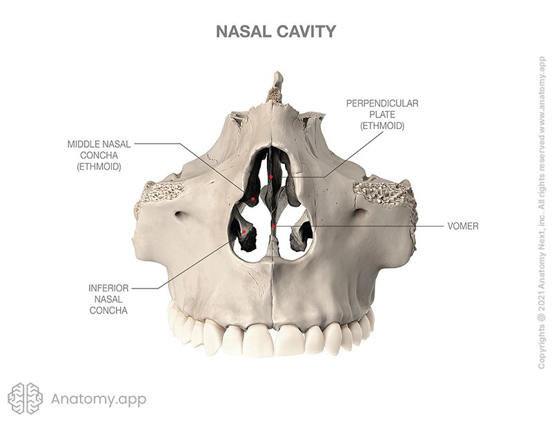
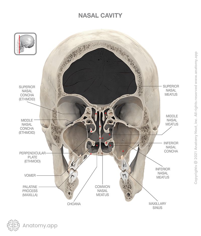
- The medial wall of each half of the nasal cavity corresponds to the nasal septum. The bony nasal septum is formed by the vomer and the perpendicular plate of the ethmoid.
- The lateral wall of the nasal cavity is formed by the nasal surface of the maxilla, perpendicular plate of the palatine bone, labyrinth of the ethmoid bone (with superior nasal concha and middle nasal concha), inferior nasal conchae, lacrimal bone, and medial plate of the pterygoid process.
- The anterior wall of the nasal cavity is formed by the nasal bone and the anterior nasal aperture.
- The posterior wall of the nasal cavity is formed by the body of the sphenoid and paired choanae.
Nasal conchae and meatuses
There are several formations in the nasal cavity that are shaped like curved shelves of bone called conchae or conchas.
There are three nasal conchae on each lateral wall of the nasal cavity:
- Superior nasal concha
- Middle nasal concha
- Inferior nasal concha
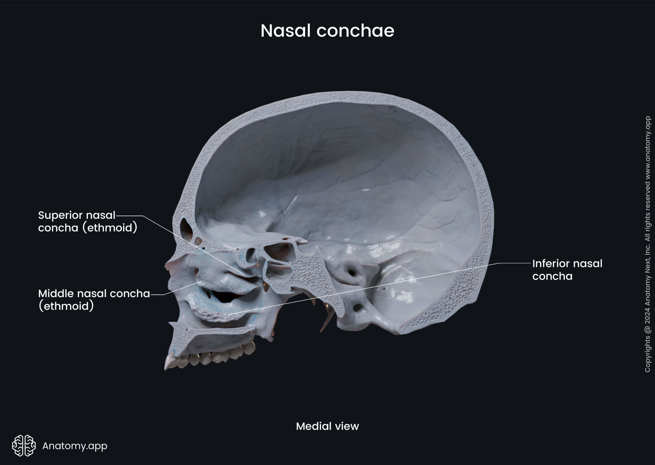
The superior nasal conchae and the middle nasal conchae are parts of the labyrinth of the ethmoid bone, while the inferior nasal conchae are separate bones of the skull.
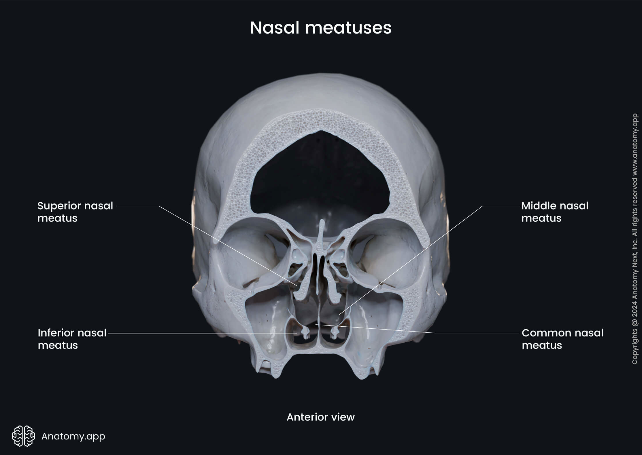
The nasal conchae project into the nasal cavity creating five pathways called nasal meatuses:
- Superior nasal meatus
- Middle nasal meatus
- Inferior nasal meatus
- Common nasal meatus
- Nasopharyngeal meatus
Parts and functions
Functionally the nasal cavity is divided into two parts. The middle nasal conchae mark the border between both parts, and they are:
- Respiratory part - below the conchae; it contains glands, the secretion of which moisturize the surface of the mucous membranes and trap dust parts and microbes, expelling them to the external environment; this part also contains venous plexus that warms the inhaled air and releases heat;
- Olfactory part - located above the conchae; it contains glands and olfactory receptors from which the olfactory nerve fibers start.
Overall, the functions of the nasal cavity are:
- Air transmission
- Air heating
- Air purification
- Olfactory function
- Resonator function
Neurovascular supply
The arterial blood supply of the nasal cavity is provided by two main arteries - the external and internal carotid arteries.
- The internal carotid artery gives rise to the ophthalmic artery, which gives off the anterior ethmoidal artery and posterior ethmoidal artery supplying the nasal cavity. Both arteries supply the roof of the nasal cavity, as well as the frontal sinus and ethmoidal air cells.
- The external carotid artery gives the maxillary artery which supplies branches to the nasal cavity, such as the sphenopalatine artery and the greater palatine artery. The first branch supplies not only the nasal cavity but also the sphenoid sinus, while the second artery provides blood supply to the region of the inferior nasal meatus.
Venous drainage is provided by the veins accompanying same-named arteries. Venous blood is carried to the pterygoid venous plexus.
The sensory nerves innervating different parts of the nasal cavity include:
- Olfactory nerves (CN I) - arise from the olfactory epithelium in the upper part of the nasal cavity and provide olfaction (sense of smell);
- Anterior ethmoidal nerve (a branch of the nasociliary nerve, originating from the CN V1) - supplies the roof of the nasal cavity and gives off a lateral internal branch to supply the anterior part of the lateral wall and a medial internal branch to supply the nasal septum;
- Nerves arising from the maxillary nerve (CN V2) or its branches:
- Infraorbital nerve - supplies the nasal vestibule;
- Anterior superior alveolar nerve - supplies part of the nasal septum, the floor of the nasal cavity near the anterior nasal spine, and the anterior part of the lateral wall of the cavity;
- Posterior superior nasal nerves and posterior inferior nasal nerves (branches from the greater palatine nerve) - supply the posterior part of the lateral wall, roof and floor of the nasal cavity, and the medial inferior part of the nasal septum;
- Branches arising from the nerve of the pterygoid canal (Vidian nerve) - supply the superior and posterior part of the roof of the nasal cavity and nasal septum.
Overall, the olfactory nerve (CN I) provides the sense of smell, while the ophthalmic (CN V1) and maxillary (CN V2) nerves are responsible for general sensations.