- Anatomical terminology
- Skeletal system
- Skeleton of trunk
- Skull
- Skeleton of upper limb
- Skeleton of lower limb
- Joints
- Muscles
- Heart
- Blood vessels
- Lymphatic system
- Nervous system
- Respiratory system
- Digestive system
- Urinary system
- Female reproductive system
- Male reproductive system
- Endocrine glands
- Eye
- Ear
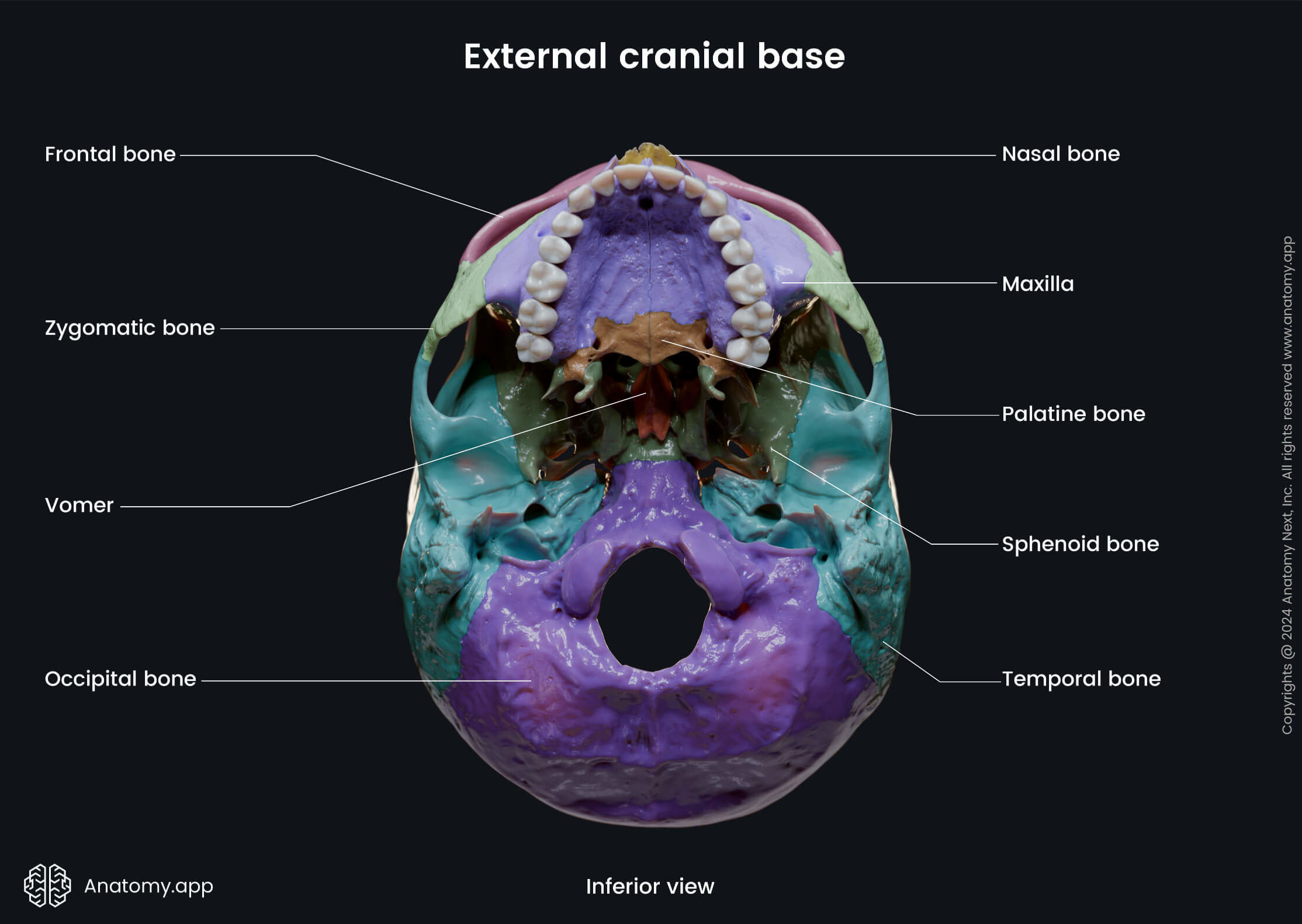
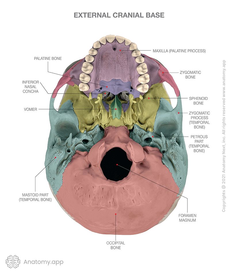
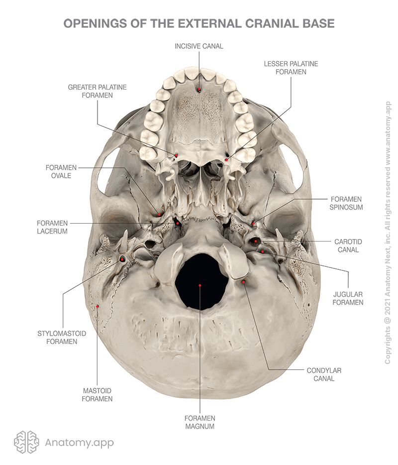
External cranial base
The external cranial base (or external surface of cranial base, Latin: basis cranii externa) is the outer or inferior aspect of the skull base. This part of the skull extends from the superior incisor teeth to the superior nuchal lines of the occipital bone. The external cranial base can be subdivided into anterior, middle, posterior and lateral (2) parts.
Anterior part of external cranial base
The anterior part of the external cranial base is made of three major structures:
- Superior alveolar arch
- Bony palate
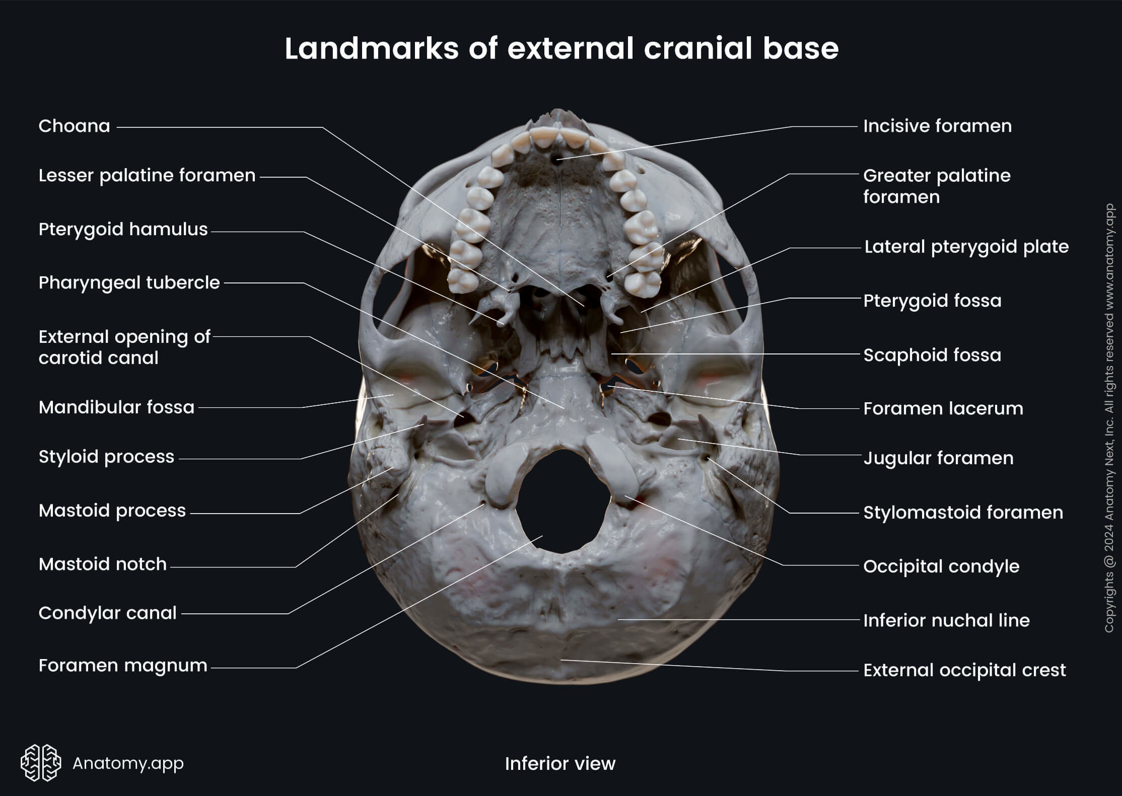
- Choanae
The superior alveolar arch is formed by the alveolar processes of the maxillae and it contains the dental alveoli or sockets for the upper teeth.
The choana (plural: choanae) is a paired posterior aperture of the nasal cavity that opens into the nasopharynx, and can be seen in the anterior portion of the external cranial base. Each choana has four borders formed by different bones of the skull:
- Anteroinferior border - horizontal plate of the palatine bone;
- Superoposterior border - sphenoid bone;
- Lateral border - medial pterygoid plates of the sphenoid;
- Medial border - vomer.
The bony palate is formed by the palatine process of the maxillae anteriorly and the horizontal plates of the palatine bones posteriorly. The bony palate is the skeleton of the hard palate, which forms the roof of the oral cavity and floor of the nasal cavity. There are several openings in the bony plate:
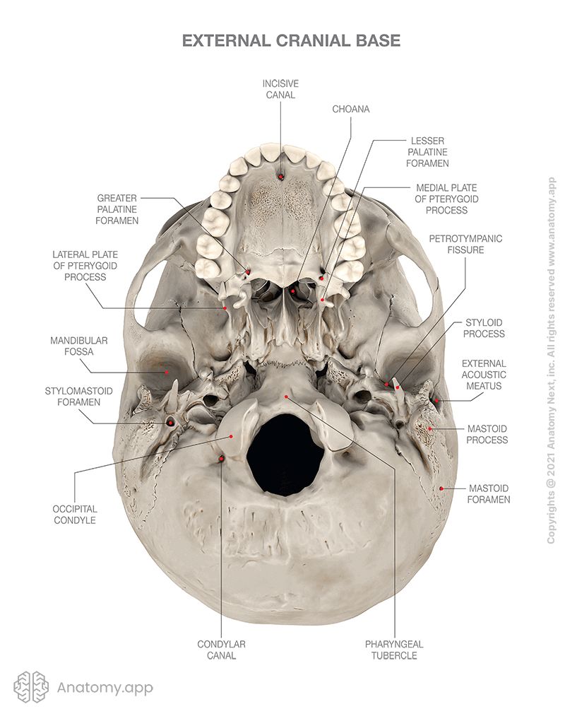
- Incisive foramen
- Greater palatine foramen
- Lesser palatine foramina
- Small foramina for ducts of palatine glands
The paired incisive foramen leads into the incisive canal which connects the external cranial base and the oral cavity to the nasal cavity. This canal transmits the nasopalatine nerve and the terminal branches of the greater palatine vessels.
The greater palatine foramen is the opening of the greater palatine canal found in the oral cavity. The greater palatine canal connects the external cranial base with the pterygopalatine fossa. It transmits the greater palatine nerve and vessels.
The lesser palatine foramina are one or more openings in the hard palate that lead into the lesser palatine canal, which is an accessory canal arising from the greater palatine canal. These foramina carry the lesser palatine nerves and vessels. The lesser palatine foramina like the greater palatine foramen are interconnections between the pterygopalatine fossa and the external cranial base.
The bony palate also contains several small foramina that accommodate secretory ducts of the palatine glands (minor salivary glands). These ducts open into the oral cavity.
Middle part of external cranial base
The middle part of the external cranial base is formed by the body of the sphenoid, petrous parts of both temporal bones, and basiocciput (basal part of the occipital bone). It extends from the choanae anteriorly to the anterior margin of the foramen magnum posteriorly.
Several openings can be seen on the middle part of the external cranial base. Most of these openings lead to the cranial cavity, and they are as follows:
- Foramen lacerum
- Pterygomaxillary fissure
- Foramen ovale
- Foramen spinosum
- Carotid canal
The foramen lacerum is located between the middle cranial fossa and the external cranial base. The foramen lacerum is filled with cartilage after birth and it transmits the artery and nerve of the pterygoid canal.
The pterygomaxillary fissure connects the external cranial base and the infratemporal fossa with the pterygopalatine fossa. It transmits the terminal part of the maxillary artery, as well as the superior alveolar nerve of the maxillary nerve (CN V2).
The foramen ovale is an opening that connects the external cranial base with the middle cranial fossa. It transmits the mandibular division of the trigeminal nerve (CN V3), lesser petrosal nerve, accessory meningeal branch of the maxillary artery, and an emissary vein.
The foramen spinosum also connects the external cranial base with the middle cranial fossa. This opening transmits the middle meningeal artery and the meningeal branch of the mandibular nerve (CN V3).
The carotid canal connects the external cranial base with the middle cranial fossa, and it carries the internal carotid artery, meningeal branches of the ascending pharyngeal artery, and emissary veins from the cavernous sinus.
Posterior part of external cranial base
The posterior part of the external cranial base is mainly formed by the occipital bone. There are several openings in the posterior part of the external cranial base, most of which lead to the posterior cranial fossa inside the cranial cavity:
- Foramen magnum
- Jugular foramen
- Hypoglossal canal
- Condylar canal
- Canaliculus for tympanic nerve
- Mastoid canaliculus
The foramen magnum is a single large opening that connects the external cranial base with the posterior cranial fossa of the internal cranial base. This opening contains the lower end of the medulla oblongata, meninges, vertebral arteries, and the accessory nerve (CN XI).
The jugular foramen also leads from the external cranial base to the posterior cranial fossa and transmits the inferior petrosal sinus (situated anteriorly), the glossopharyngeal (CN IX), vagus (CN X) and accessory (CN XI) cranial nerves (in middle), and the internal jugular vein (posteriorly).
The hypoglossal canal connects the external cranial base with the posterior cranial fossa. It transmits the hypoglossal nerve (CN XII), a meningeal branch of the ascending pharyngeal artery and an emissary vein from the basilar plexus.
The condylar canal is another connection between the external cranial base and the posterior cranial fossa. It conducts an emissary vein from the sigmoid sinus.
The canaliculus for the tympanic nerve leads from the external cranial base to the posterior cranial fossa and transmits the tympanic nerve, which is a branch of the glossopharyngeal nerve (CN IX).
The mastoid canaliculus is a small canal located on the external cranial base, in the lateral part of the jugular fossa of the temporal bone. It serves as the entrance for the auricular branch of the vagus nerve (CN X) into the mastoid process.
Lateral part of external cranial base
The lateral part forms each side of the external cranial base and is made from the zygomatic arch and the infratemporal fossa anteriorly, and mandibular fossa, tympanic plate of the temporal bone, styloid and mastoid processes of the temporal bone posteriorly. The lateral part of the external cranial base has the following openings on each side:
- Petrotympanic fissure
- Stylomastoid foramen
- External acoustic opening (leads into the external acoustic meatus)
- Musculotubal canal
- Petrosal fossula
- Cochlear canaliculus (transmits a small vein from the cochlea to the inferior petrosal sinus)
The petrotympanic fissure is located on the temporal bone and connects the external cranial base with the tympanic cavity. It transmits the chorda tympani of the facial nerve (CN VII) from the cranial cavity to the infratemporal fossa.
The stylomastoid foramen is the opening of the facial canal found on the external surface of the cranial base. It transmits the facial nerve (CN VII) and stylomastoid artery.
The external acoustic opening leads into a canal - external acoustic meatus - that begins on the external cranial base and leads to the tympanic cavity. The external acoustic meatus is where sound waves are conducted to the tympanic membrane.
The musculotubal canal connects the external cranial base with the tympanic cavity. It is a double canal consisting of two semicanals - one for the auditory tube and the other for the tensor tympani muscle.
The petrosal fossula is the opening of the tympanic canaliculus (that leads to the tympanic cavity) on the external cranial base. It accommodates the inferior ganglion of the glossopharyngeal nerve (CN IX).
The cochlear canaliculus is a small opening that connects the lateral part of the external cranial base with the cochlea of the inner ear. The cochlear canaliculus contains the cochlear aqueduct, which is a tubular prolongation of the dura mater that establishes communication between the perilymphatic space and the subarachnoid space. It transmits a vein from the cochlea to join the internal jugular vein.
Anatomy.app
Contact information
- For questions regarding business inquiries. Please contact:
- info@anatomy.app