- Anatomical terminology
- Skeletal system
- Joints
- Muscles
- Heart
- Blood vessels
- Lymphatic system
- Nervous system
- Respiratory system
- Digestive system
- Urinary system
- Female reproductive system
- Male reproductive system
- Endocrine glands
- Eye
- Ear
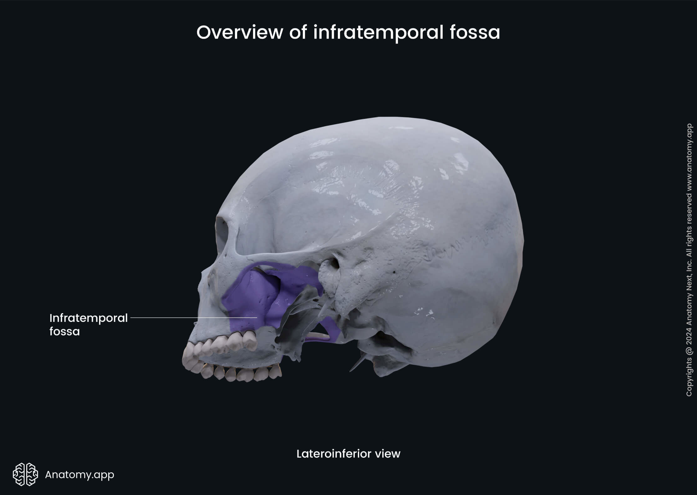
Infratemporal fossa
The infratemporal fossa (Latin: fossa infratemporalis) is an irregularly shaped space on the side of the skull below the zygomatic arch and deep to the ramus of the mandible. The infratemporal fossa is located below the temporal fossa and is continuous with it.
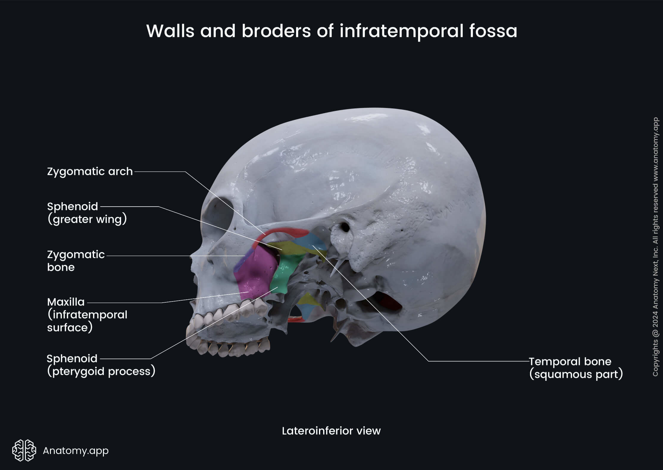
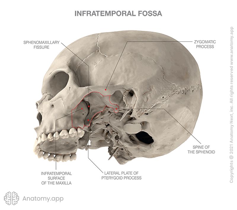
Borders of infratemporal fossa
The borders or walls of the infratemporal fossa are formed by bones and soft tissue structures, and they are as follows:
- Superior (roof): greater wing of the sphenoid;
- Inferior (floor): medial pterygoid muscle;
- Lateral: medial surface of ramus of the mandible;
- Medial: lateral pterygoid plate of the pterygoid process (of the sphenoid);
- Posterior: carotid sheath.
Contents of infratemporal fossa
The infratemporal fossa serves as a passageway for neurovascular structures that travel between the cranial cavity, temporal fossa and pterygopalatine fossa. It also contains several muscles of mastication. The structures found in the infratemporal fossa are as follows:
- Nerves and ganglia: mandibular nerve (CN V3) and its major branches - auriculotemporal, buccal, lingual, and inferior alveolar nerves, chorda tympani - a branch of the facial nerve (CN VII), lesser petrosal nerve, and otic ganglion;
- Blood vessels: maxillary artery, and pterygoid venous plexus;
- Muscles: medial and lateral pterygoid muscles, tendon of the temporal muscle.
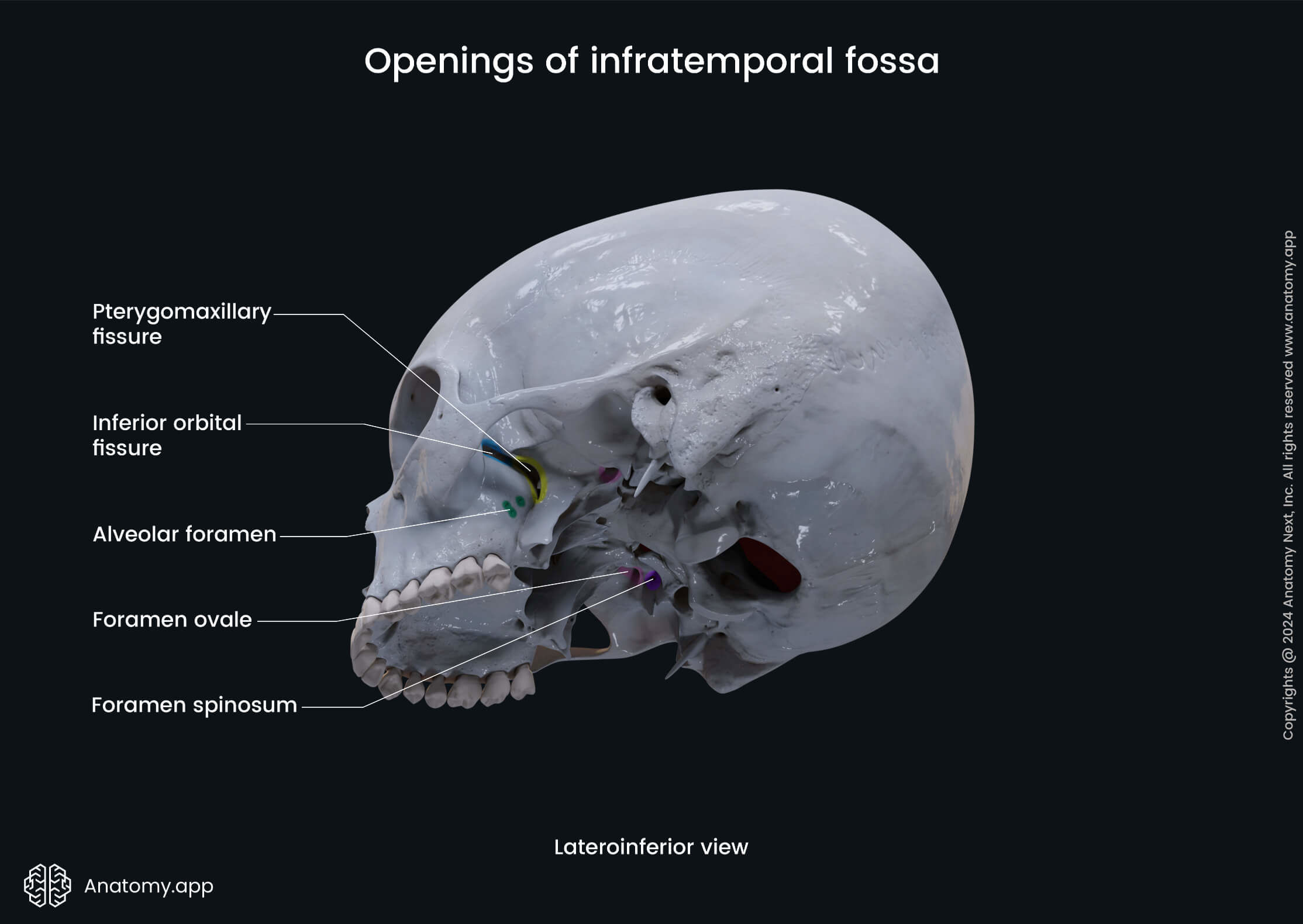
Openings of infratemporal fossa
There are several openings in the walls of the infratemporal fossa that connect it with different cavities and fossae of the skull. These openings transmit nerves and blood vessels. The openings of the infratemporal fossa include the following:
- Foramen ovale
- Foramen spinosum
- Petrotympanic fissure
- Pterygomaxillary fissure
The foramen ovale connects the infratemporal fossa with the middle cranial fossa. It transmits the mandibular nerve (CN V3), lesser petrosal nerve, the accessory meningeal branch of the maxillary artery, and an emissary vein.
The foramen spinosum also connects the infratemporal fossa with the middle cranial fossa of the internal cranial base. This opening serves as a passage for the meningeal branch of the mandibular nerve (CN V3), as well as the middle meningeal artery and vein.
The petrotympanic fissure is located on the temporal bone and serves as a passage for the chorda tympani (a branch of the facial nerve (CN VII)) between the tympanic cavity and the infratemporal fossa.
The pterygomaxillary fissure connects the infratemporal fossa with the pterygopalatine fossa and transmits the terminal part of the maxillary artery, as well as the superior alveolar nerve, which is a branch of the maxillary nerve (CN V2).
Anatomy.app
Contact information
- For questions regarding business inquiries. Please contact:
- info@anatomy.app