- Anatomical terminology
- Skeletal system
- Joints
- Muscles
- Heart
- Blood vessels
- Lymphatic system
- Nervous system
- Respiratory system
- Digestive system
- Urinary system
- Female reproductive system
- Male reproductive system
- Endocrine glands
- Eye
- Ear
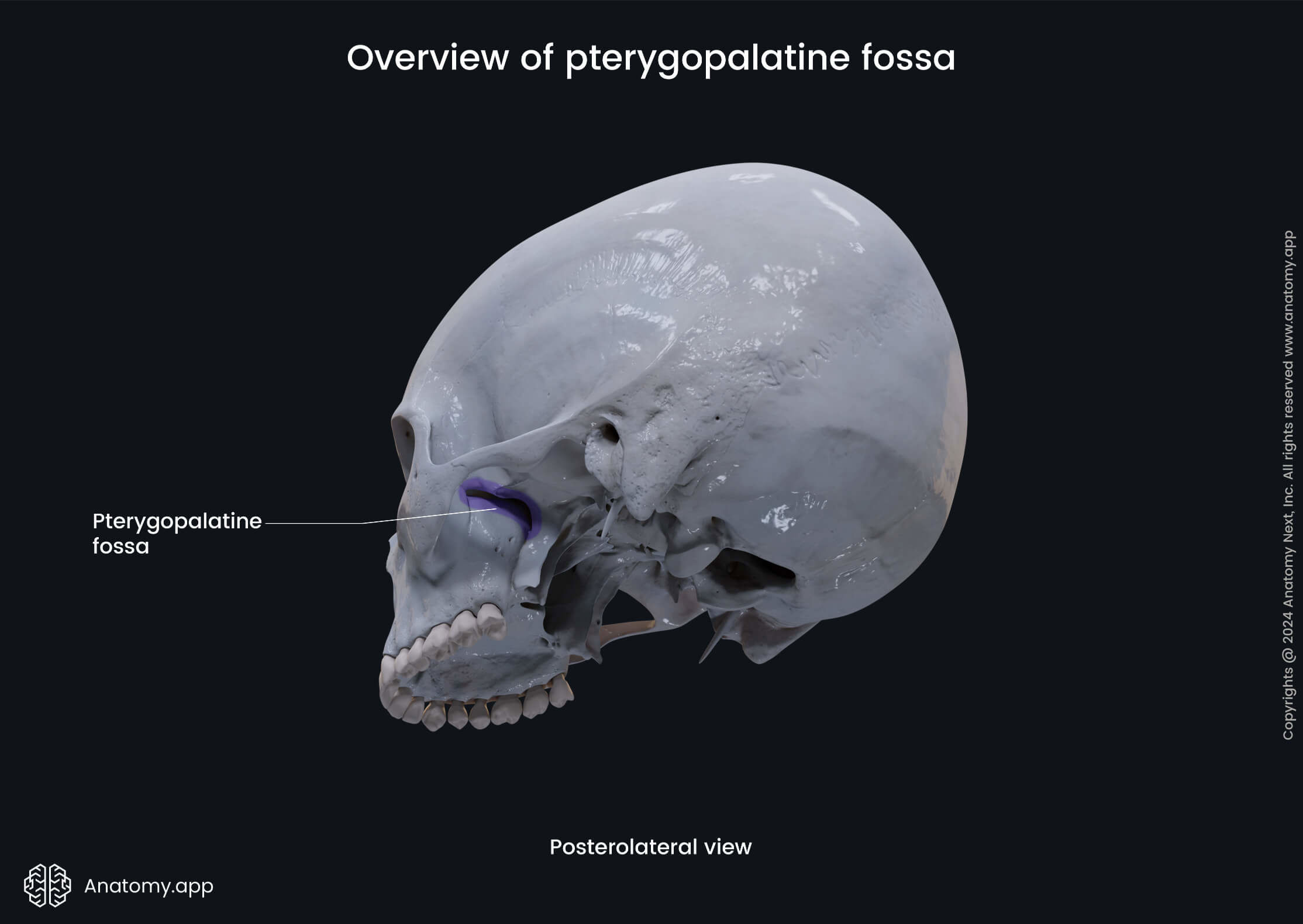
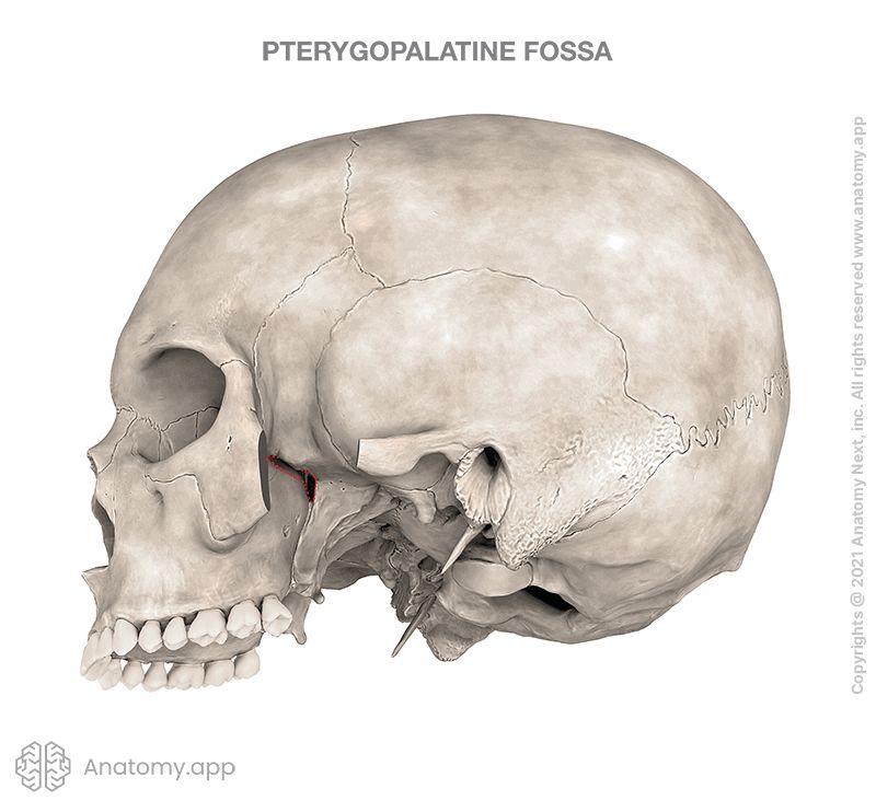
Pterygopalatine fossa
The pterygopalatine fossa (Latin: fossa pterygopalatina) is a cone-shaped, bilateral space on the lateral side of the skull below the apex of the orbit. This space is formed between the maxilla, sphenoid bone and palatine bone.
Borders of pterygopalatine fossa
The borders forming the pterygopalatine fossa are formed by the following structures:
- Anterior - superomedial part of the maxilla's infratemporal surface;
- Posterior: root of the pterygoid process and adjacent anterior surface of the greater wing of the sphenoid bone;
- Inferior: palatine bone;
- Superior: inferior orbital fissure;
- Medial: perpendicular plate of the palatine bone, orbital and sphenoidal processes of the palatine bone;
- Lateral: pterygomaxillary fissure.
Contents of pterygopalatine fossa
The pterygopalatine fossa houses one of the parasympathetic ganglia - the pterygopalatine ganglion. It also serves as a passageway for other neurovascular structures. The contents of the pterygopalatine fossa include the following:
- Nerves and ganglia: maxillary nerve (CN V2) and many of its branches, including the infraorbital, zygomatic, nasopalatine, superior alveolar, pharyngeal and the greater and lesser palatine nerves, and pterygopalatine nerves (that communicate with the pterygopalatine ganglion), and the pterygopalatine ganglion;
- Blood vessels: terminal part of the maxillary artery and its branches, including the sphenopalatine artery, descending palatine artery, infraorbital artery, and posterior superior alveolar artery.
Openings of pterygopalatine fossa
The walls of the pterygopalatine fossa feature canals and foramina that connect this space with other regions of the skull such as the orbit, nasal cavity and oral cavity, middle cranial fossa, and infratemporal fossa. These openings mostly transmit nerves and blood vessels, and they are as following:
- Sphenopalatine foramen
- Inferior orbital fissure
- Pterygomaxillary fissure
- Greater palatine canal
- Foramen rotundum
- Pterygoid canal
- Pharyngeal canal
The sphenopalatine foramen is found in the medial wall of the pterygopalatine fossa, and it connects this region with the nasal cavity to transmit the nasopalatine nerve, as well as the sphenopalatine artery and vein.
The inferior orbital fissure is a space between the sphenoid bone and maxilla that forms the superior boundary of the pterygopalatine fossa. It connects this fossa with the orbit and transmits the zygomatic branch of the maxillary nerve (CN V2), as well as the infraorbital artery and vein.
The pterygomaxillary fissure is an opening forming the lateral boundary of the pterygopalatine fossa and connecting it with the infratemporal fossa. The pterygomaxillary fissure transmits the terminal part of the maxillary artery and the posterior superior alveolar nerve of the maxillary nerve (CN V2).
The greater palatine foramen is an opening located in the inferior wall of the pterygopalatine fossa. It connects the pterygopalatine fossa with the oral cavity and transmits the greater palatine and lesser palatine nerves, and the descending palatine artery and vein.
The posterior wall of the pterygopalatine fossa has several openings such as the foramen rotundum, the pterygoid canal and the pharyngeal canal.
The foramen rotundum forms a communication between the middle cranial fossa and the pterygopalatine fossa. It conducts only one structure - the maxillary nerve (CN V2), which is the second branch of the trigeminal nerve (CN V).
The pterygoid canal connects the pterygopalatine fossa with the middle cranial fossa and transmits the nerve of the pterygoid canal (Vidian nerve), as well as the artery and vein of the pterygoid canal.
The pharyngeal canal forms a connection between the pterygopalatine fossa and the nasal cavity. This canal carries the pharyngeal branches of the maxillary nerve and artery.
Anatomy.app
Contact information
- For questions regarding business inquiries. Please contact:
- info@anatomy.app