- Anatomical terminology
- Skeletal system
- Joints
- Muscles
- Heart
- Blood vessels
- Lymphatic system
- Nervous system
- Respiratory system
- Digestive system
- Urinary system
- Female reproductive system
- Male reproductive system
- Endocrine glands
- Eye
- Ear
Nasal cavity
The nasal cavity (Latin: cavitas nasi) is an irregular-shaped paired air-filled space located above the roof of the oral cavity. It forms the internal part of the nose. The nasal cavity is also an initial part of the respiratory system, and it lodges the olfactory receptors providing the sense of smell.
Most of the nasal cavity is lined with the respiratory mucosa, as it is a part of the upper airways. The nasal cavity is connected to all paranasal sinuses, including the ethmoidal air cells, frontal, sphenoidal and maxillary sinuses. The openings of mentioned sinuses are located within the nasal meatuses.
The bony framework of the nasal cavity is formed by several bones of the skull. The nasal cavity is bounded by the nasal conchae laterally, cribriform plate of the ethmoid bone superiorly, and the palatine processes of the maxilla and horizontal portion of the palatine bone inferiorly.
The nasal cavity is subdivided into the nasal vestibule and nasal cavity proper, and it has six walls. The nasal vestibule is the most anterior part of the nasal cavity covered by the nasal cartilages and lined with the same epithelium as the skin. The walls of the nasal cavity are formed by the bones of the skull, and the posterior and anterior walls contain large openings.
Anteriorly, the nasal cavity opens with the anterior nasal aperture (or the pyriform aperture) and connects with the external environment. Posteriorly, the cavity communicates with the nasopharynx via a pair of oval openings called the choanae. The nasal cavity in the midline is separated by the nasal septum that also is the medial wall of each half of the cavity.
Walls of nasal cavity
The nasal cavity is formed by six walls, and they include the following:
- Superior wall (roof)
- Inferior wall (floor)
- Medial wall
- Lateral wall
- Anterior wall
- Posterior wall
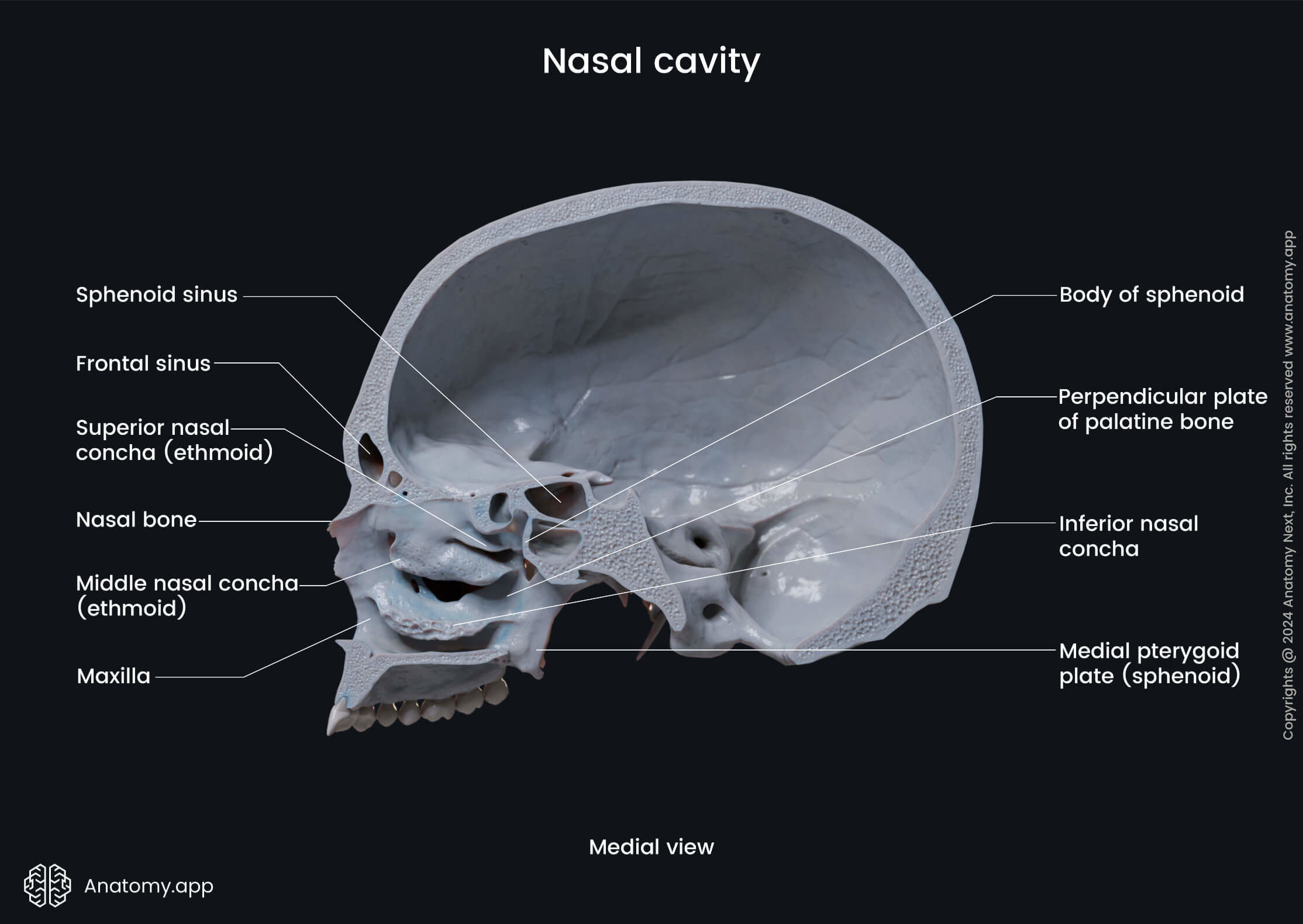
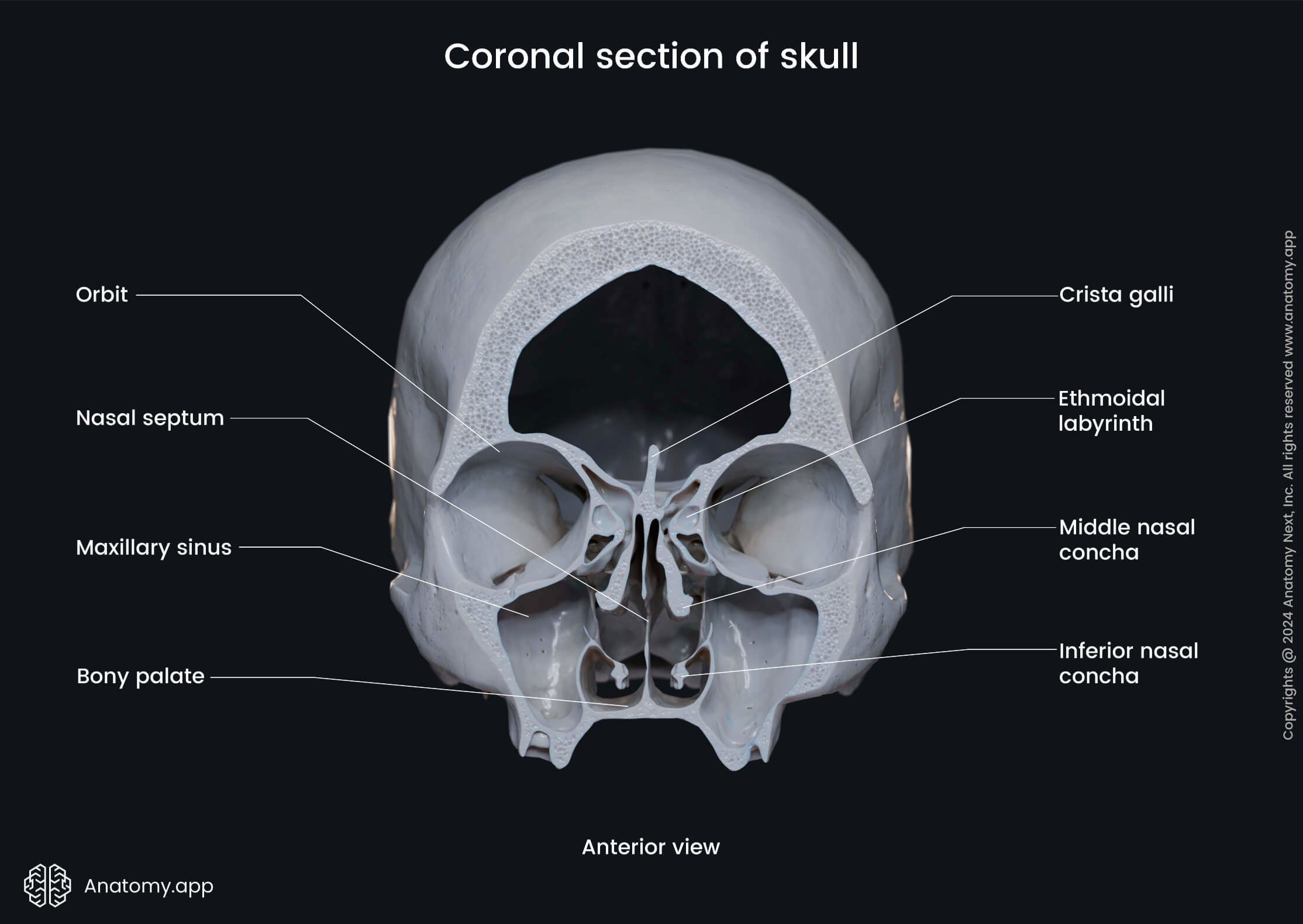
The roof of the nasal cavity is the superior wall formed by the ethmoid and sphenoid bones. The anterior slope of the roof is formed by the cribriform plate of the ethmoid bone, while the posterior slope is formed by the anterior aspect of the body of the sphenoid.
The floor is the inferior wall, and it is broader than the roof. It is formed by the maxillae and palatine bones. Anteriorly are the palatine processes of the maxillae, while posteriorly - the horizontal plates of the palatine bones.
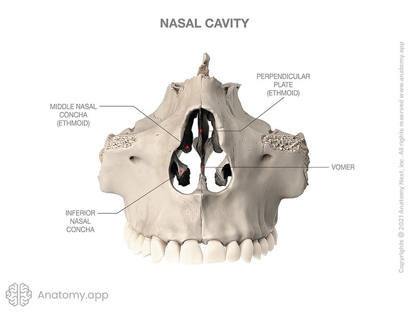
The medial wall corresponds to the nasal septum, which divides the cavity into two symmetrical parts. The anterior part is made of cartilages, while the posterior of bones. The bony nasal septum is formed by the vomer and the perpendicular plate of the ethmoid bone.
The lateral wall is formed by the nasal surface of the maxilla, perpendicular plate of the palatine bone, the labyrinth of the ethmoid bone, lacrimal bone, the medial plate of the pterygoid process (sphenoid bone), and the inferior nasal concha.
The anterior wall of the nasal cavity is formed by the nasal bone and a pair of anterior nasal apertures. In contrast, the posterior wall is formed by the body of the sphenoid bone and paired openings called choanae.
Nasal conchae and meatuses
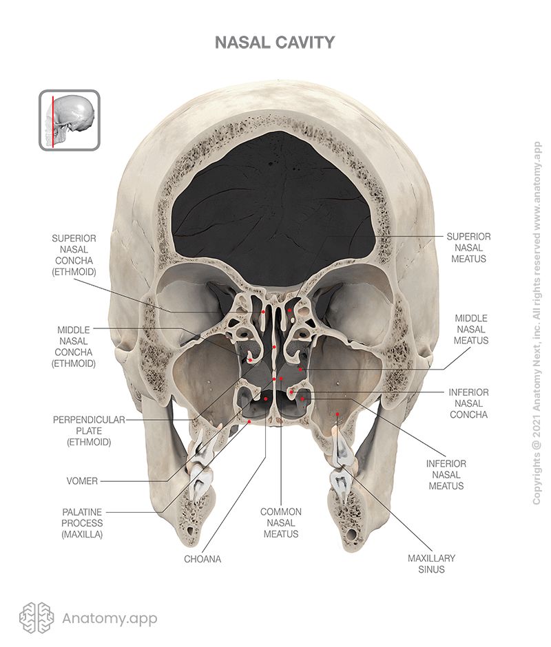
The nasal cavity presents several long, narrow and paired formations on its lateral wall called conchae or conchas. These formations are shaped like curved shelves of bone. Each lateral wall of the nasal cavity contains three nasal conchae:
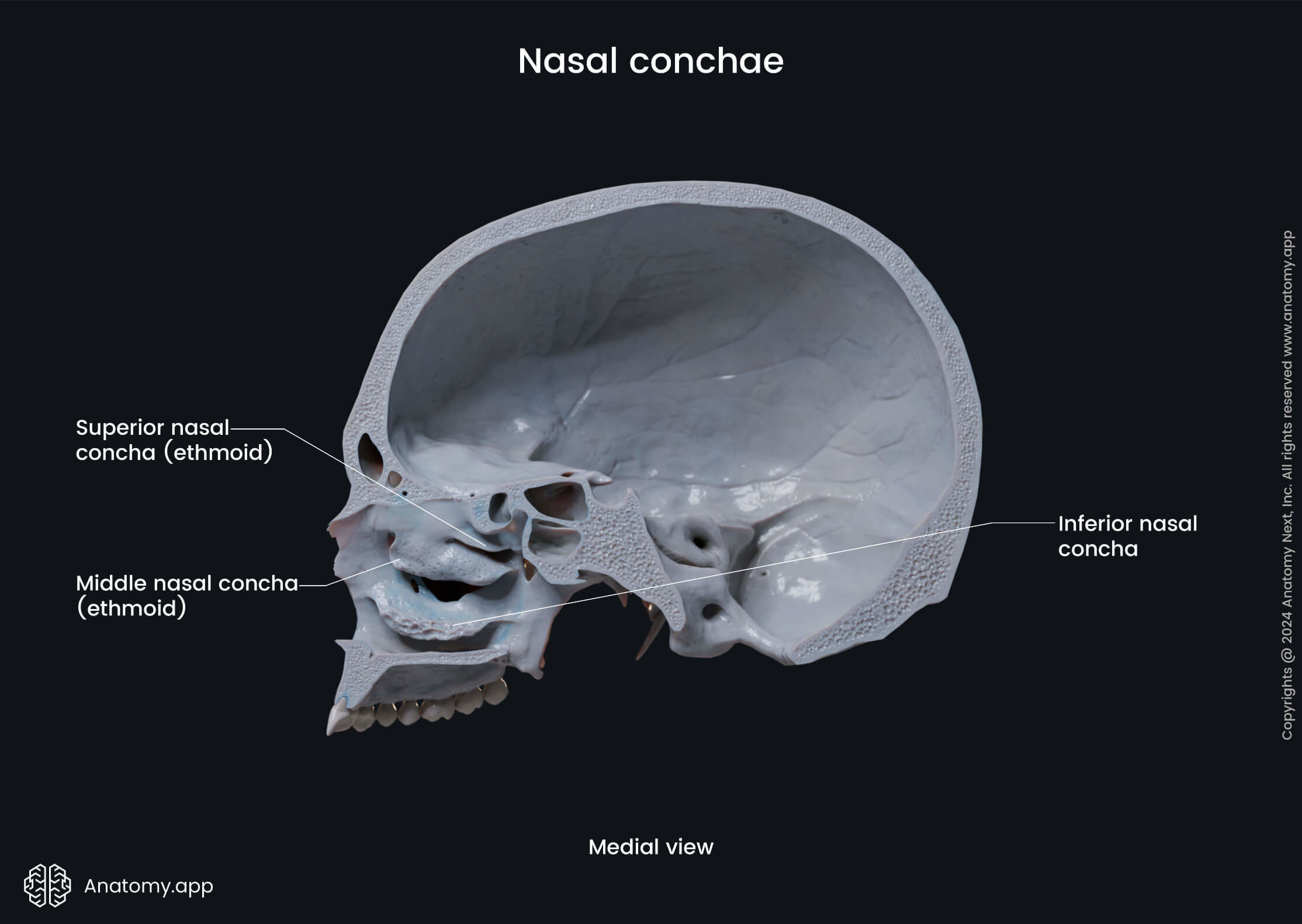
The superior and middle nasal conchae are parts of the labyrinth of the ethmoid bone, while the inferior nasal conchae are separate bones of the skull. Sometimes, the nasal cavity contains one more paired concha - supreme nasal concha - located above the superior nasal concha.
The nasal conchae project into the nasal cavity creating several pathways between them along the lateral wall called nasal meatuses. In total, the nasal cavity has five nasal meatuses, and they are known as follows:
- Superior nasal meatus - passage between the middle and superior nasal conchae;
- Middle nasal meatus - pathway between the middle and inferior nasal conchae;
- Inferior nasal meatus - between the inferior nasal concha and floor of the nasal cavity;
- Common nasal meatus - space between the medial aspects of conchae and nasal septum;
- Nasopharyngeal meatus - space behind the nasal conchae at the superior, middle and inferior nasal meatuses fusion site.
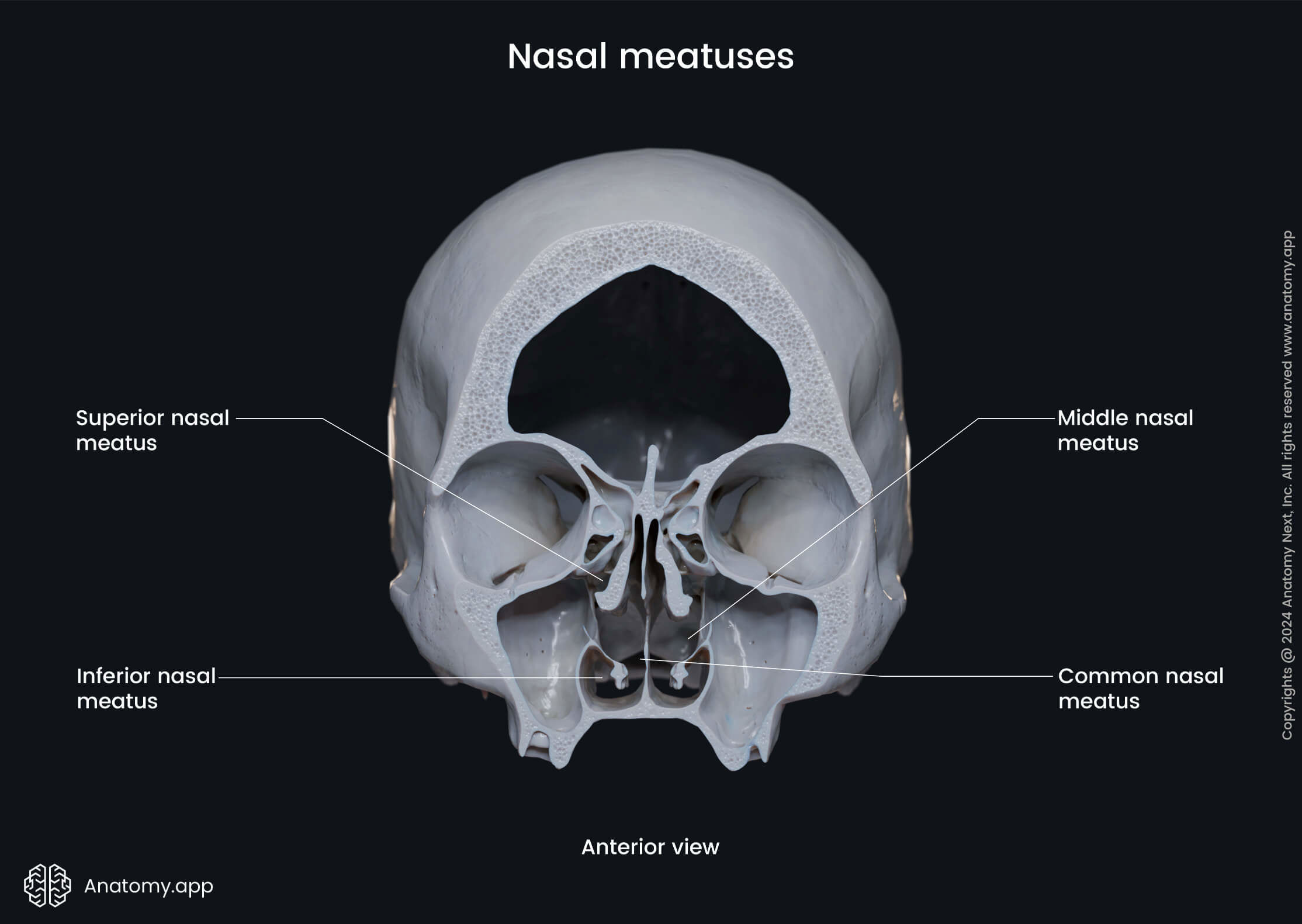
If the supreme nasal concha is present, the nasal cavity has one more meatus called the supreme nasal meatus located between the supreme and superior nasal conchae. The first four meatuses are paired, while the nasopharyngeal meatus is a single pathway.
Openings of nasal cavity
The nasal cavity is interconnected with other parts of the skull via numerous openings. These openings connect each nasal meatus either with a different cavity or region of the skull. The openings of the nasal cavity are known as follows:
- Openings to the ethmoidal air cells
- Opening of the frontal sinus
- Opening of the maxillary sinus
- Opening of the sphenoidal sinus
- Nasolacrimal canal
- Cribriform foramina
- Incisive canal
- Sphenopalatine foramen
- Pyriform aperture
- Choanae
The opening to the posterior ethmoidal cells is located within the superior nasal meatus, while the openings to the anterior and middle ethmoidal cells can be found within the middle nasal meatus. Besides these openings, the middle nasal meatus is also connected with the frontal and maxillary sinuses.
The openings to the anterior ethmoidal air cells, frontal and maxillary sinuses are marked with the semilunar hiatus - a deep crescent-shaped groove on the lateral wall of the middle nasal meatus. The middle ethmoidal air cells drain anterior to the semilunar hiatus via the ethmoidal bulla.
The opening of the sphenoid sinus can be found within the superior nasal meatus. The sphenoid sinus opens into a small space within the meatus called the sphenoethmoidal recess, which lies superior and posterior to the superior nasal concha.
The inferior meatus has an opening for the nasolacrimal canal that connects the nasal cavity with the orbit and drains tears from the eyes. The cribriform foramina connect the common nasal meatus with the anterior cranial fossa and transmit the olfactory nerves.
The incisive canal connects the common nasal meatus with the oral cavity, where it opens via the incisive foramen. It transmits the nasopalatine nerve and greater palatine artery. The sphenopalatine foramen connects the nasal cavity with the pterygopalatine fossa, and it transmits the sphenopalatine artery, nasopalatine and superior nasal nerves.
The pyriform aperture or anterior nasal aperture opens on the external aspect of the face, connecting the nasal cavity with the external environment. Two posterior openings are called the choanae, and they connect the nasal cavity with the external cranial base and nasopharynx.
Blood supply of nasal cavity
The arterial blood supply to the nasal cavity is provided by several arteries and their branches, including:
- Anterior and posterior ethmoidal branches of the ophthalmic artery - supply the roof of the nasal cavity;
- Sphenopalatine branch of the maxillary artery - provides arterial blood supply to the nasal mucosa;
- Greater palatine branch of the maxillary artery - supplies the region of the inferior nasal meatus.
All mentioned arteries ramify to form several anastomotic plexuses located within the nasal mucosa. One of the plexuses is the submucosal cavernous plexus situated in the posterior part of the septum and the middle and inferior nasal conchae. Numerous arteriovenous anastomoses are positioned in the deep layer of the mucosa.
Nerve supply of nasal cavity
Different parts of the nasal cavity are innervated by sensory nerves, and these nerves are as follows:
- Olfactory nerves (CN I) - arise from the olfactory epithelium in the upper part of the nasal cavity and provide olfaction (sense of smell);
- Anterior ethmoidal nerve - a branch of the nasociliary nerve; supplies the roof; gives off a lateral internal branch to supply the anterior part of the lateral wall and a medial internal branch to supply the nasal septum;
- Infraorbital nerve - supplies the nasal vestibule;
- Anterior superior alveolar nerve - supplies part of the nasal septum, floor of the nasal cavity near the anterior nasal spine, and the anterior part of the lateral wall;
- Posterior superior and inferior nasal nerves - branches of the greater palatine nerve; supply the posterior part of the lateral wall, roof and floor, and the inferior medial part of the nasal septum;
- Branches from the nerve of the pterygoid canal - supply the superior and posterior part of the roof and nasal septum.
Anatomy.app
Contact information
- For questions regarding business inquiries. Please contact:
- info@anatomy.app