- Anatomical terminology
- Skeletal system
- Joints
- Muscles
- Heart
- Blood vessels
- Lymphatic system
- Nervous system
- Respiratory system
- Digestive system
- Urinary system
- Female reproductive system
- Male reproductive system
- Endocrine glands
- Eye
- Ear
Tympanic membrane
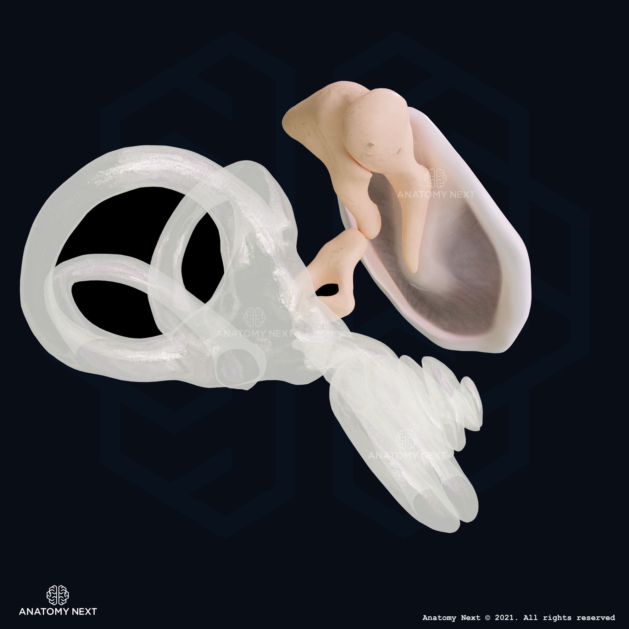
The tympanic membrane (also known as eardrum, myringa, membranous wall of tympanic cavity, latin: membrana tympanica) is a cone-shaped thin, transparent membrane at the end of the external acoustic meatus, which separates the external ear from the middle ear.

Structure of tympanic membrane
The tympanic membrane contains fibrous connective tissue that is covered with skin externally and lined by mucosa internally, where the membrane projects to the tympanic cavity of the middle ear, forming the membranous or lateral wall of the tympanic cavity. Attached to the inner surface of the eardrum is one of the auditory ossicles, the malleus, which is connected on the other side with the next ossicle, the incus. The tympanic membrane is located at a 45-degree angle with a diameter of 9 x 11 mm.
There are two areas of the tympanic membrane called the pars flaccida and the pars tensa.
The pars flaccida is the smaller, more flaccid part of the tympanic membrane in its upper region, while the pars tensa is the largest part of the tympanic membrane situated within a fibrocartilaginous ring, called the tympanic ring. The center of the pars flaccida is drawn inward and is called the umbo. The umbo of the tympanic membrane is situated at the tip of the manubrium of the malleus, which is fused with the tympanic membrane.
Superior to the umbo is a stripe - malleolar stria. It is the impression made by the malleus handle. The superior end of the malleolar stria has a structure called the malleolar prominence. The impression is made by the lateral process of the malleus. The parts of the tympanic membrane moving away from the lateral process are called anterior and posterior malleolar folds.
The tympanic membrane has 3 layers: the outer layer covered with the skin and a thin layer of the cerumen from the external acoustic canal side; the middle layer, which has two types of connective tissue fiber; the inner layer is covered with mucosa and completely convexed towards the middle ear. Here, around the border between the pars tensa and pars flaccida, the chorda tympani is located. Below the chorda tympani is the chorda tympani nerve that is a branch of the facial nerve.
The translucency of the tympanic membrane allows the structures within the middle ear to be seen during otoscopy.
Function of the tympanic membrane
The main function of the tympanic membrane is to transfer sound waves from the air from outside, which reach the membrane through the external acoustic meatus, to the auditory ossicles in the middle ear, which then conduct the vibrations to the oval window transferring them to the fluid and membranes of the cochlea of the inner ear. Thus, the eardrum participates in converting and amplifying the vibrations in the air to fluid-membrane vibrations.
Vasculature and innervation of tympanic membrane
Blood supply and venous drainage
The blood to the tympanic membrane is supplied through:
- The deep auricular artery and anterior tympanic arteries, branches of the maxillary artery
- The stylomastoid artery, a branch of the posterior auricular artery
- The inferior tympanic artery, a branch of the ascending pharyngeal artery
Venous drainage of the tympanic membrane happens through the external jugular veins from the superficial portion of the membrane, while from the deep portion - through veins that drain into the transverse sinus and dural veins.
Lymphatic drainage
The lymph from the tympanic membrane is drained to the periauricular lymph nodes.
Innervation
The sensory innervation of the tympanic membrane is provided by several cranial nerves. The auriculotemporal nerve, arising from the mandibular nerve (CN V3), supplies the external surface of the tympanic membrane, which also receives fibers from the auricular branch of the vagus nerve (CN X), and the facial nerve (CN VII). The inner surface of the tympanic membrane is supplied by the glossopharyngeal nerve (CN IX).