- Anatomical terminology
- Skeletal system
- Joints
- Muscles
- Heart
- Blood vessels
- Lymphatic system
- Nervous system
- Respiratory system
- Digestive system
- Urinary system
- Female reproductive system
- Male reproductive system
- Endocrine glands
- Eye
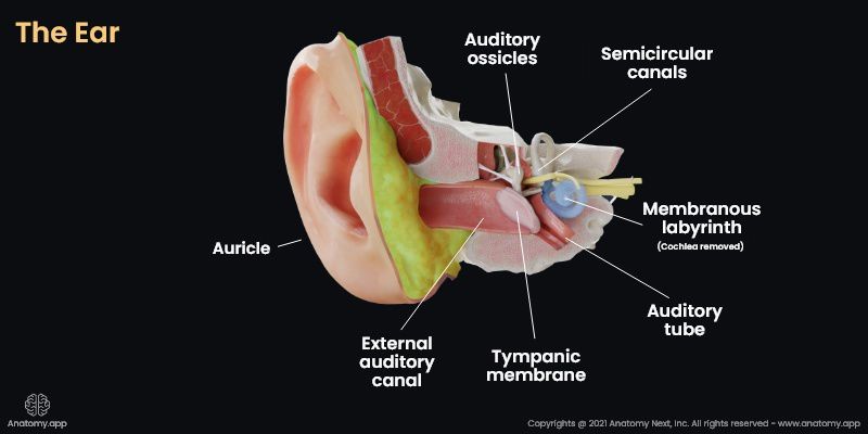
- Ear
External acoustic meatus
The external acoustic meatus (also external auditory canal, external auditory meatus or ear canal, Latin: meatus acusticus externus) is an air-filled tubular space that extends from the auricle of the external ear into the temporal bone to the tympanic membrane. The external acoustic meatus conducts sound waves to the tympanic membrane.
Structure of external acoustic meatus
The external acoustic meatus is a partly cartilaginous and partly bony S-shaped canal, about 2.5-3.5 cm in length with a diameter of about 9 mm, leading to the tympanic membrane, which separates the external ear from the middle ear.
The outer or lateral third of the external auditory canal is the cartilaginous part. In contrast, the inner and posterior walls of the canal are fibrous and lie within the temporal bone, forming the bony part.
The anterior and inferior walls of the bony part are built by the tympanic part of the temporal bone, while the posterior and superior walls - by the squamous part of the temporal bone. Anatomically the posterior wall is related to the mastoid cells from the mastoid process, meanwhile the anterior and inferior walls - to the temporomandibular joint.
The lateral part of the external acoustic canal is directed posteriorly and superiorly, but the middle part is directed anteriorly and inferiorly.
During the ear examination, these directions are important to know for better visualization of the tympanic membrane. In order to gain better visualization, the auricle has to be pulled backward, upwards, and slightly lateral. This action will help to align the lateral and medial parts.
The cartilage of the external acoustic meatus together with the cartilage of the auricle forms a groove that opens superiorly and posteriorly. The canal is lined with skin that has rarely located hairs. Within the canal are glands responsible for the production of the wax.
Cerumen
Medically known as cerumen, in a common language known as earwax, is a brown, yellowish, gray substance that is secreted in the external acoustic canal. Cerumen is produced in the cartilaginous part of the canal. It is created by two different glands - sebaceous and apocrine sweat glands. Sebaceous glands produce viscous secrete, while the apocrine glands produce less viscous secrete.
The main components of the earwax are shed layers of skin, hair, long-chain fatty acids, cholesterol, and squalene.
The main function of the earwax is to protect the skin of the canal, take part in cleaning and lubrication, as well as provide protection against water, bacteria, and fungi.
Vasculature and innervation of the external acoustic meatus
Arterial supply and venous drainage
The anterior part of the external acoustic meatus is supplied by the superficial temporal artery, while the posterior part of the ear canal is supplied by the posterior auricular artery.
Venous drainage happens through the superficial temporal vein, the external jugular vein, and the pterygoid plexus.
Lymphatic drainage
The lymph from the external acoustic meatus is drained into the superficial and deep cervical lymph nodes.
Innervation
The sensory innervation of the external auditory meatus is provided by branches of several cranial nerves. The auriculotemporal branch of the mandibular nerve (CN V3) with the nerve to external acoustic meatus supplies the anterior and superior wall of the canal with sensory innervation. The auricular branch of the vagus nerve (CN X) supplies the posterior and inferior walls of the canal, and the facial nerve (CN VII) may also supply it due to its connection with the vagus nerve.
Anatomy.app
Contact information
- For questions regarding business inquiries. Please contact:
- info@anatomy.app