- Anatomical terminology
- Skeletal system
- Skeleton of trunk
- Skull
- Skeleton of upper limb
- Skeleton of lower limb
- Joints
- Muscles
- Heart
- Blood vessels
- Lymphatic system
- Nervous system
- Respiratory system
- Digestive system
- Urinary system
- Female reproductive system
- Male reproductive system
- Endocrine glands
- Eye
- Ear
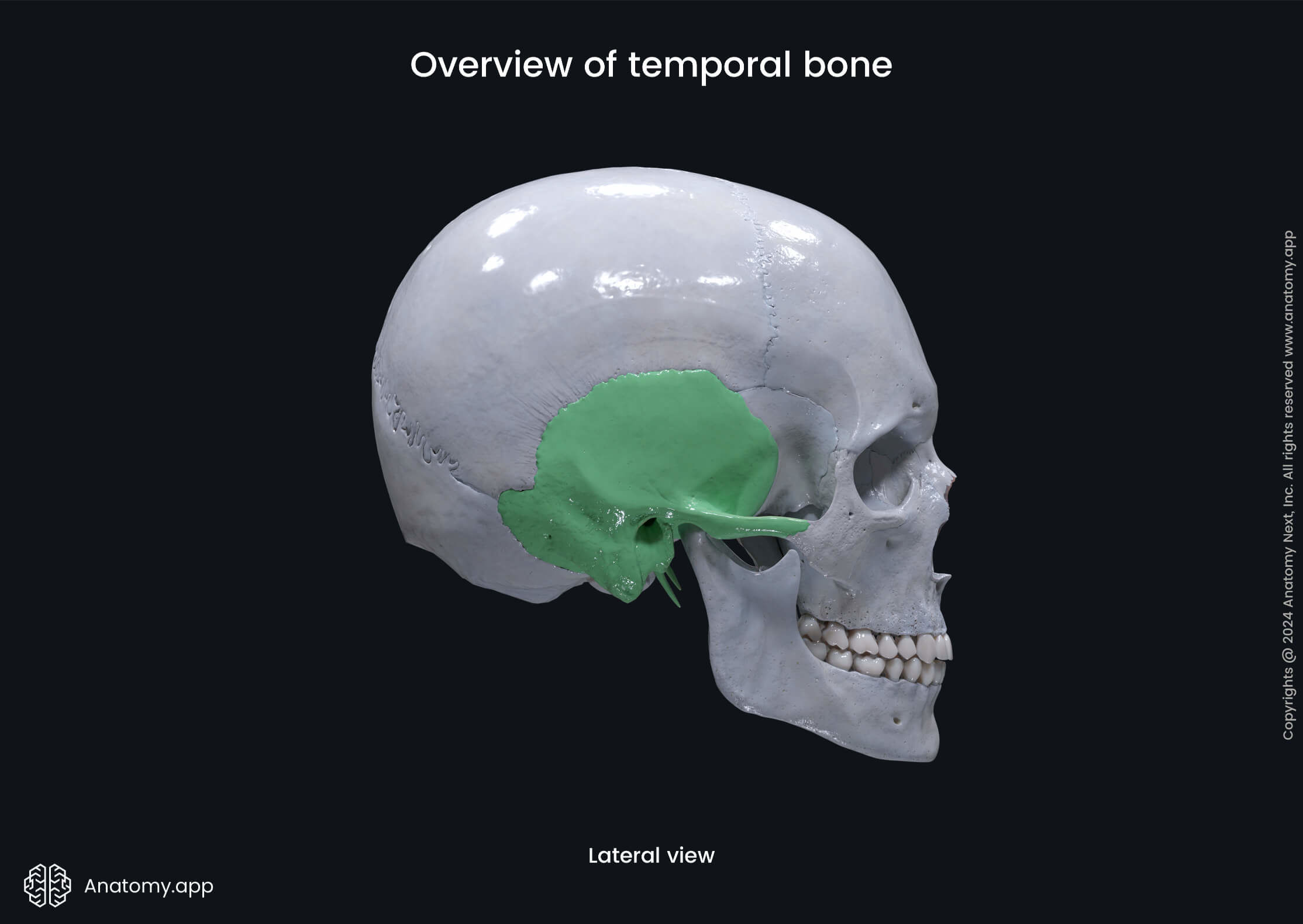
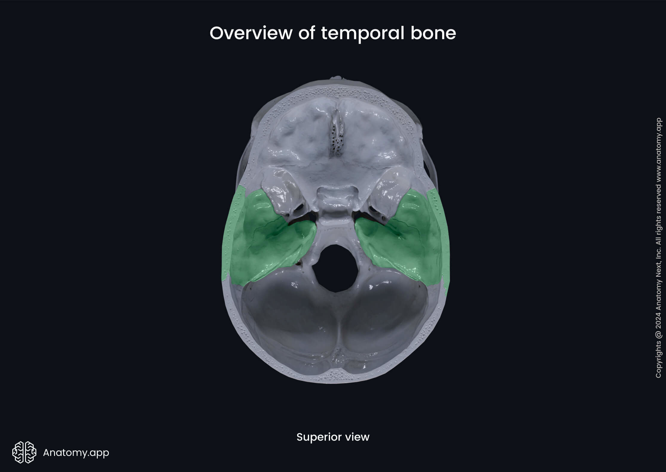
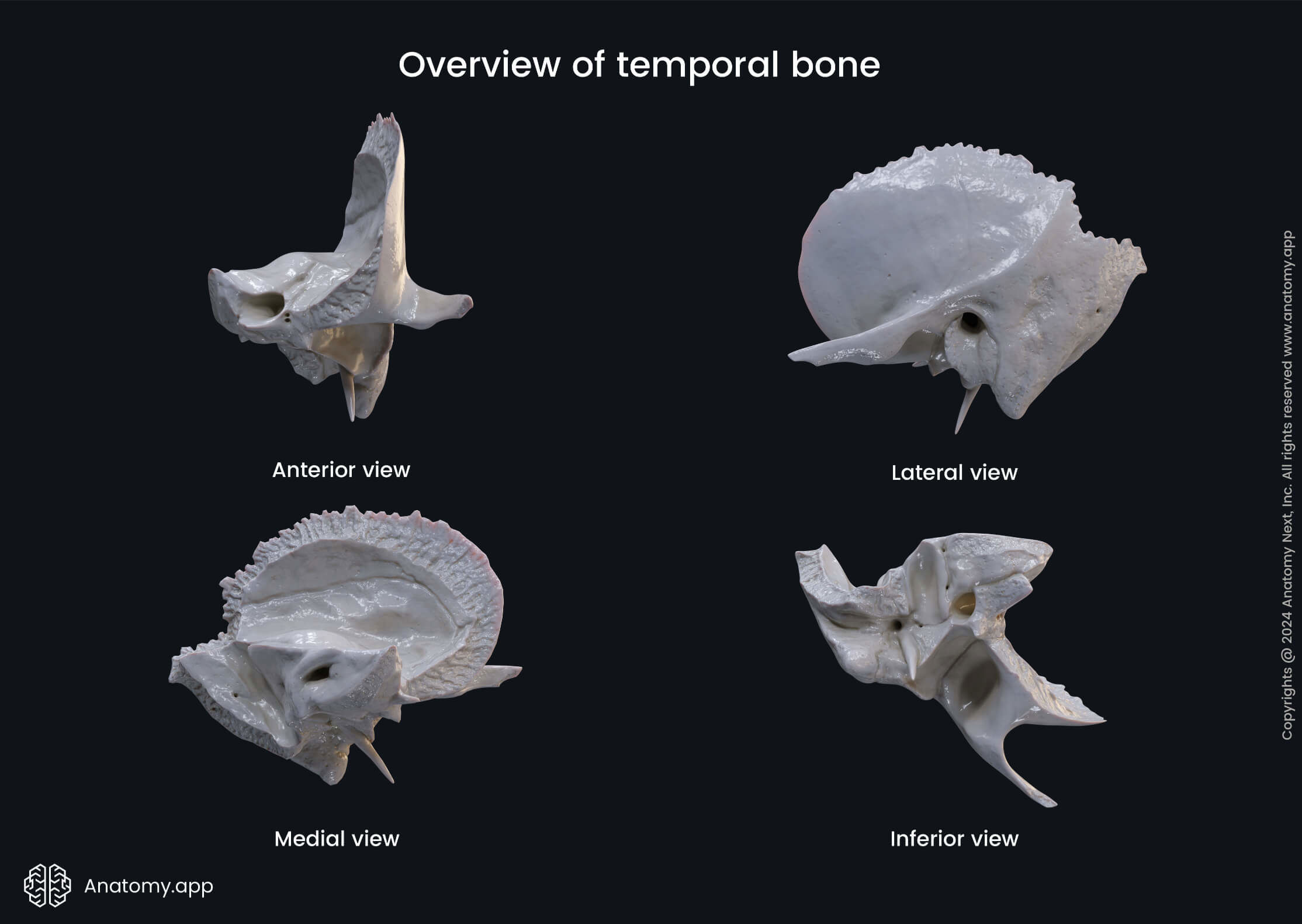
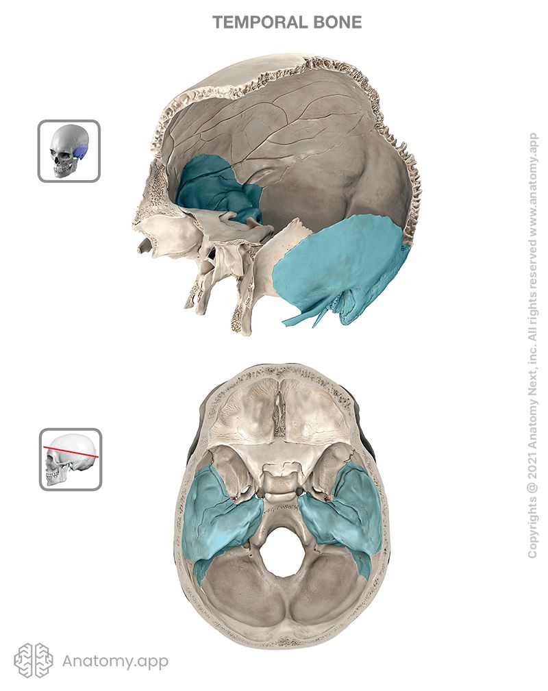
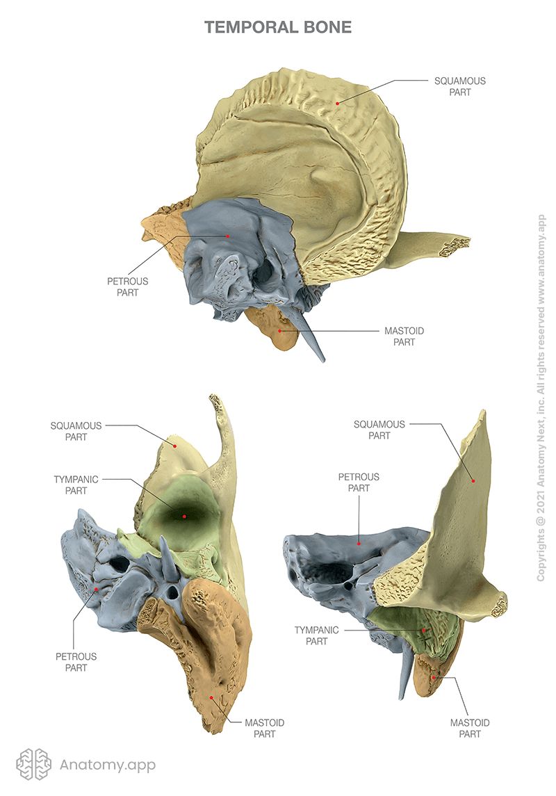
Temporal bone
The temporal bone (Latin: os temporale) is a paired bone situated at the lateral side and base of the skull. This bone features important structures of the vestibulocochlear apparatus, including the external acoustic meatus, the tympanic cavity and the structures forming the inner ear.
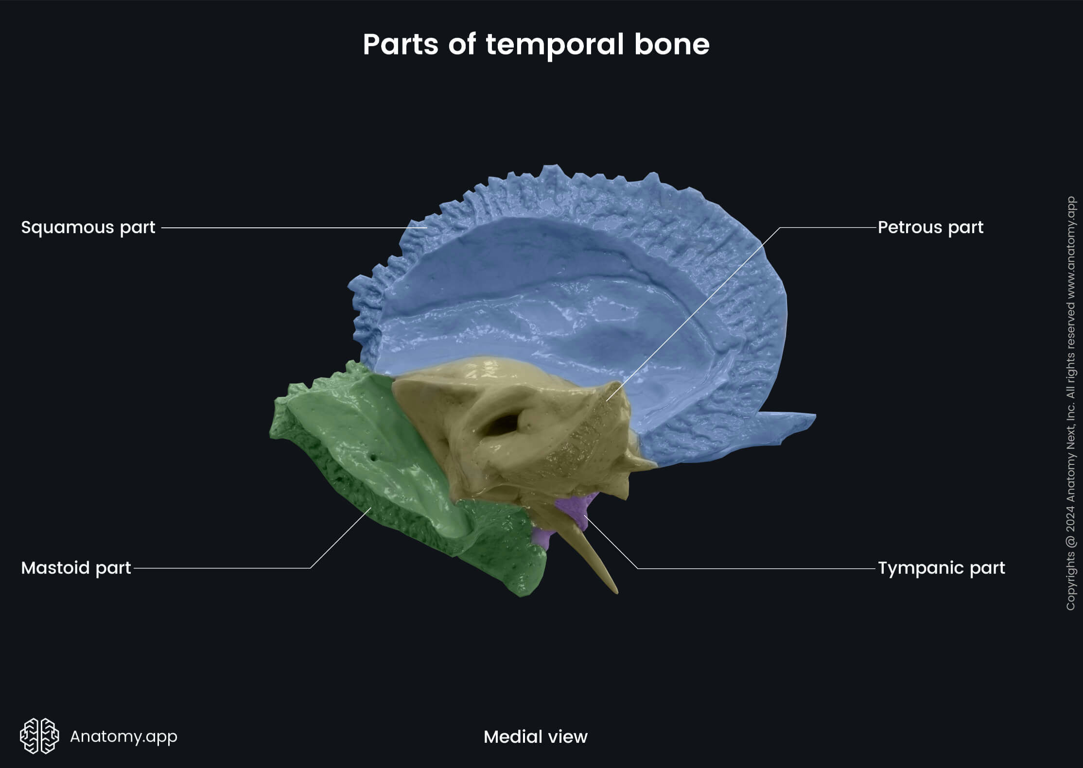
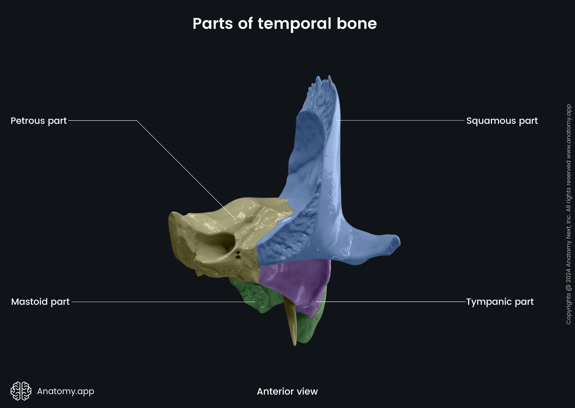
Parts of temporal bone
Each temporal bone consists of four parts:
- Petrous part (Read more!)
- Mastoid part (Read more!)
- Tympanic part (Read more!)
- Squamous part (Read more!)
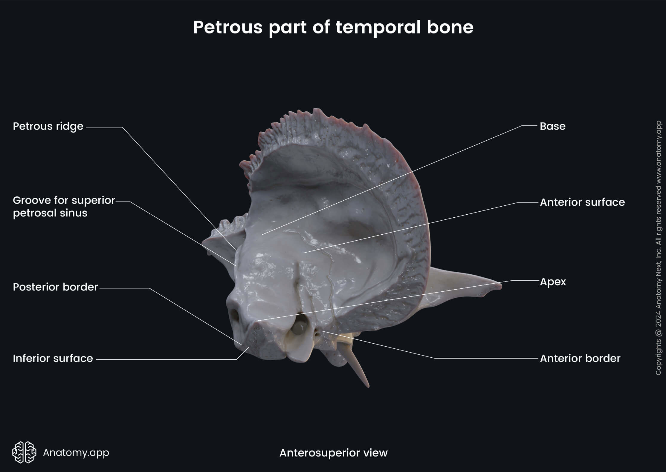
Petrous part of temporal bone
The petrous part (also called the pyramid) is the part of the temporal bone which houses the inner ear. It is located in the base of the skull between the sphenoid and occipital bones. The petrous part of the temporal bone is pyramid shaped, it has an apex and three surfaces (anterior, posterior, inferior) and three margins (anterior, superior, posterior).
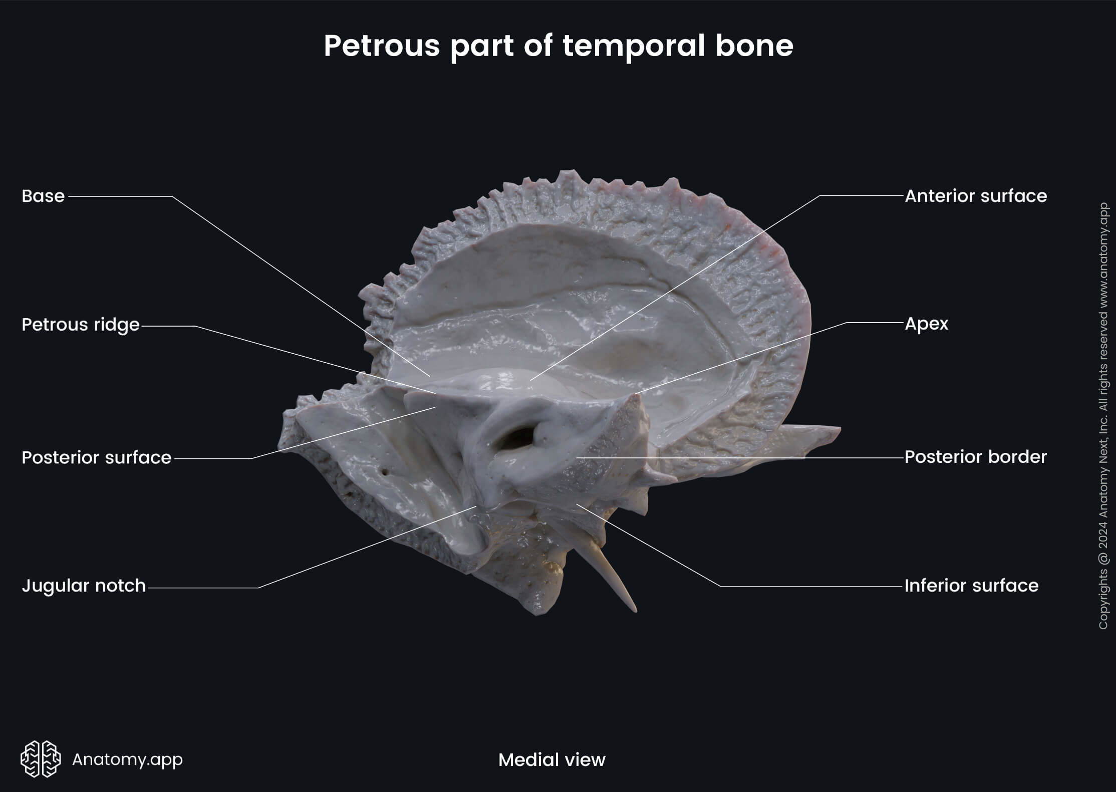
Apex of petrous part
The apex of the petrous part of the temporal bone features the anterior or internal opening of the carotid canal, and forms the posterolateral margin of the foramen lacerum.
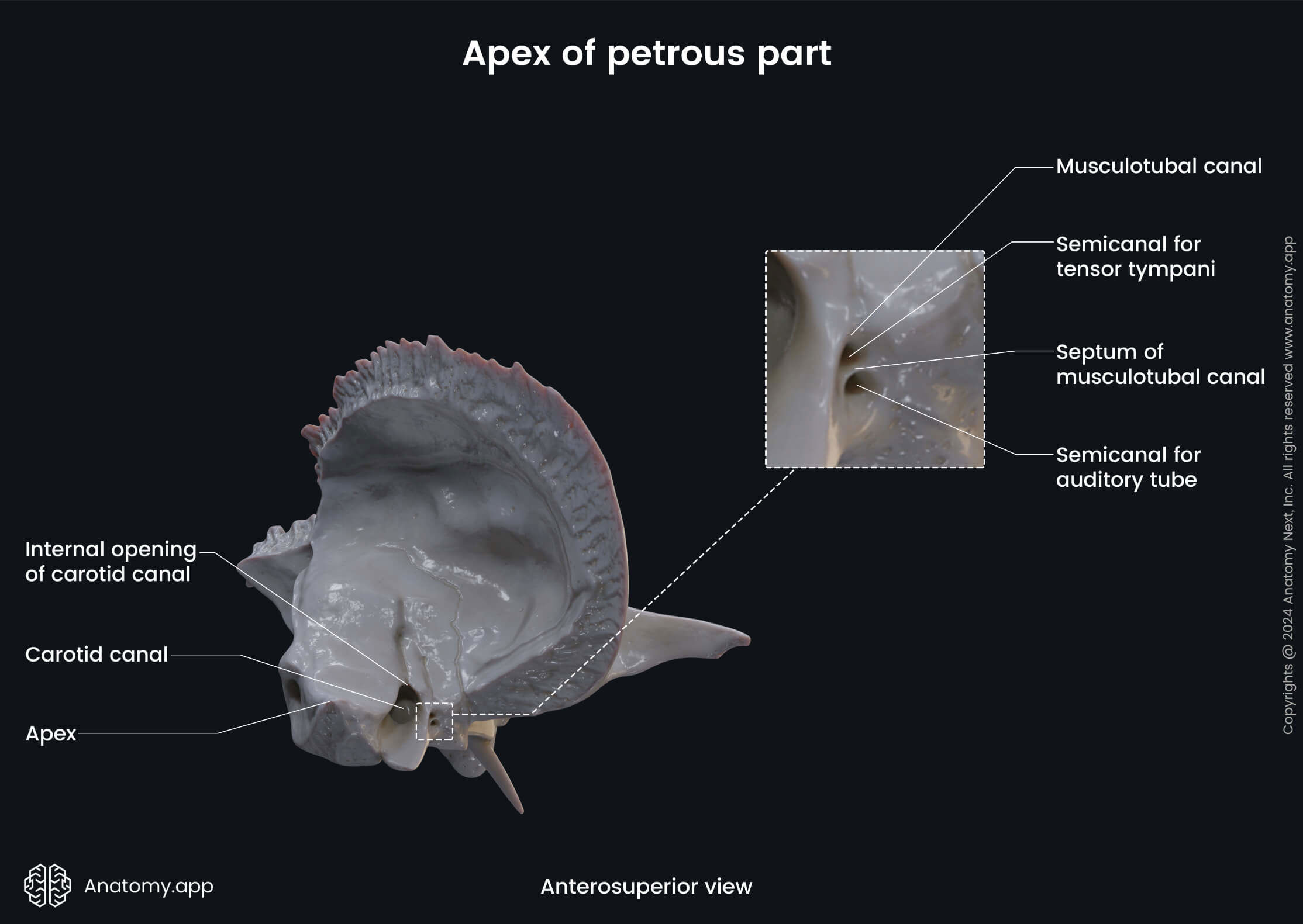
The carotid canal is a bony passage for the internal carotid artery. The canal is located in the apex of the petrous part of the temporal bone. Through it the mentioned artery reaches the middle cranial fossa from the neck region.
The foramen lacerum is a triangular-shaped hole in the base of the skull, located between the sphenoid bone, the apex of the petrous part of the temporal bone, and the basilar part of the occipital bone. The foramen lacerum fills with cartilage after birth.
Surfaces of petrous part
The anterior surface of the petrous part of the temporal bone features the following anatomical landmarks:
- Tegmen tympani
- Arcuate eminence
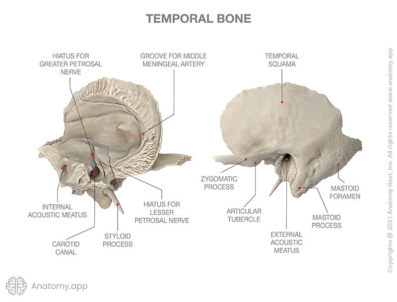
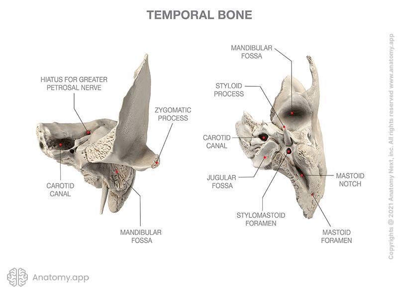
- Hiatus for greater petrosal nerve
- Hiatus for lesser petrosal nerve
- Trigeminal impression
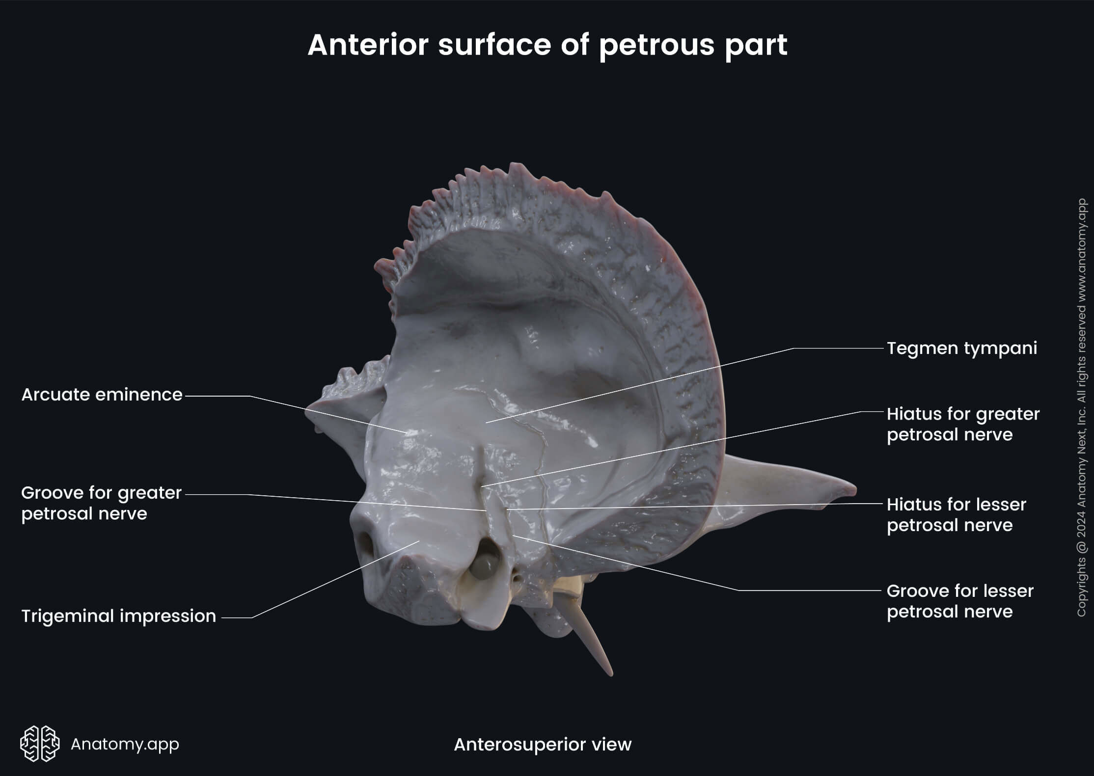
The tegmen tympani is the roof of the tympanic cavity. It is a thin bony plate found on the pyramid of the temporal bone anterolateral to the arcuate eminence.
The arcuate eminence is an elevation on the anterior surface of the pyramid. This eminence is produced by the underlying anterior semicircular canal.
The hiatus for the greater petrosal nerve is a shallow groove in the petrous part leading to an oblique opening. It connects the facial canal to the middle cranial fossa. The opening serves for the passage of the greater petrosal nerve and the petrosal branch of the middle meningeal artery.
The hiatus for the lesser petrosal nerve is an opening that serves as a passage for the lesser petrosal nerve as it separates from the glossopharyngeal nerve (CN IX). This opening connects the posterior cranial fossa to the external surface of the skull.
The trigeminal impression is a shallow, oval depression on the anterior surface of the petrous part of the temporal bone. It is produced by pressure from the trigeminal ganglion located here.
The posterior surface of the petrous part of the temporal bone has two openings:
- Internal acoustic opening (leads into the internal acoustic meatus)
- External opening of vestibular aqueduct
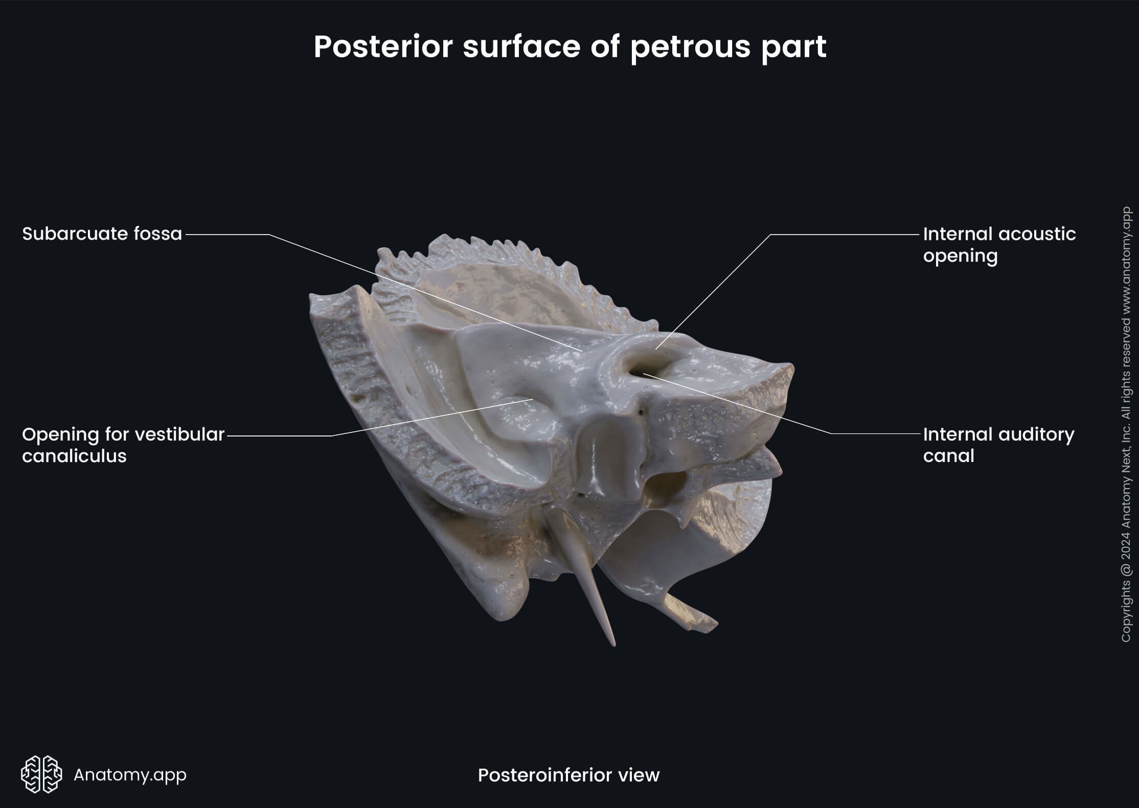
The internal acoustic opening is a small opening at the beginning of the internal acoustic meatus (or internal auditory canal). The opening can be seen is in the posterior cranial fossa of the skull. The internal acoustic meatus runs laterally into the petrous part of the temporal bone. Its content includes components of the facial nerve (CN VII) and vestibulocochlear nerve (CN VIII), the vestibular ganglion and the labyrinthine artery.
There are five nerves that run through the internal auditory canal: the intermediate nerve (sensory component of CN VII), facial motor root (motor component of CN VII), cochlear nerve (component of CN VIII), inferior vestibular nerve (component of CN VIII) and the superior vestibular nerve (component of CN VIII).
A horizontal ridge called the falciform crescent divides the internal auditory canal into superior and inferior portions. The facial nerve and the superior vestibular nerve go in the superior portion of the canal. The cochlear nerve and inferior vestibular nerve run inferior to the falciform crescent with the cochlear nerve situated more anteriorly.
The vestibular aqueduct is a narrow canal extending from the endolymphatic space of the internal ear to the posterior wall of the petrous part. Its external opening can be seen on the posterior surface of the petrous part of the temporal bone.
The inferior surface of the petrous part of the temporal bone features the following strucures:
- Styloid process
- Stylomastoid foramen (external opening of the facial canal)
- Jugular fossa
- Petrosal fossula (featuring the tympanic canaliculus)
- Carotid canal
- Musculotubal canal, divided into two semicanals:
- Semicanal for tensor tympani muscle
- Semicanal for auditory tube
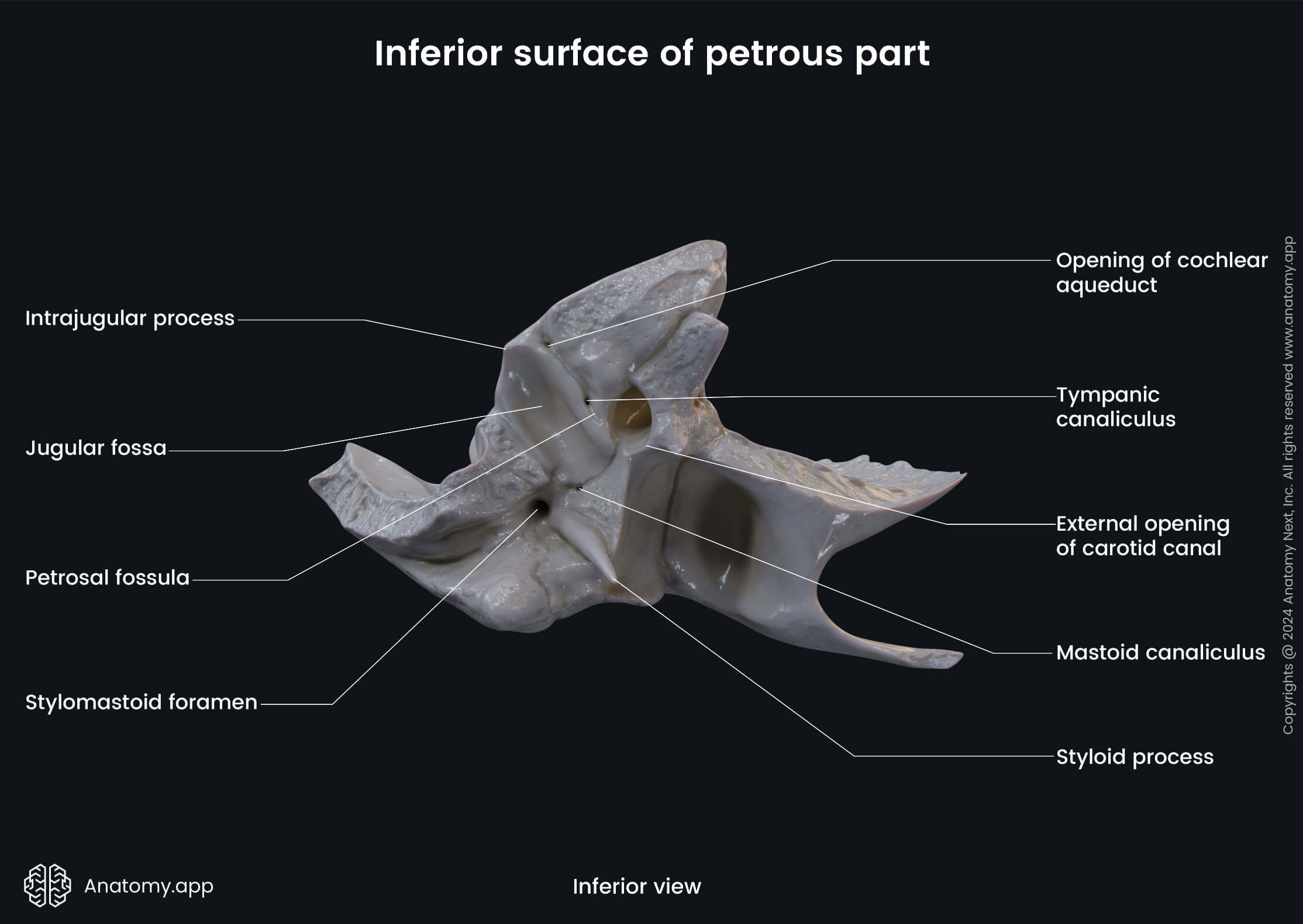
The styloid process is a long process extending from the inferior surface of the petrous part of the temporal bone. It is located in front of the jugular fossa. The stylomastoid foramen is a round opening located on the external surface of the skull between the styloid and the mastoid processes. It is the external opening of the facial canal and transmits the facial nerve (CN VII) and the stylomastoid artery.
The facial canal is a bony canal running through the temporal bone containing the facial nerve (CN VII). It is the longest canal (around 3 cm) for a nerve in the human body. The inner opening of the canal is located in the internal acoustic meatus. And its outer opening is the stylomastoid foramen presenting on the external cranial base, as mentioned before. There is also a small canal arising from the facial canal called the canaliculus for chorda tympani.
The canaliculus for chorda tympani is a small canal in the temporal bone, which starts at the descending part of the facial canal and ends at the tympanic cavity. It serves as a passage for the chorda tympani (a branch of the facial nerve) into the tympanic cavity.
The jugular fossa is a deep depression on the external base of the skull, specifically, on the inferior surface of the temporal bone posterior to the carotid canal. The jugular fossa houses the superior bulb of the internal jugular vein.
The petrosal fossula is a slight depression in the bony ridge between the carotid canal and the jugular fossa. It is occupied by the otic ganglion. The petrosal fossula presents the opening of the tympanic canaliculus. This small canal serves as a passage of the tympanic nerve and the inferior tympanic artery.
The musculotubal canal is a double canal within the petrous part of the temporal bone for the auditory tube and the tensor tympani muscle located in front of the carotid canal and leading into the tympanic cavity. The canal is divided into two semicanals - for the tensor tympani muscle and for the auditory tube.
The semicanal for the tensor tympani muscle is the superior division of the musculotubal canal for the tensor tympani muscle. The semicanal for the auditory tube is the inferior division of the musculotubal canal that forms the bony part of the auditory (pharyngotympanic) tube.
Margins of petrous part
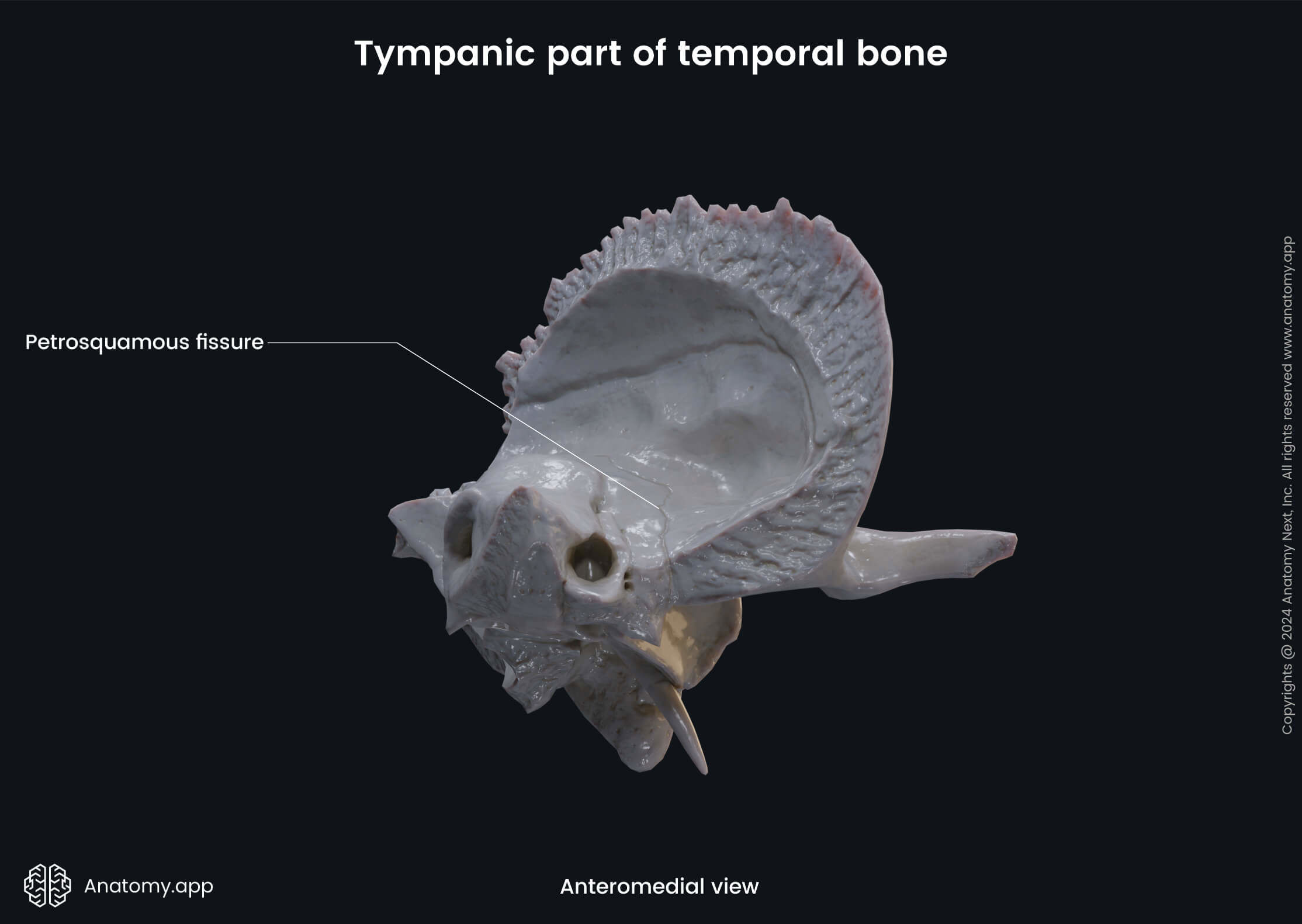
The anterior margin (or anterior angle) of the petrous part of the temporal bone can be divided into two parts: lateral and medial. The lateral part joins the squamous part of the temporal bone with a petrosquamous fissure. The medial part of the anterior margin is free and articulates with the sphenoid bone.
The superior margin (or superior angle) of the petrous part presents the groove for the superior petrosal sinus, and is the attachment site for the tentorium cerebelli. The groove for superior petrosal sinus is grooved into the superior border of the petrous part of the temporal bone accommodating the superior petrosal sinus.
The posterior margin (or posterior angle) of the petrous part presents the following features:
- Groove for inferior petrosal sinus
- Jugular notch (with external opening of cochlear canaliculus)
The groove for inferior petrosal sinus is formed by the junction of the petrous part of the temporal bone with the basilar part of the occipital bone. It houses the inferior petrosal sinus - one of the dural venous sinuses.
The jugular notch is an indentation on the posterior margin of the petrous part forming the anterior margin of the jugular foramen. It also features the external opening of the cochlear canaliculus, which is a small bony canal for the cochlear aqueduct.
Other structures of petrous part
The petrous part of the temporal bone also houses the tympanic cavity and the internal ear structures. The tympanic cavity is a narrow, irregular space situated between the external and internal ear. Therefore, it is a structure of the middle ear.
The internal ear is an essential part of the auditory and vestibular system, where sound waves are converted into electrical impulses. It consists of the bony labyrinth and the membranous labyrinth, which lies within the bony labyrinth and forms small sacs and tubules.
Note that there are many important canals located within the petrous part of the temporal bone described earlier. To sum up, the petrous part has the following canals:
- Carotid canal
- Facial canal
- Canaliculus for chorda tympani
- Tympanic canaliculus
- Musculotubal canal
- Cochlear canaliculus
- Vestibular aqueduct
- Internal acoustic meatus
Squamous part of temporal bone
The squamous part is the largest and most superiorly situated part of the temporal bone. The squamous part has two surfaces: temporal (outer) and cerebral (inner) surface.
The temporal surface of the squamous part contributes to forming the temporal fossa. Also, this part features the following three anatomical structures:
- Zygomatic process
- Mandibular fossa
- Articular tubercle
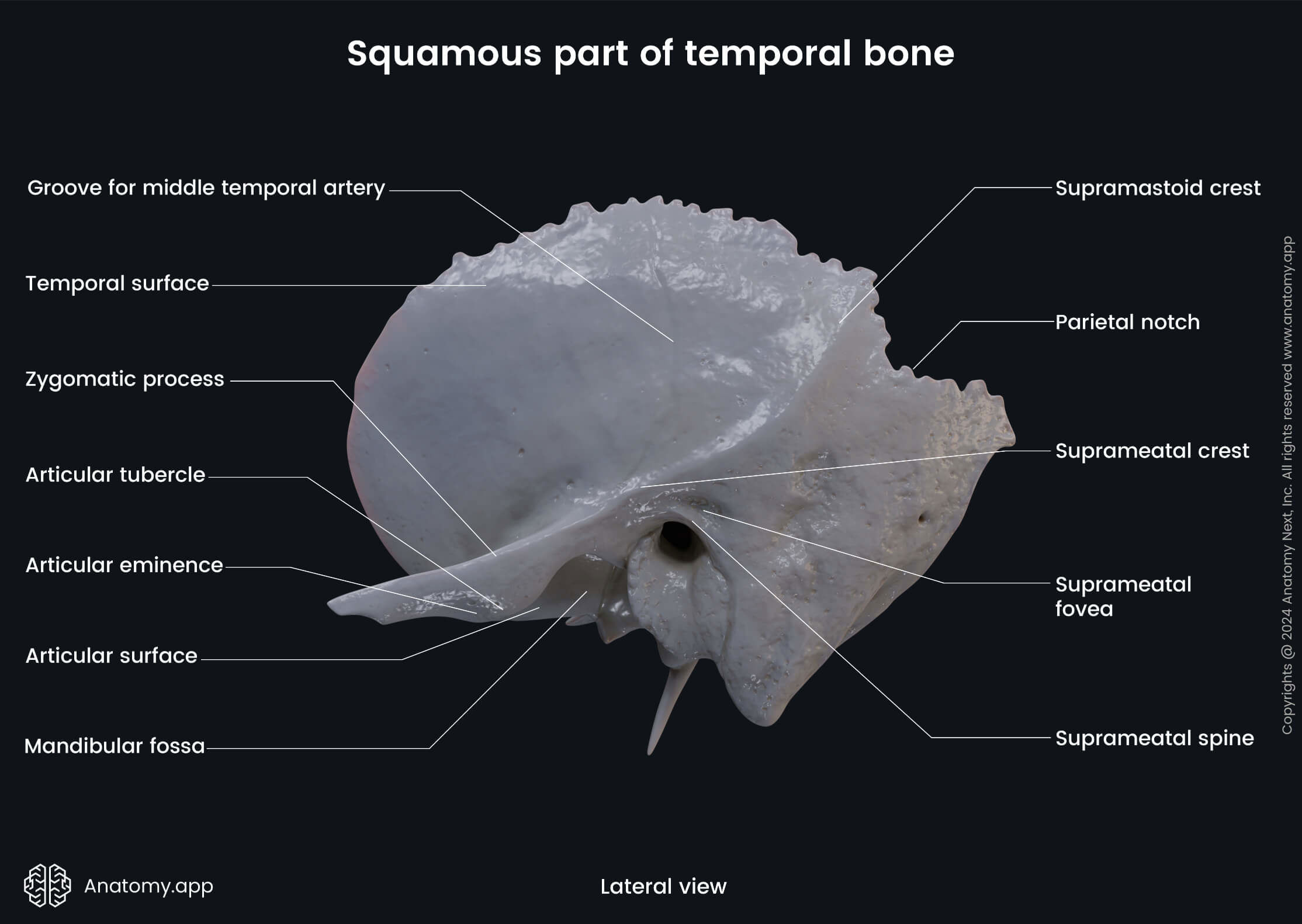
The zygomatic process of the temporal bone is an extension of the bone that contributes to the formation of the zygomatic arch. Any zygomatic process refers to a process of a bone which articulates with the zygomatic bone.
The mandibular fossa is a depression for the head of the mandible. In front of the mandibular fossa is the articular tubercle - a cylindrical elevation on the temporal fossa of the squamous part of the temporal bone.
The cerebral surface of the squamous part features typical landmarks of the inner surface of the skull, which are:
- Impressions of cerebral gyri
- Arterial grooves
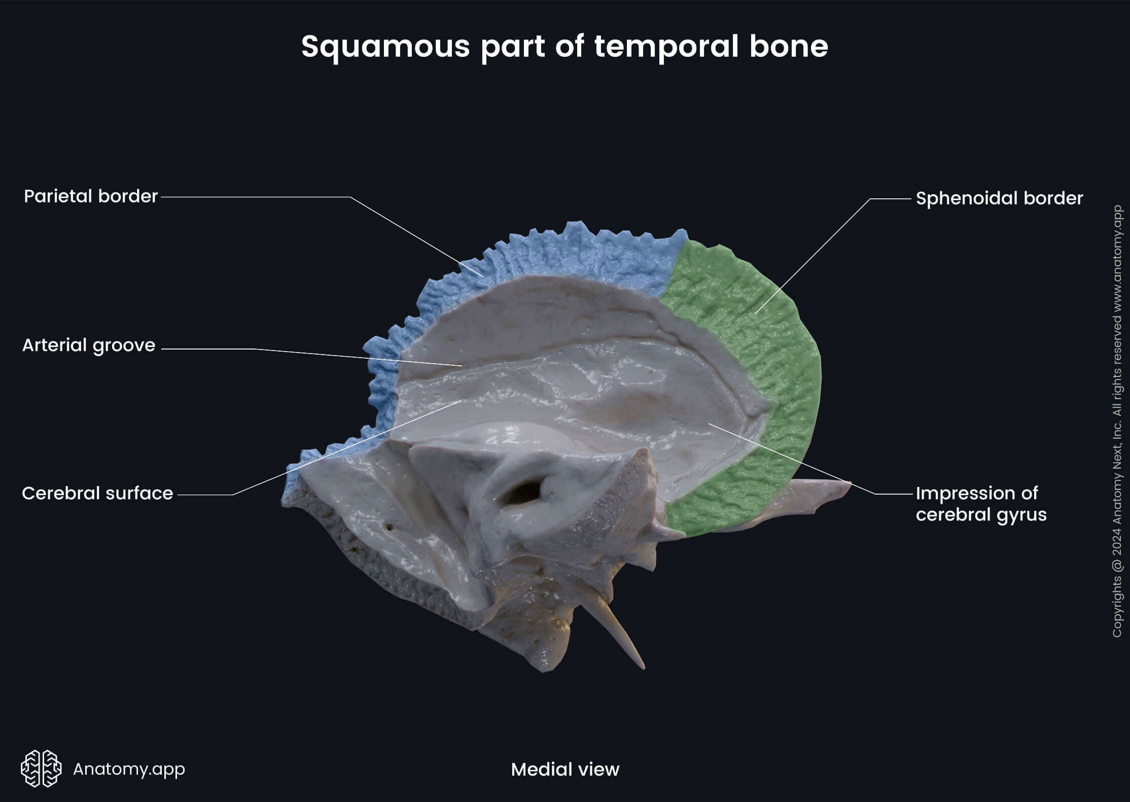
The impressions of cerebral gyri (also called the digital impressions) are flat indentations corresponding to the cerebral gyri which produce them. And the arterial grooves are produced by pressure from arteries, primarily the middle meningeal artery and its branches.
Mastoid part of temporal bone
The mastoid part of the temporal bone is situated postero-inferior to the squamous part. This part features the following anatomical landmarks:
- Mastoid process, containing
- Mastoid cells
- Mastoid antrum
- Mastoid notch
- Groove for occipital artery
- Mastoid foramen
- Groove for sigmoid sinus
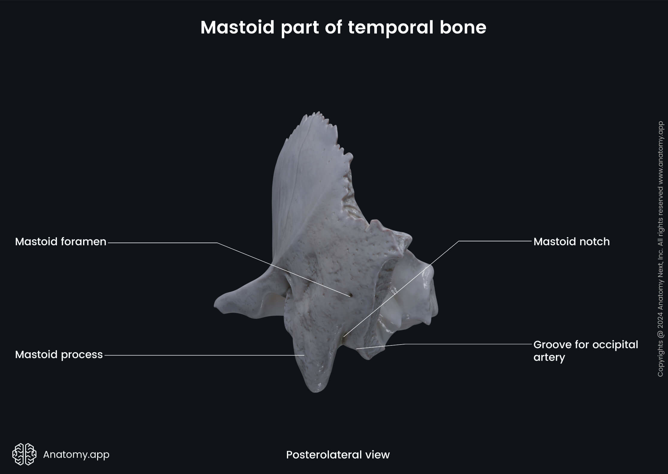
The mastoid process is an extension of the temporal bone located behind the ear, just posterior to the external acoustic opening. It is somewhat conical in shape. Many muscles of the neck attach to this process. Also, the mastoid process contains the mastoid cells and mastoid antrum, and the mastoid notch.
The mastoid cells are a number of air-filled spaces within the mastoid process, variable in size and number. They become pneumatized during the first year of life. The mastoid antrum is a closed space within the mastoid process. Its posterosuperior part is continuous with the tympanic cavity, and its inferior part communicates with the mastoid cells.
The mastoid notch is a deep groove on the medial aspect of the mastoid process. It is the site of origin to the posterior belly of the digastric muscle. Medial to this notch is the groove for the occipital artery or occipital groove which houses the occipital artey.
Behind the mastoid process on the mastoid part of the temporal bone is the mastoid foramen. It serves for additional venous drainage from the cranial cavity, as it transmits an emissary vein that connects the sigmoid sinus with the suboccipital venous sinus.
The groove for the sigmoid sinus is located in the posterior cranial fossa, found on three bones: beginning on the lateral part of the occipital bone, then curving around the jugular process on the mastoid part of the temporal bone, finally, turning sharply on the inner surface of the parietal bone. Here, it continues as the transverse groove.
Tympanic part of temporal bone
The tympanic part of the temporal bone is a relatively small part situated inferior to the squamous part, anterior to the mastoid part, and superior to the styloid process. The tympanic part features the petrotympanic fissure.
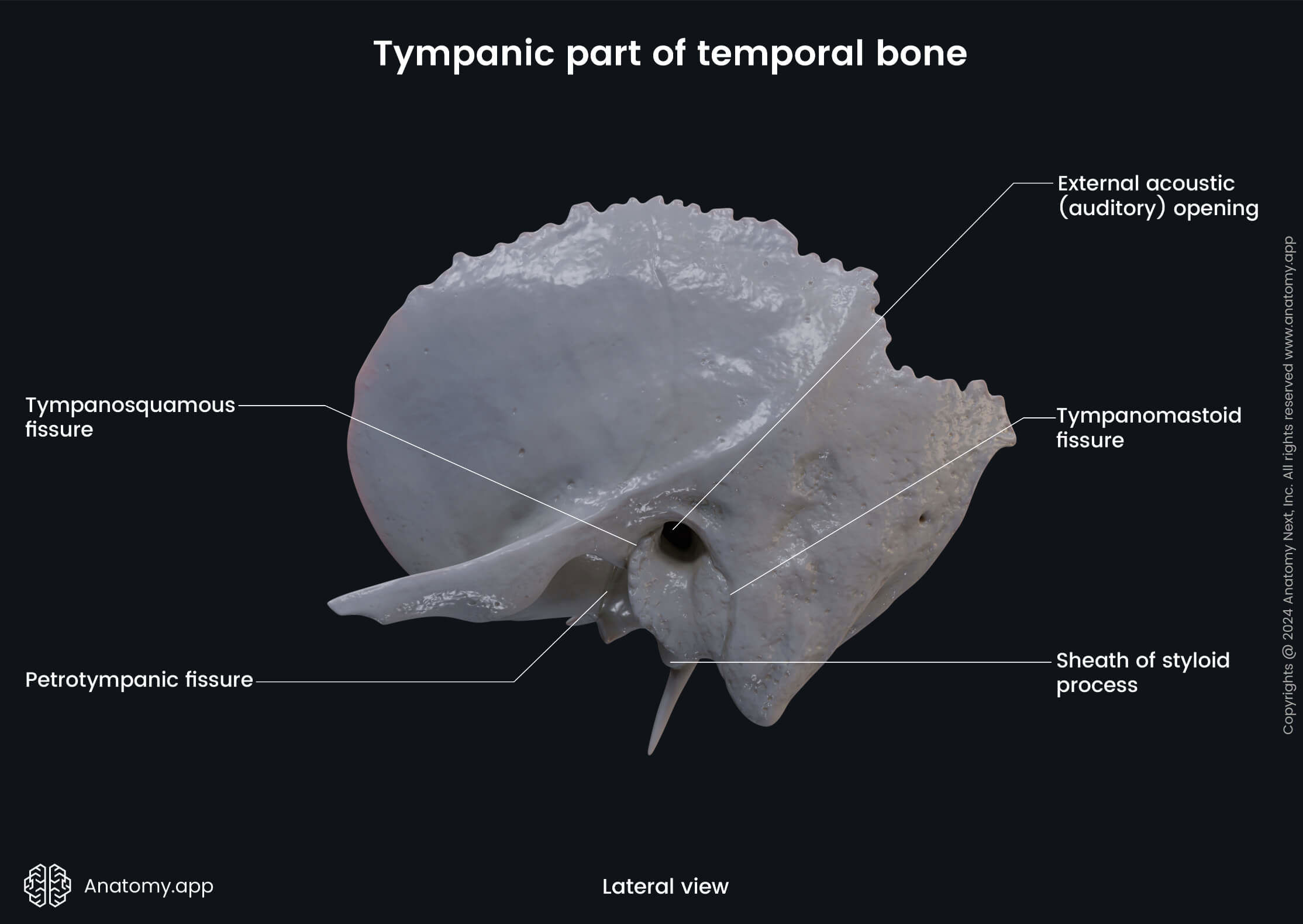
The petrotympanic fissure is a gap which stretches from the temporomandibular joint to the tympanic cavity. It serves as passage for the facial nerve (CN VII) to the infratemporal fossa, and the chorda tympani which passes through and joins the lingual nerve.
External acoustic meatus
There is also an important opening located on the outer surface of the temporal bone between its parts called the external acoustic opening (also external auditory opening), that leads into the external acoustic meatus.
The external acoustic meatus (or external auditory meatus) is an air-filled tubular space in the temporal bone extending from the auricle of the external ear. One of its main functions is to conduct sounds to the tympanic membrane.
Anatomy.app
Contact information
- For questions regarding business inquiries. Please contact:
- info@anatomy.app