- Anatomical terminology
- Skeletal system
- Skeleton of trunk
- Skull
- Skeleton of upper limb
- Skeleton of lower limb
- Joints
- Muscles
- Heart
- Blood vessels
- Lymphatic system
- Nervous system
- Respiratory system
- Digestive system
- Urinary system
- Female reproductive system
- Male reproductive system
- Endocrine glands
- Eye
- Ear
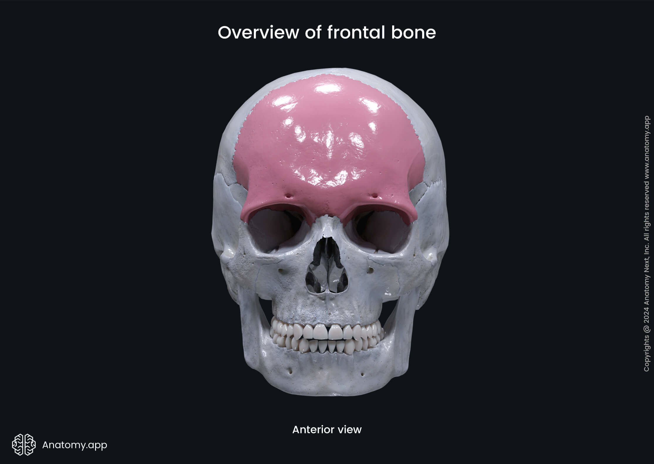
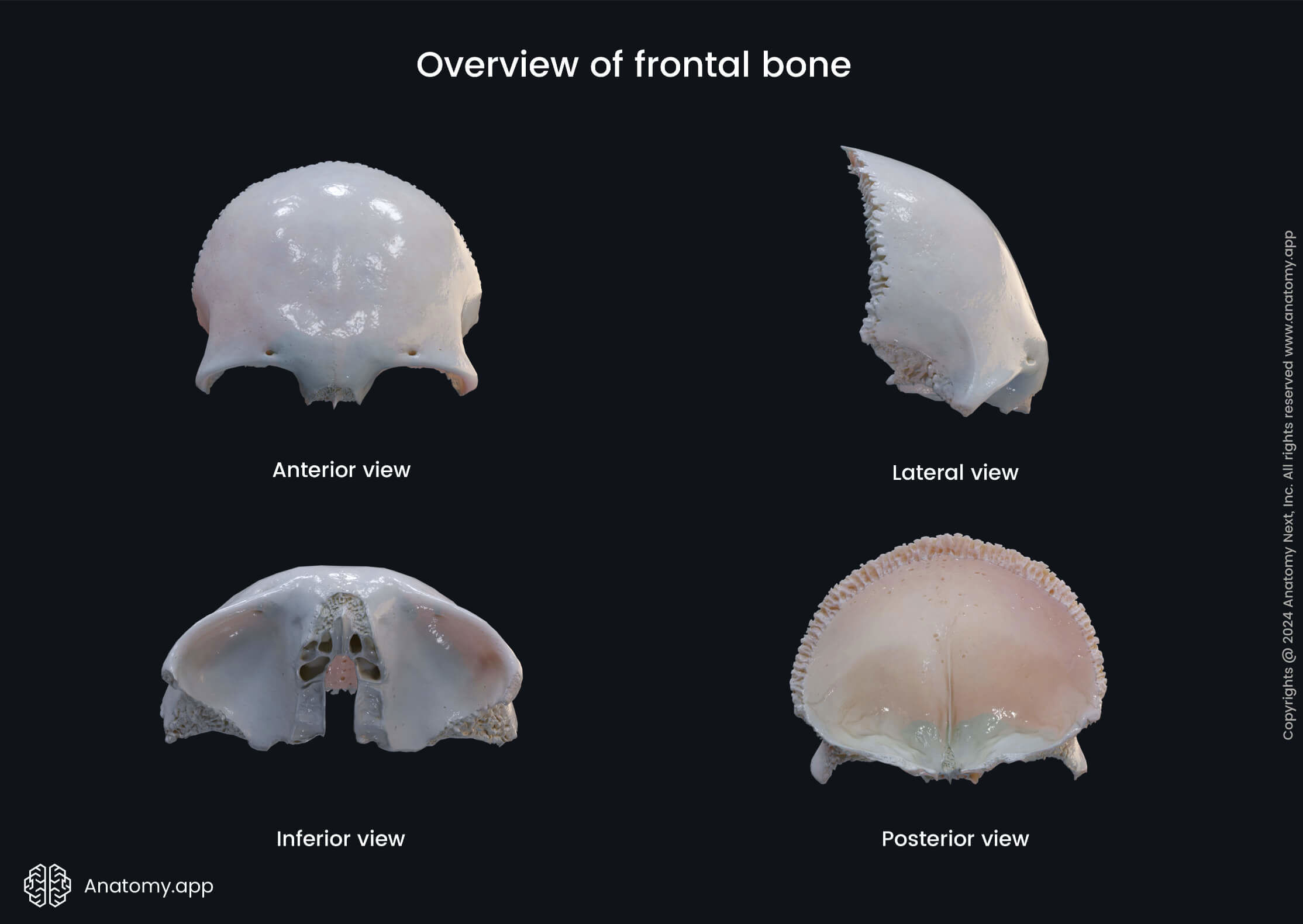
Frontal bone
The frontal bone (Latin: os frontale) is an unpaired bowl-shaped bone located in the forehead region. It contributes in forming the anterior part of the skull. The frontal bone lies superior to the nasal bones and anterior to the parietal bones.
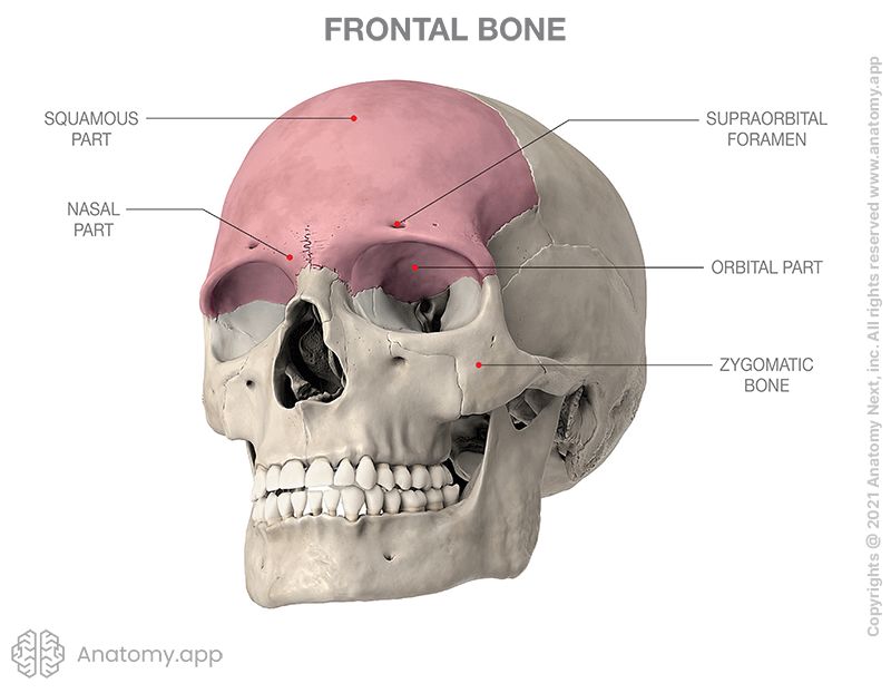
Parts of frontal bone
The frontal bone has four parts:
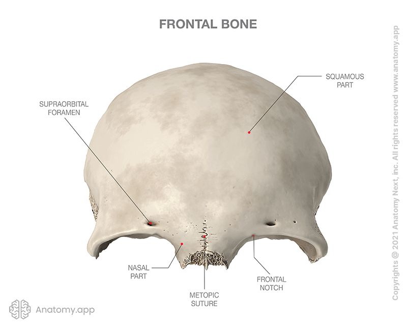
The squamous part of the frontal bone is the largest part of the bone, and it forms the area of the forehead. The squamous part houses the frontal sinuses. The pair of sinuses are separated by a septum and are located above the orbits.
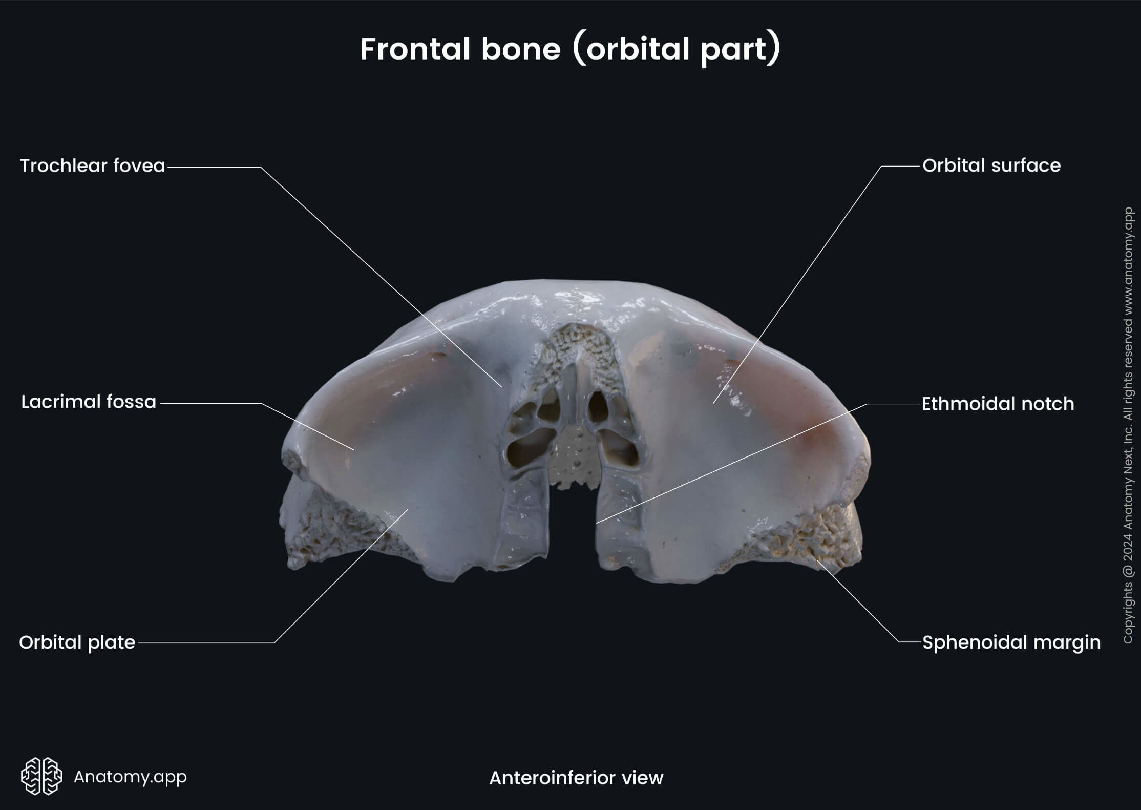
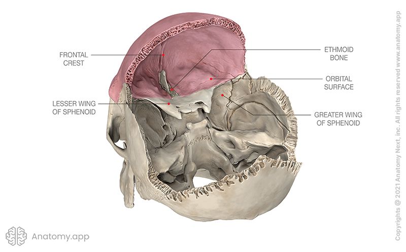
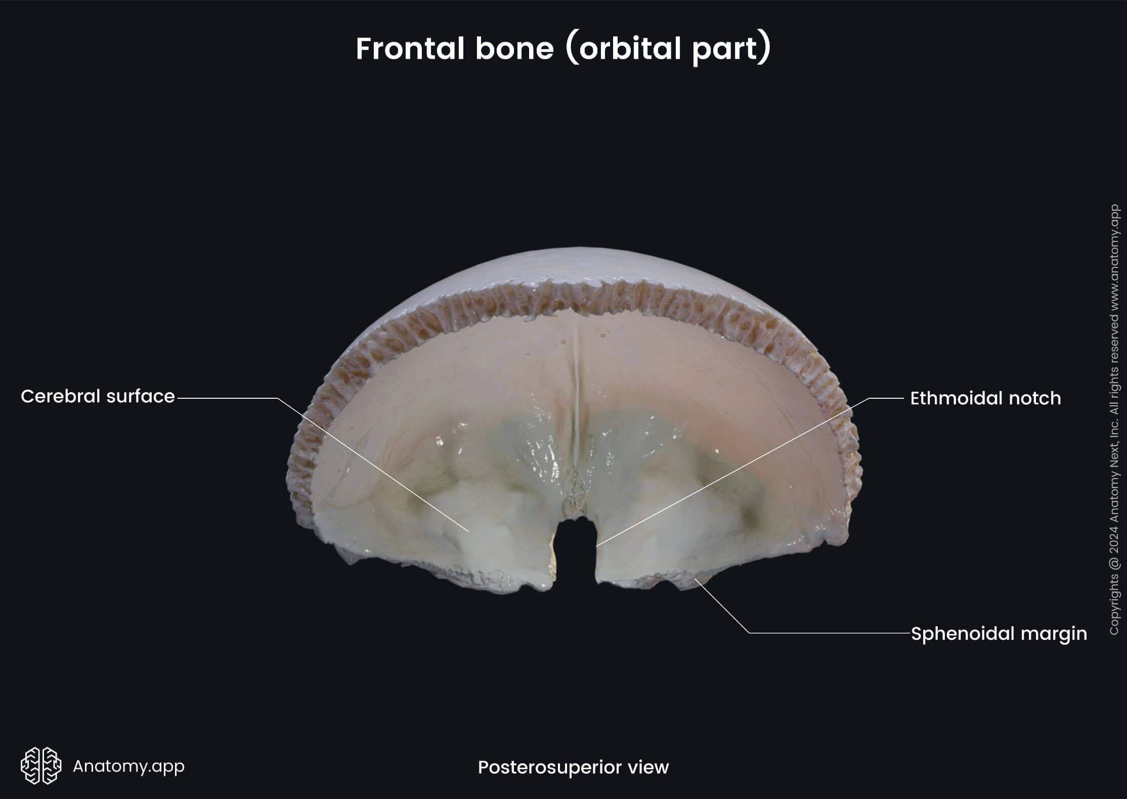
The orbital (or horizontal) part of the frontal bone forms the roof of the orbit and ethmoidal sinuses. The orbital part consists of two orbital plates separated by a gap - the ethmoidal notch.
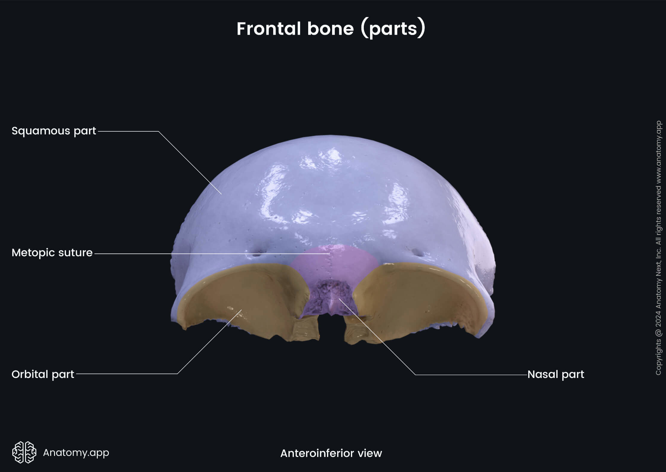
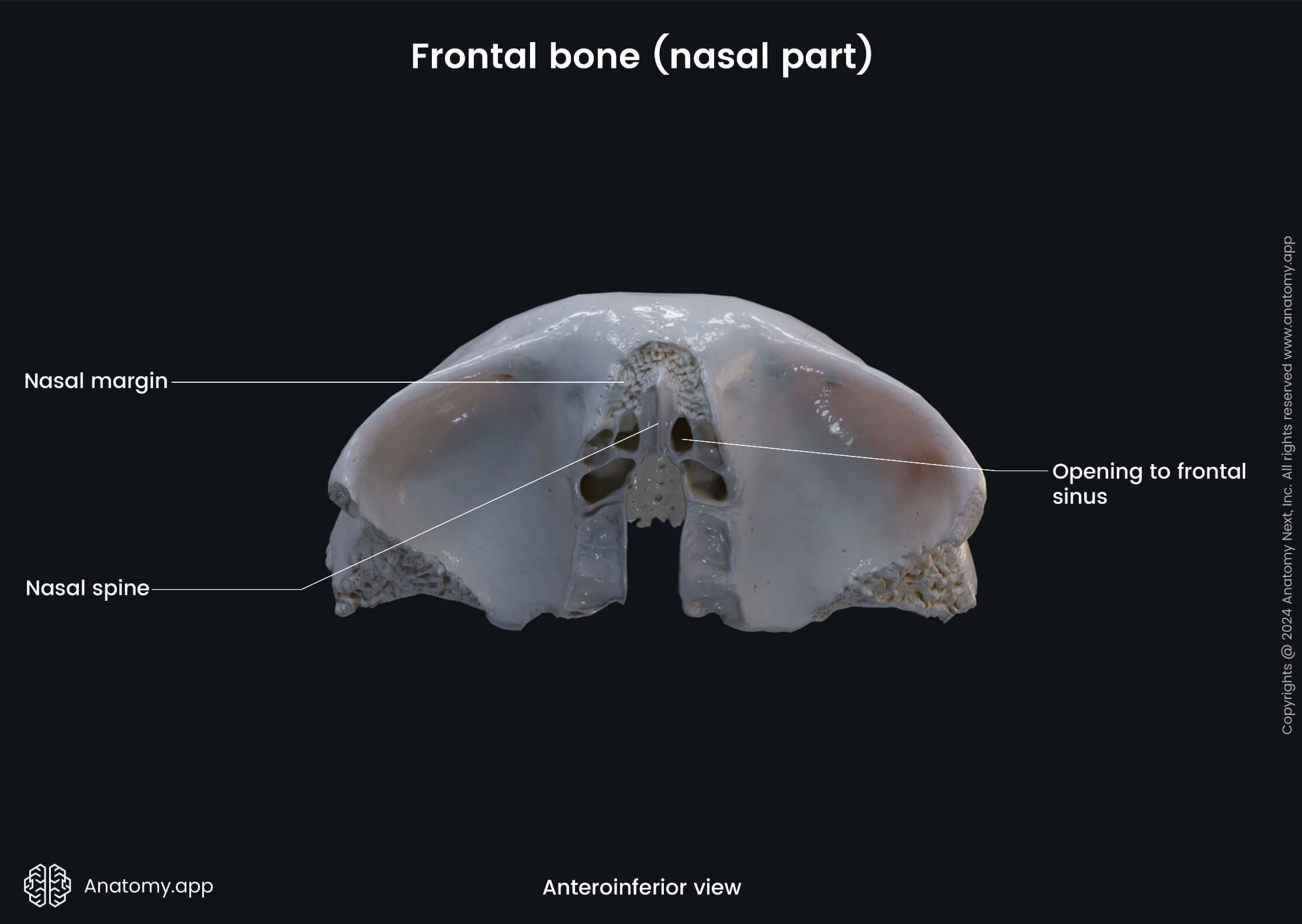
Finally, the nasal part is located anteriorly between the two orbital parts of the frontal bone. The nasal part of the frontal bone forms a portion of the nasal cavity's roof.
Surfaces and landmarks of frontal bone
The frontal bone has an external and an internal surface. Several anatomical landmarks and structures can be found on each surface. The external surface of the frontal bone features the following landmarks:
- Frontal eminence (2)
- Supraorbital margin (2), featuring:
- Frontal notch (or foramen)
- Supraorbital notch (or foramen)
- Superciliary arch (2)
- Glabella
- Temporal line
- Temporal surface
- Zygomatic process (2)
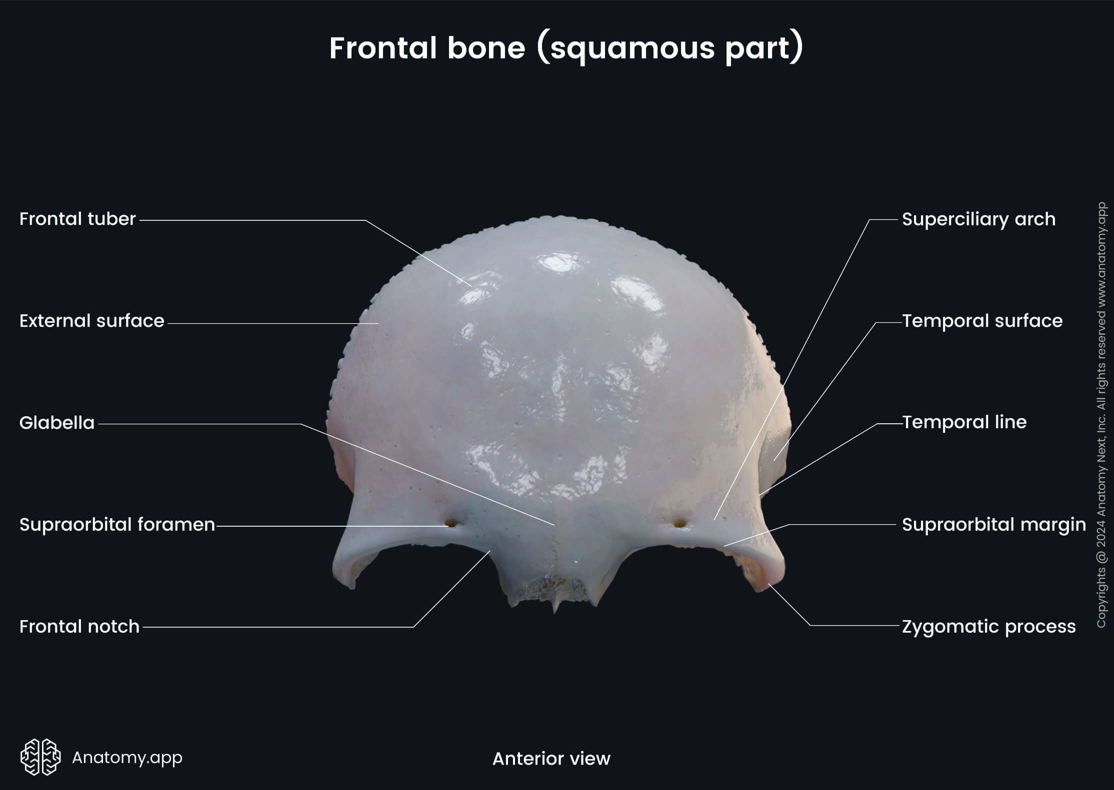
The frontal eminence is a round elevation on each side of the frontal bone. It is located above the supraorbital margin on both sides of the frontal suture. The supraorbital margin is the upper orbital margin of the frontal bone. It features the superciliary arch and the frontal notch.
The frontal notch (sometimes manifests as the frontal foramen) is an opening on each of the supraorbital margins of the frontal bone medial to the supraorbital notch (or foramen). The frontal notch or foramen serves as a passage for the supratrochlear artery (a branch of the ophthalmic artery) and the medial branch of the supraorbital nerve.
The supraorbital notch (sometimes presents as the supraorbital foramen) is an opening that looks like a notch or a hole located on each of the supraorbital margins. It is the passage for the supraorbital artery (another branch of the ophthalmic artery) and the lateral branch of the supraorbital nerve (a branch of the frontal nerve that arises from the ophthalmic nerve (CN V1)).
The superciliary arch is an elevation on the frontal bone above the upper margin of each orbit. Both superciliary arches are joined together by the glabella. The glabella is a smooth elevation on the outer surface of the frontal bone. It is found above and between the eyebrows.
The temporal line of the frontal bone is the continuation of a line formed by the union of the superior and inferior temporal lines of the parietal bone. Laterally from the temporal line, there is the temporal surface of the frontal bone. It contributes to the formation of the temporal fossa, forming the anterosuperior part of the fossa.
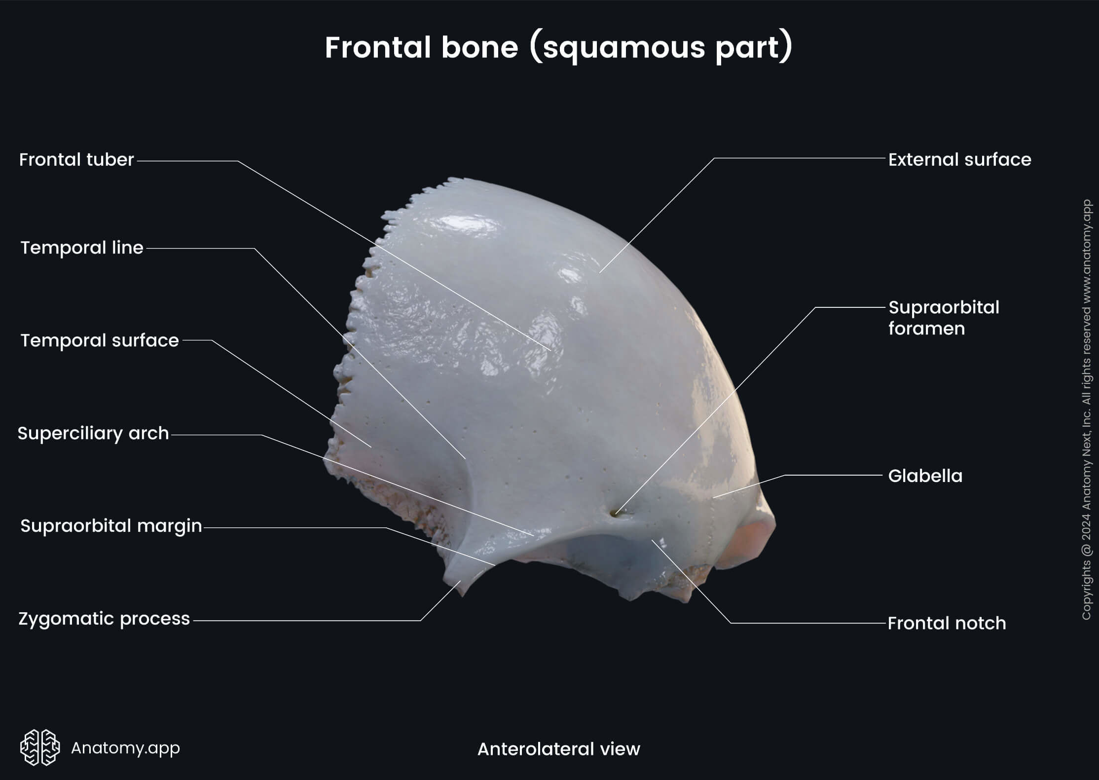
Finally, the external aspect of the frontal bone features one process - they zygomatic process. A zygomatic process refers to a part of a bone that articulates with the zygomatic bone. The zygomatic process of the frontal bone is an extension of the bone lateral to the orbit. It is the process for frontal bone's articulation with the zygomatic bone.
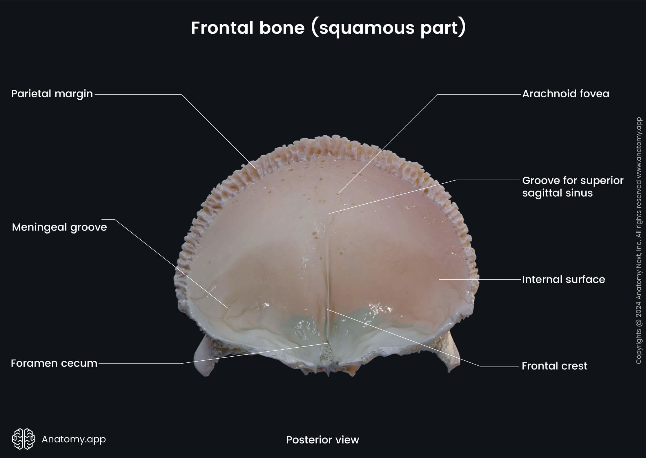
The internal surface of the frontal bone features two important landmarks:
The frontal crest is a median ridge on the internal surface of the frontal bone for attachment of the falx cerebri. And the groove for the superior sagittal sinus is a shallow depression found on the frontal, parietal, and occipital bones forming a channel for the superior sagittal sinus. As the groove passes downward, its margins come together and become continuous with the frontal crest.
Anatomy.app
Contact information
- For questions regarding business inquiries. Please contact:
- info@anatomy.app