- Anatomical terminology
- Skeletal system
- Skeleton of trunk
- Skull
- Skeleton of upper limb
- Skeleton of lower limb
- Joints
- Muscles
- Heart
- Blood vessels
- Lymphatic system
- Nervous system
- Respiratory system
- Digestive system
- Urinary system
- Female reproductive system
- Male reproductive system
- Endocrine glands
- Eye
- Ear
Occipital bone
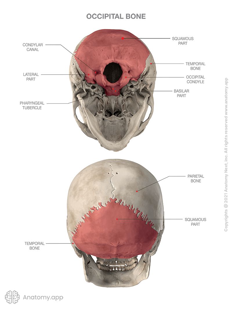
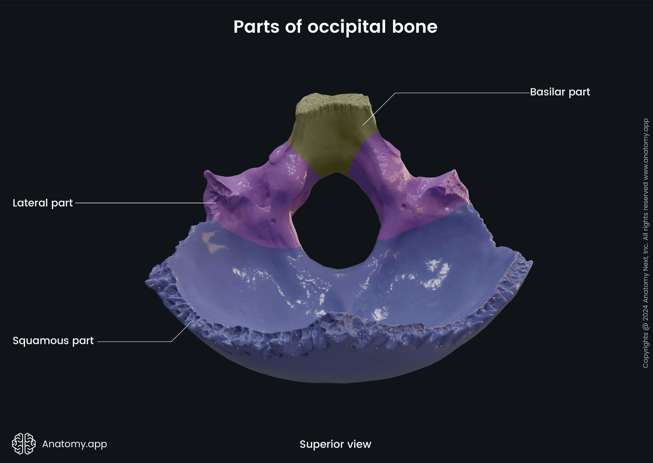
The occipital bone (Latin: os occipitale) is a single bone of the skull that consists of four parts surrounding the foramen magnum. It forms a large part of the cranial base and the postero-inferior part of the neurocranium.
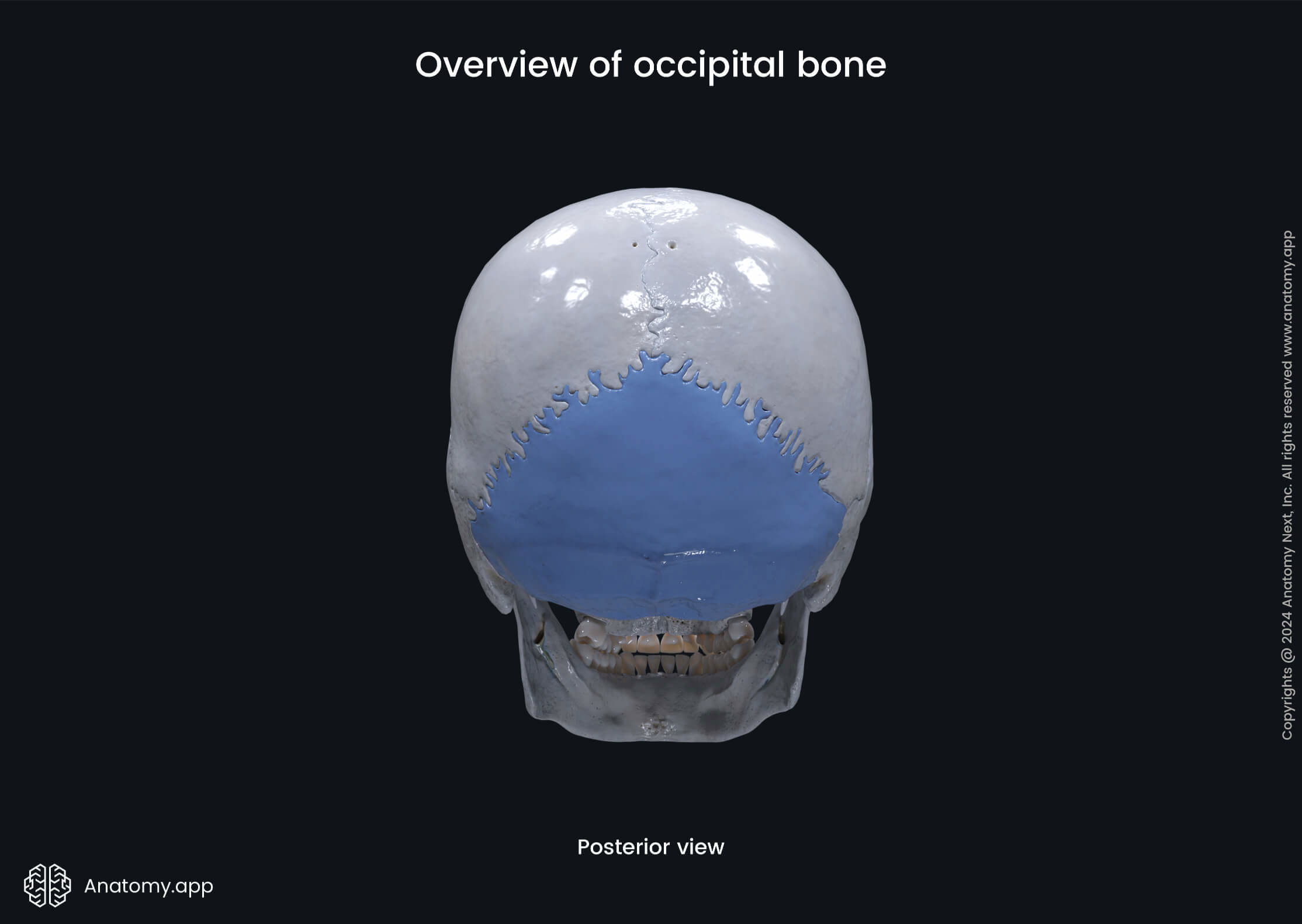
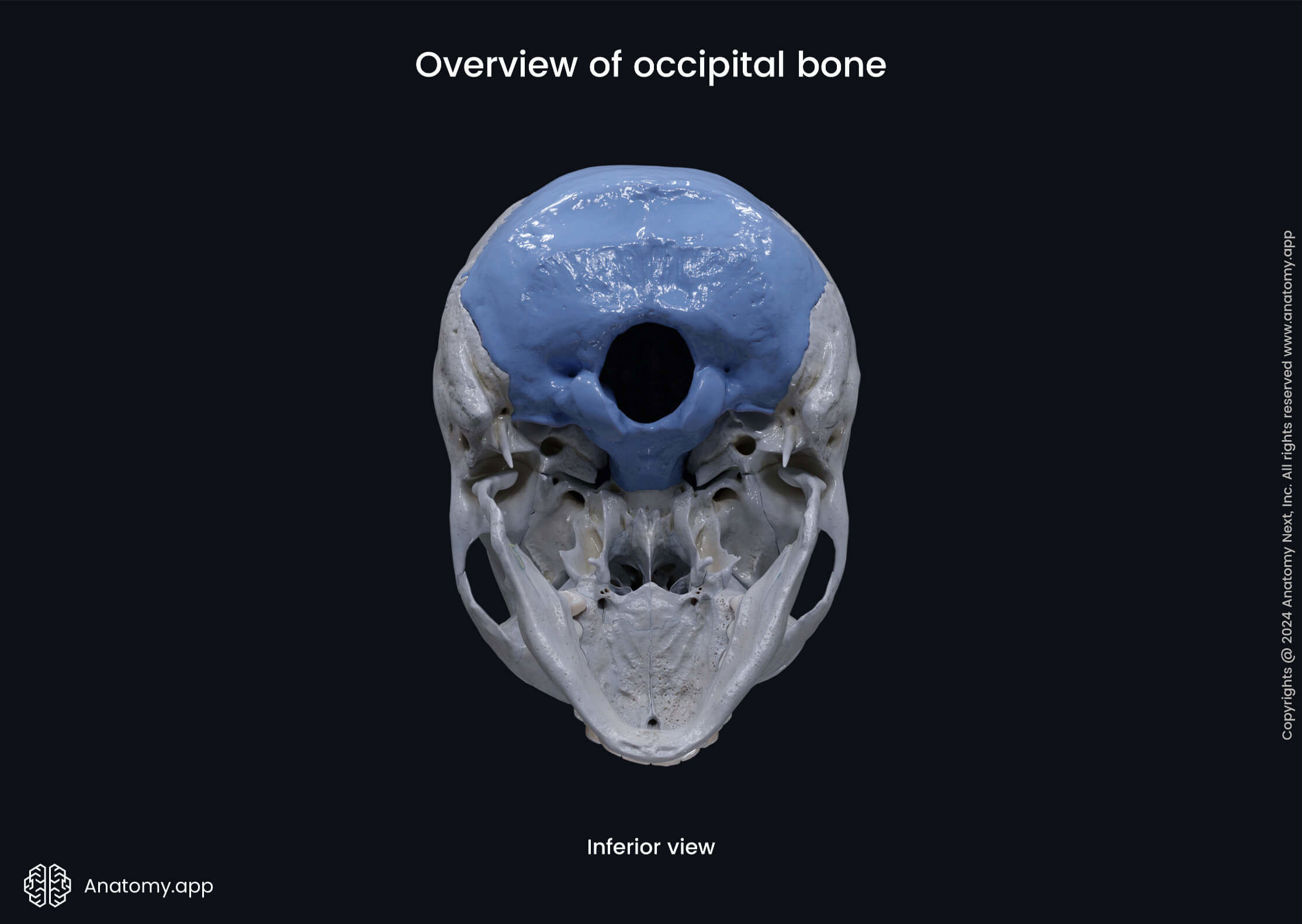
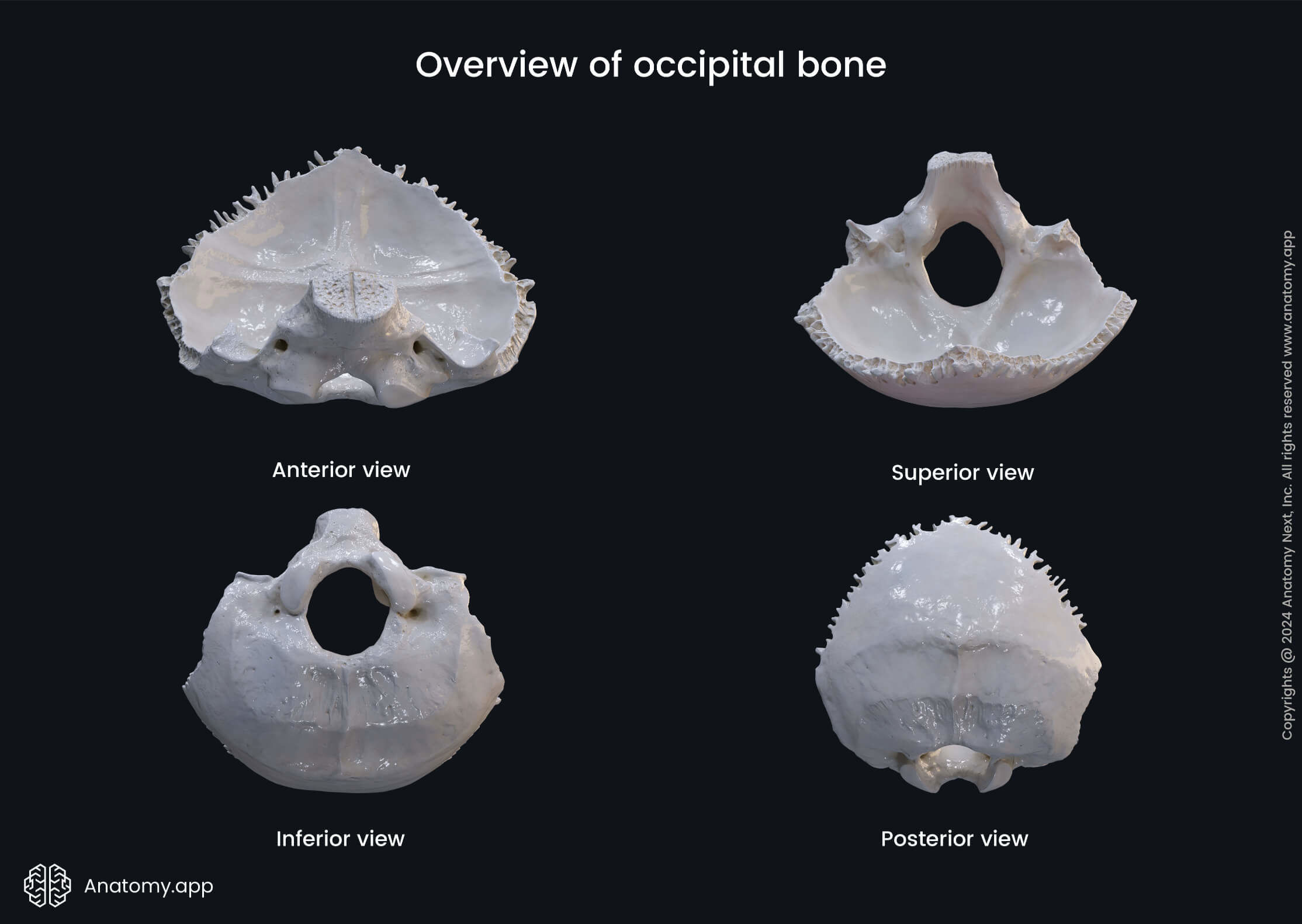
Parts of occipital bone
The four parts forming the occipital bone are as following:
Basilar part of occipital bone
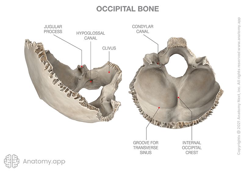
The basilar part is the portion of the occipital bone extending anteriorly from the foramen magnum and joining with the body of the sphenoid bone. This part of the occipital bone is also known as the basiocciput. It has two surfaces - internal and external.
The internal surface of the basiocciput features the following anatomical landmarks:
- Clivus
- Groove for inferior petrosal sinus (2)
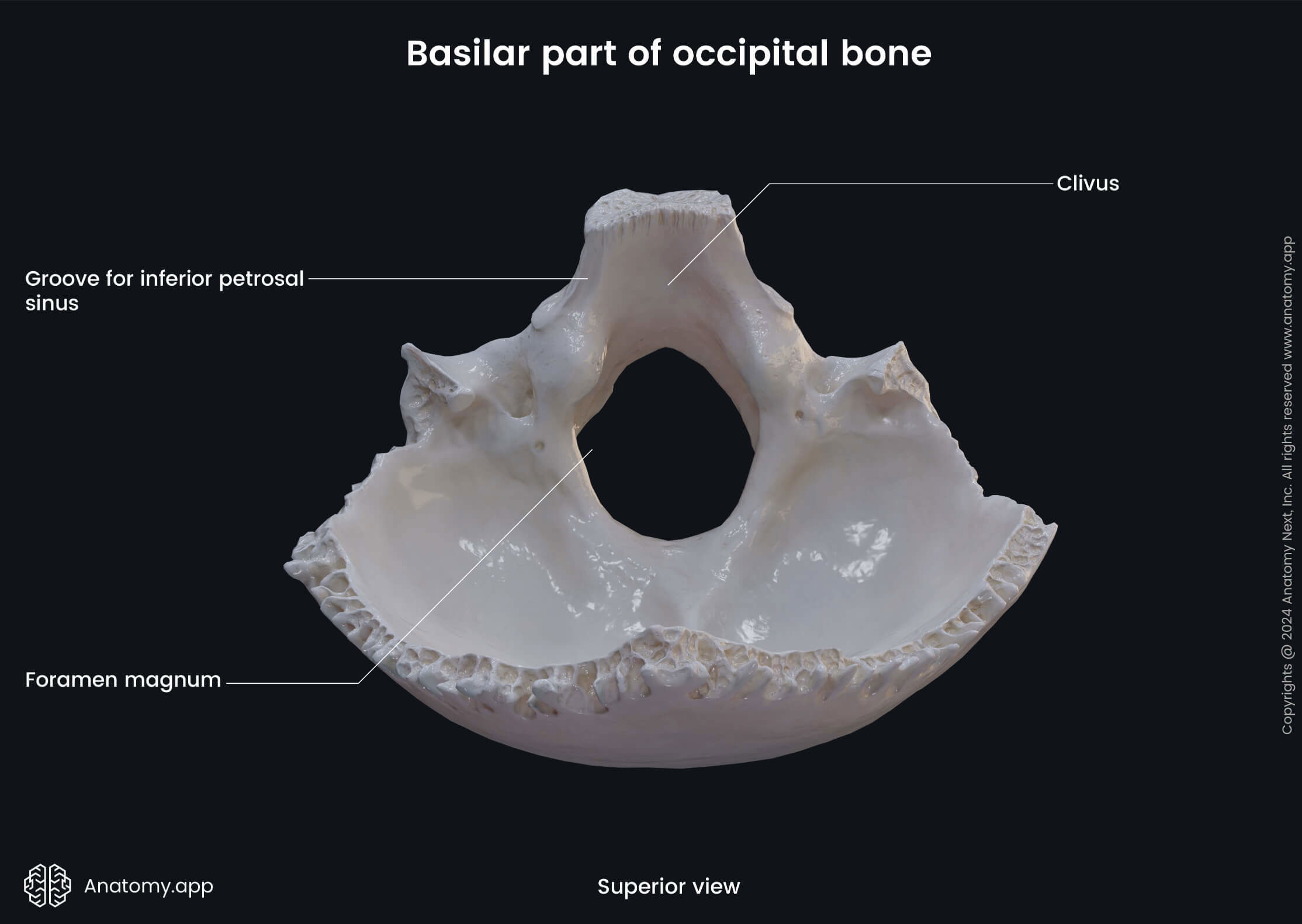
The clivus is a sloping medial surface of the basal part of the occipital bone behind the dorsum sellae of the sphenoid. The clivus slopes downward to the foramen magnum. It is occupied by the medulla oblongata and pons.
The groove for the inferior petrosal sinus is formed by the junction of the petrous part of the temporal bone with the basilar part of the occipital bone. As its name suggests, this groove lodges one of the dural venous sinuses - the inferior petrosal sinus.
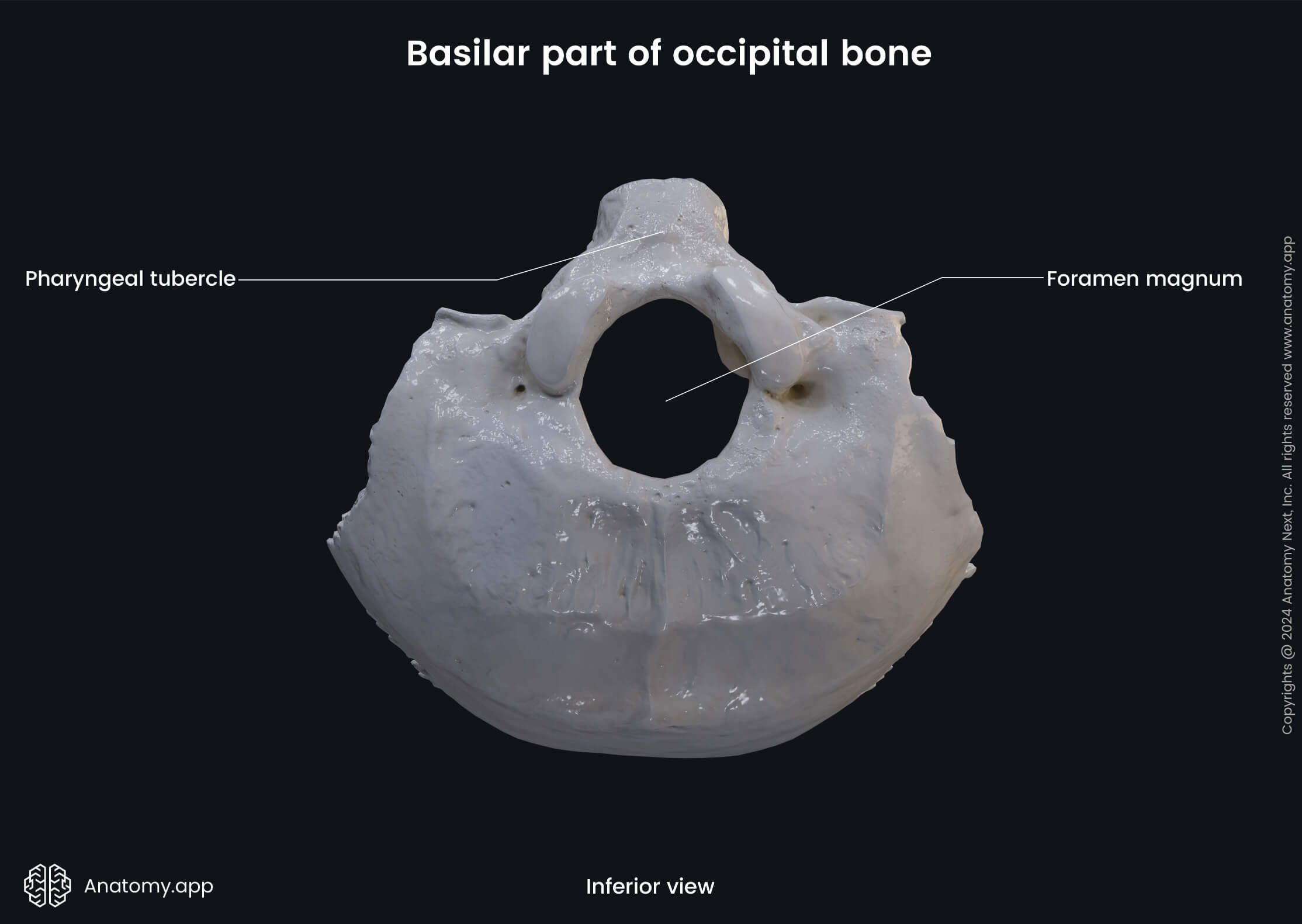
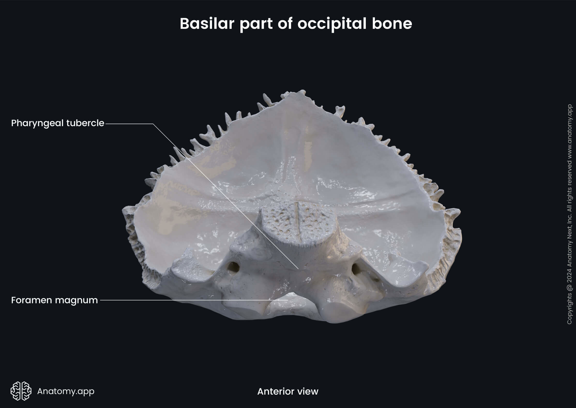
The external surface of the basiocciput features the pharyngeal tubercle, also known as pharyngeal eminence. It is a prominence on the inferior aspect of the occipital bone that serves for attachment of the pharyngeal raphe.
Lateral part of occipital bone
There are two lateral parts of the occipital bone located on each side of the foramen magnum. Each lateral part has an internal and an external surface that feature some important anatomical landmarks.
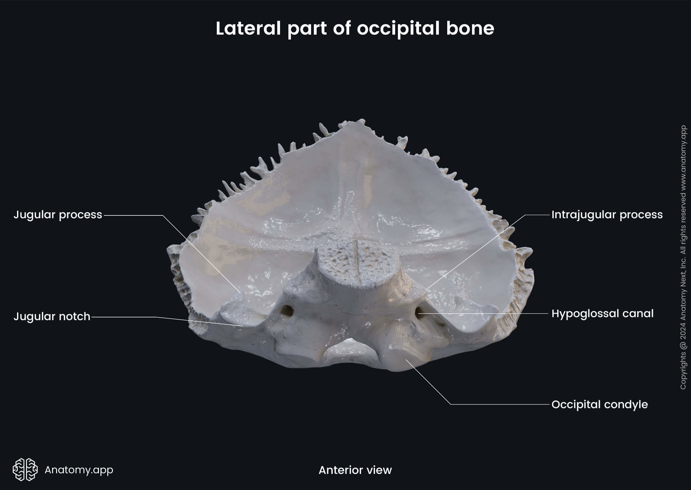
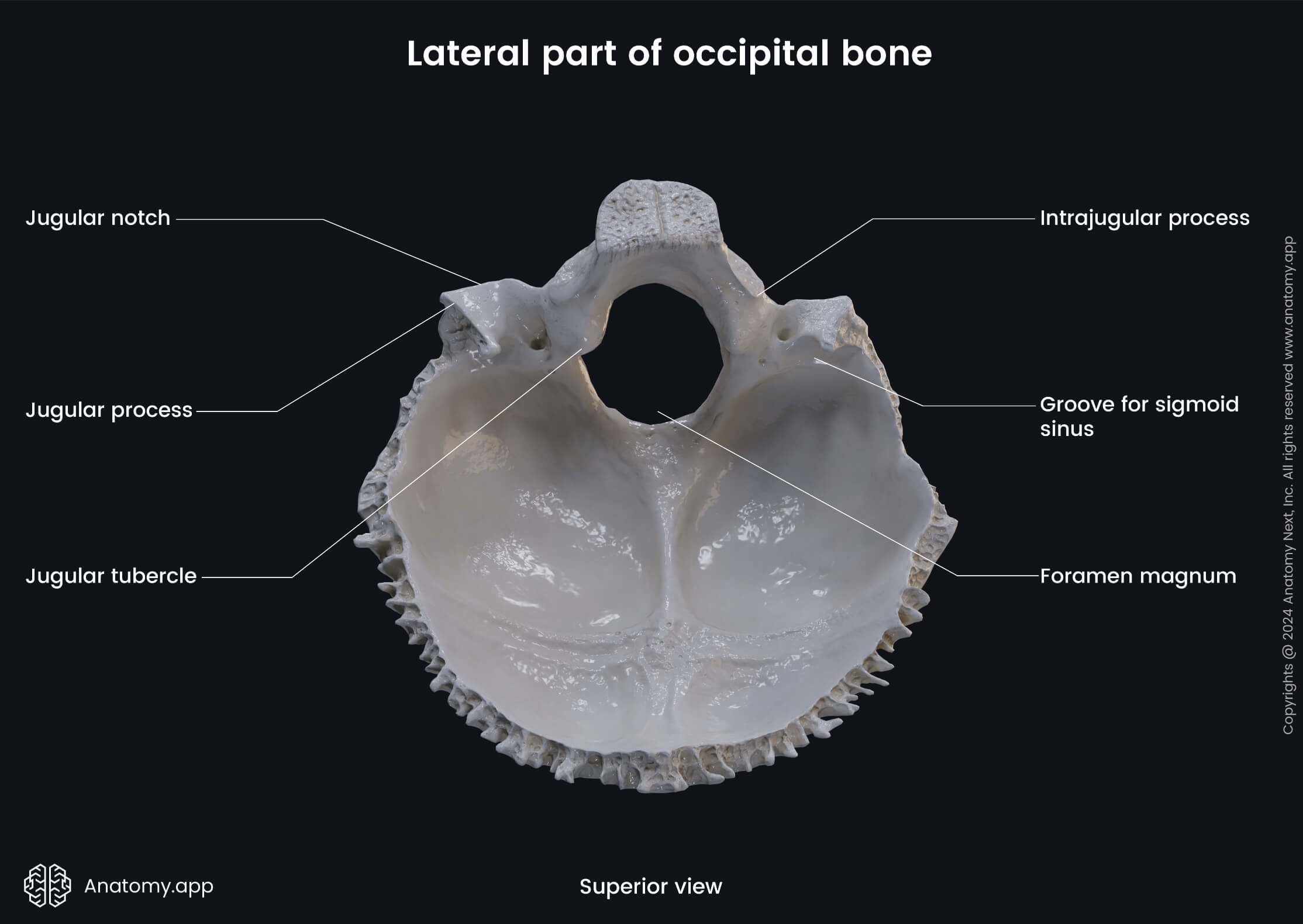
The internal surface of the lateral part features the groove for the sigmoid sinus. This groove can be seen in the posterior cranial fossa, found on three bones: occipital, temporal, parietal. It begins from the lateral part of the occipital bone, curves around the jugular process on the mastoid part of the temporal bone. Finally, turns sharply on the inner surface of the parietal bone continuing as the transverse groove.
The external surface of the lateral part of the occipital bone features the following structures:
- Occipital condyle
- Condylar canal
- Hypoglossal canal
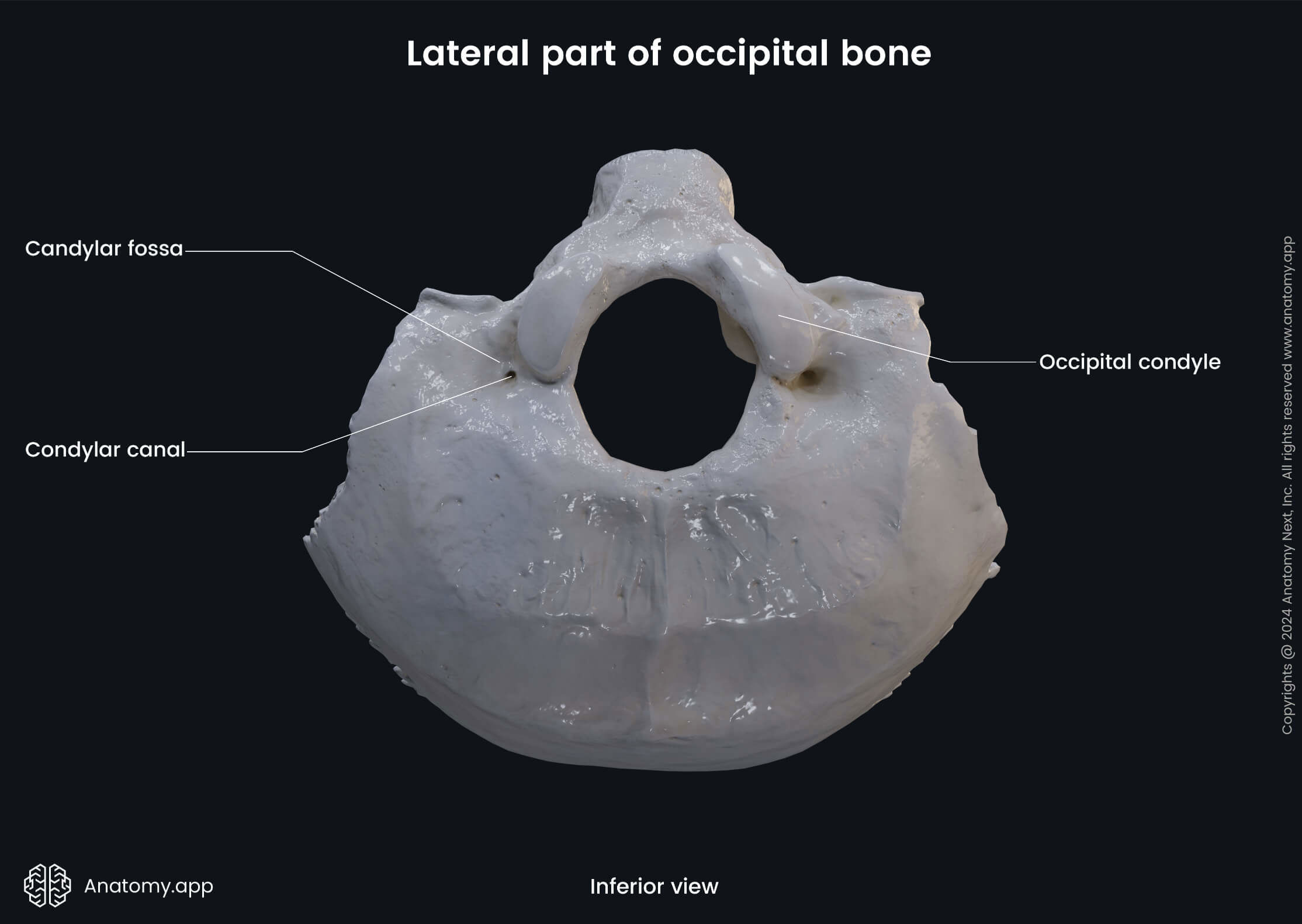
The occipital condyle is a process on the occipital bone for articulation with the 1st cervical vertebra - the atlas (C1). Posterior to the occipital condyle is the condylar canal. It is a bony passage in the lateral part of the occipital bone that transmits a vein from the sigmoid sinus.
The hypoglossal canal is a bony passage that originates from the lateral part of the occipital bone anterior to the foramen magnum and ends on the outer surface anterior to the occipital condyle. This canal transmits the hypoglossal nerve (CN XII) and a venous plexus.
Squamous part of occipital bone
The squamous part of the occipital bone is also called squama of the occipital bone or occipital squama. Like other parts of the occipital bone, it has an internal and an external surface.
The internal surface of the occipital squama features the following landmarks:
- Cruciform eminence
- Internal occipital protuberance
- Internal occipital crest
- Groove for superior sagittal sinus
- Groove for transverse sinus (2)
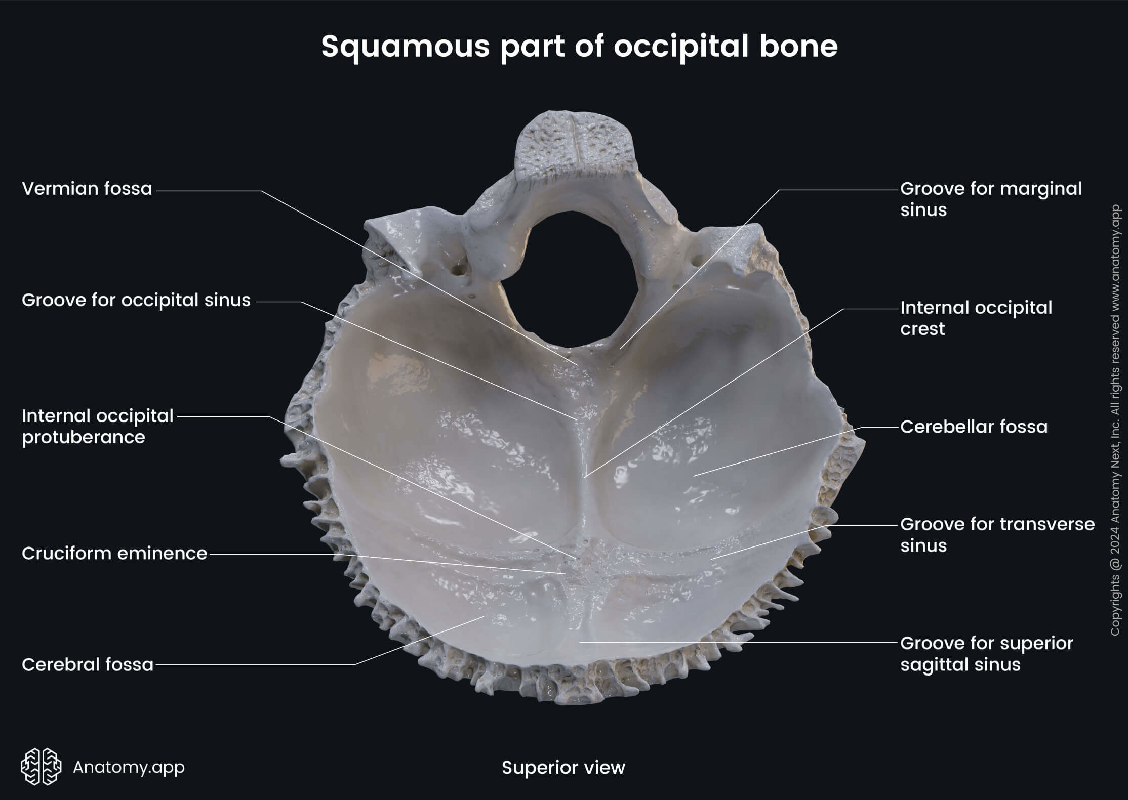
The internal occipital protuberance is a prominent midpoint of the cruciform eminence - a cross-shaped bony prominence found on the internal surface of the occipital bone. Occasionally, from the internal occipital prominence to the foramen magnum extends a thick bony ridge called the internal occipital crest.
The groove for the superior sagittal sinus is a shallow depression on the frontal, parietal, and occipital bones forming a channel for the superior sagittal sinus. The margins of this groove come together as it passes downward and become continuous with the frontal crest (a ridge on the internal surface of the frontal bone).
The groove for the transverse sinus is located bilaterally on the internal surface of the occipital squama extending from the internal occipital protuberance to the lateral angles of the occipital bone. As its name suggests, this groove lodges the transverse sinus, which is one of the dural venous sinuses.
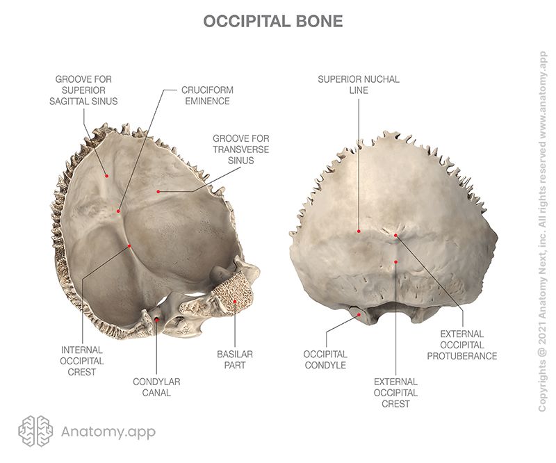
The external surface of the occipital squama features the following anatomical landmarks:
- External occipital protuberance
- External occipital crest
- Superior nuchal line
- Inferior nuchal line
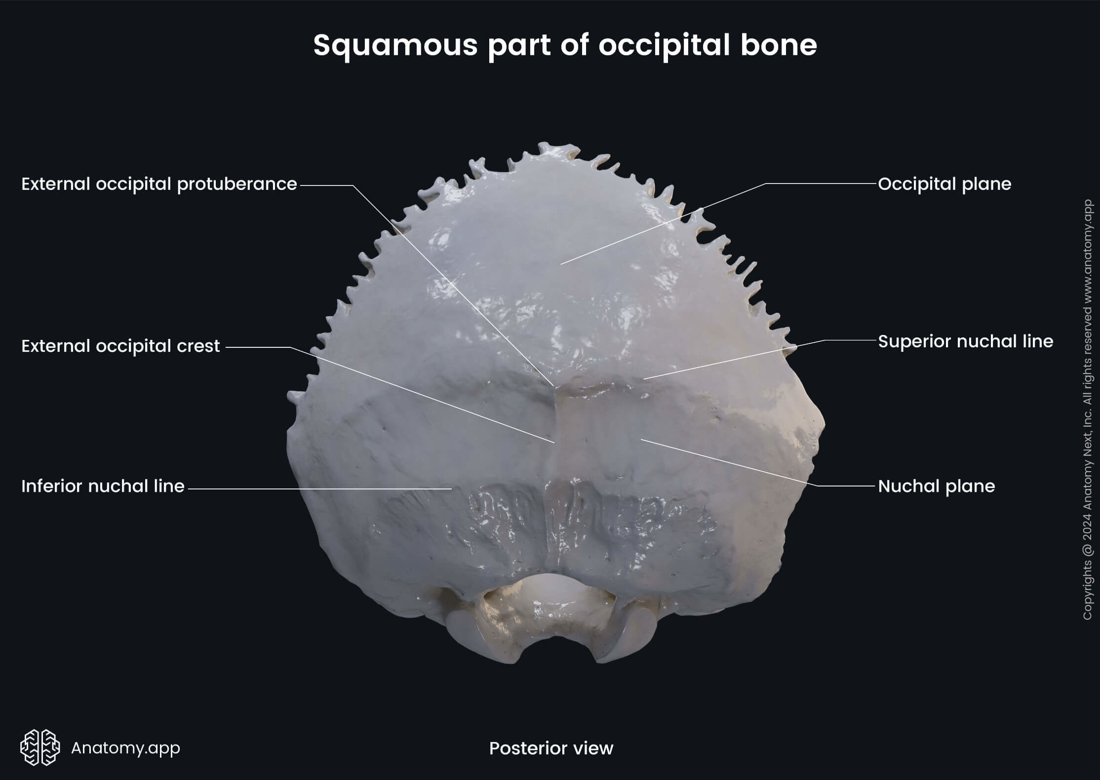
The external occipital protuberance is a palpable bony projection in the middle of the occipital bone. Occasionally, extending from the external occipital protuberance to the foramen magnum is a bony ridge called the external occipital crest.
The superior nuchal line is a transverse ridge located on the external surface of the occipital bone at the level of the external occipital protuberance. It is the attachment site for the occipital belly of the occipitofrontalis muscle.
The inferior nuchal line is another transverse ridge on the external surface of the occipital squama. It extends between the superior nuchal line and the foramen magnum. The semispinalis capitis muscle attaches between the inferior and the superior nuchal lines.