- Anatomical terminology
- Skeletal system
- Joints
- Muscles
- Heart
- Blood vessels
- Lymphatic system
- Nervous system
- Respiratory system
- Digestive system
- Urinary system
- Female reproductive system
- Male reproductive system
- Endocrine glands
- Eye
- Ear
Membranous labyrinth
The membranous labyrinth (Latin: labyrinthus membranaceus) is a system of ducts and dilatations located within the bony labyrinth of the internal ear, and it contains the receptors for hearing and balance. The membranous labyrinth is made of connective tissue. The membranous labyrinth is composed of two functional parts: vestibular labyrinth and cochlear labyrinth.
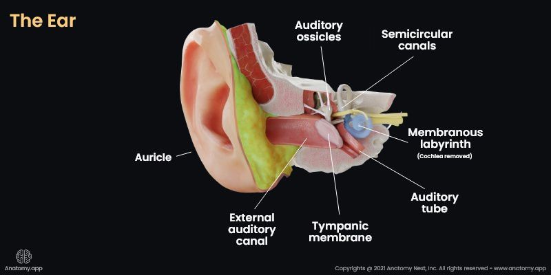
The vestibular labyrinth includes two sacs (utricle and saccule) and three semicircular ducts (anterior, lateral, and posterior semicircular ducts). The utricle and saccule are enclosed by the bony vestibule, while the semicircular ducts are lodged in the semicircular canals of the bony labyrinth. The vestibular labyrinth contains balance receptors for appreciation of the impact of gravitation (static balance) - located in the utricle and saccule, and of the impact of acceleration (kinetic balance) - located in the semicircular ducts
The cochlear labyrinth is the content of the osseous cochlea, including the cochlear duct. The cochlear labyrinth is a fluid-filled membrane housing the Corti organ with receptors for detecting sound stimulus.
The space inside the membranous labyrinth is filled with endolymphatic fluid. In contrast, the outside of the membranous labyrinth is filled with perilymph, separating the membranous labyrinth from the walls of the bony labyrinth. This space separating both labyrinths is called the perilymphatic space. In some places, the structures of the membranous labyrinth are fixated to the walls of the bony capsule.
The structures of the membranous labyrinth are connected with each other. The saccule is connected to the cochlear duct by the reuniens duct. The saccule and the utricle are connected by the utriculosaccular duct, which changes into the endolymphatic duct, leaves the temporal bone through its petrous part, and ends as the endolymphatic sac. With the help of the endolymphatic duct and sac, endolymph travels from the internal ear to the venous sinuses.
The walls of the membranous labyrinth house the branches of the vestibulocochlear nerve - the vestibular nerve and the cochlear nerve. The innervation and vasculature of the membranous labyrinth and its structures are discussed under the article on the internal ear as the vascularization and innervation of many structures are the same and overlapping.
Anatomy.app
Contact information
- For questions regarding business inquiries. Please contact:
- info@anatomy.app