- Anatomical terminology
- Skeletal system
- Joints
- Muscles
- Heart
- Blood vessels
- Lymphatic system
- Nervous system
- Respiratory system
- Digestive system
- Urinary system
- Female reproductive system
- Male reproductive system
- Endocrine glands
- Eye
- Ear
Auricle
The auricle (or pinna, pinna of ear, auricle of ear, auricula, latin: auricula) is the external, visible component of the ear around the outer opening of the ear canal. The auricle is musculocutaneous tissues attached to the skull. The main function of the auricle is to collect, amplify and direct sound waves into the external auditory canal.
Structure of auricle
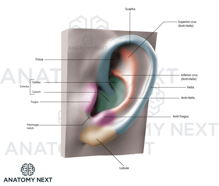
The characteristic shape of the auricle is determined by elastic cartilage forming a structural framework of the auricle, called the auricular cartilage. The auricular cartilage makes up the upper 2/3 of the auricle that is covered by skin. The lower 1/3 is the lobule of the auricle (also known as ear lobe) - skin duplicate, which has no cartilage in it and is made of fat tissue. The lobule of the auricle has no major practical significance. The auricular cartilage is not flat. The auricle is comprised of the following anatomical features:
- Helix, including:
- Crus of helix
- Spine of helix
- Tail of helix
- Antihelix, including:
- Triangular fossa,
- Crura of antihelix
- Scapha
- Concha of auricle, including:
- Cymba conchae
- Cavity of concha
- Tragus
- Antitragus
- Intertragic notch
- Lobule of auricle
The helix is the external, curved margin of the auricle. Inferiorly it ends as soft tissue, also known as the lobule of the auricle. The crus of the helix is the site of origin of the helix situated in the cavity (concha) of the auricle. The spine of the helix is a small cartilaginous prominence projecting forward from the crus. It is the thick, superior part of the helix. The tail of the helix is the posterior, inferior end of the helix separated from the antihelix by an indentation. The tail of the helix is continuous with the lobule.
The antihelix is an arched projection seen on the auricle and situated in front of the posterior part of the helix. The antihelix is parallel to the helix. The triangular fossa (or fossa triangularis) is an anterosuperiorly located fossa enclosed by the two crura of the antihelix. The crura of the antihelix are formed by a bifurcation of the antihelix superiorly. It is the widening of the antihelix that is directed posteriorly toward the helix.
The scapha (or scaphoid fossa) is a fossa located posteriorly between the helix and antihelix.
The concha of the auricle is a cavity of the auricle enclosed by the antihelix, antitragus, and tragus. It is continuous with the external acoustic meatus. The cymba conchae of the auricle is the upper, slit-like part of the concha between the crura of the helix and antihelix. The cavity of the concha of the auricle is the main part of the concha located below the crus of the helix and behind the tragus.
The tragus is a flat, triangular, cartilaginous projection in front of the external acoustic opening that starts as the concha of the auricle. The base of the tragus is attached to facial skin, while the apex is the one that partially covers the external acoustic opening.
The antitragus is a small, cartilaginous elevation above the lobule, situated on the inferior continuation of the antihelix and separated from the tragus by the intertragic notch.
The intertragic notch (also called intertragic incisure or intertragal incisure) is a notch between the tragus and antitragus.
Vasculature and innervation of the auricle
Arterial supply and venous drainage
The auricle is supplied by three arteries:
- The posterior auricular artery, a branch of the external carotid artery
- The anterior auricular arteries, a branch of the superficial temporal artery
- Minor branches of the occipital artery
The veins responsible for the drainage mirror the arteries.
Lymphatic drainage
The lymph from the auricle is drained into the superficial parotid, mastoid, deep cervical, and superficial cervical lymph nodes.
Innervation
The sensory innervation of the auricle is provided by the great auricular nerve (originating from cervical plexus) supplying the helix, antihelix and lobule of the auricle; the lesser occipital nerve (also from cervical plexus) supplying the superior part of the cranial surface of the auricle; the auriculotemporal nerve (a branch of the mandibular nerve) innervating the tragus, the crus and the adjacent part of the helix; and the auricular branch of the vagus nerve (CN X) together with the posterior auricular branch of the facial nerve (CN VII) supplying small areas of the auricle, innervating the concha, as well as the posterior and superior parts of the eminentia of the concha, which is present on the posterior surface of the auricle.
Anatomy.app
Contact information
- For questions regarding business inquiries. Please contact:
- info@anatomy.app