- Anatomical terminology
- Skeletal system
- Skeleton of trunk
- Skull
- Skeleton of upper limb
- Skeleton of lower limb
- Joints
- Muscles
- Heart
- Blood vessels
- Lymphatic system
- Nervous system
- Respiratory system
- Digestive system
- Urinary system
- Female reproductive system
- Male reproductive system
- Endocrine glands
- Eye
- Ear
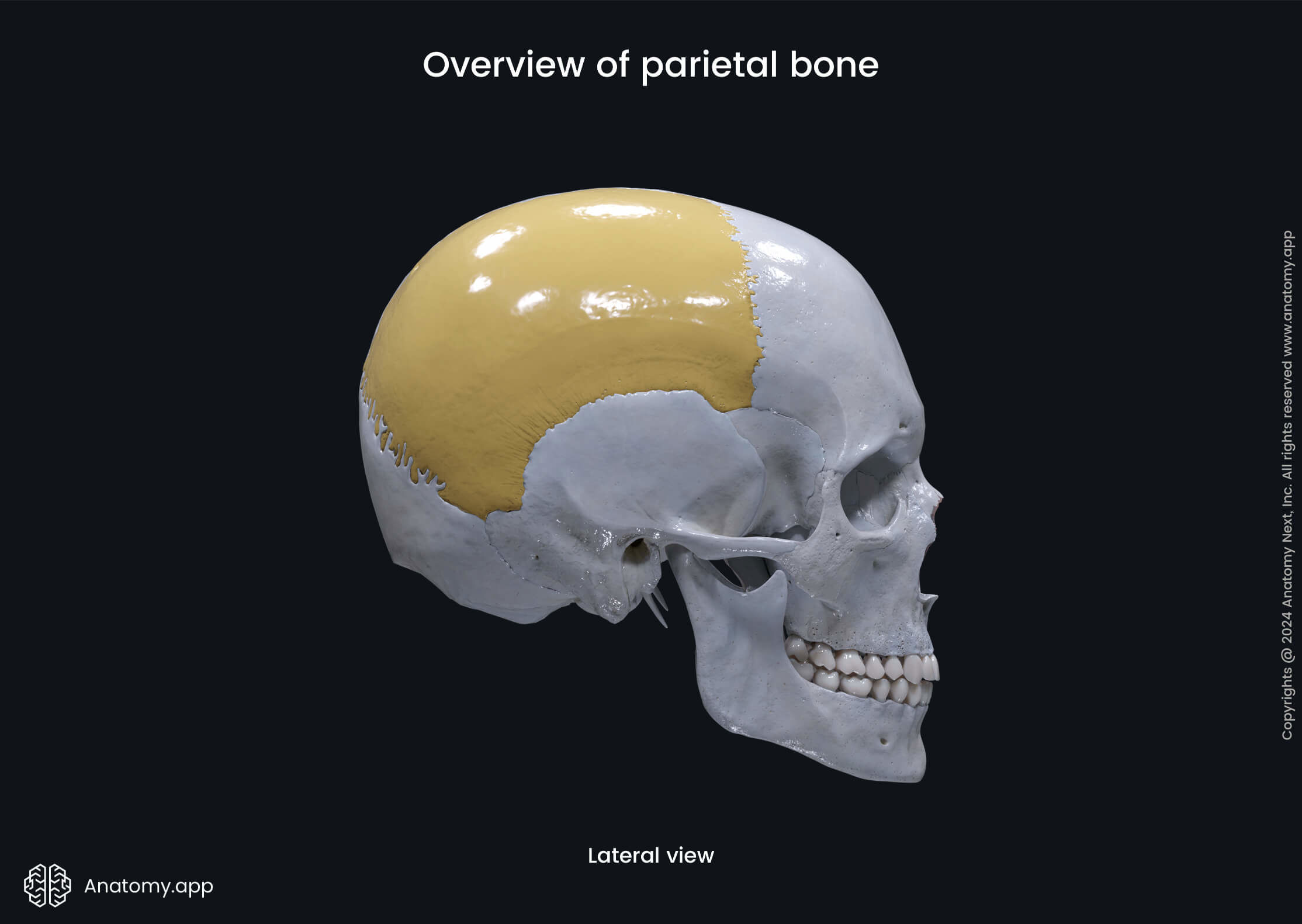
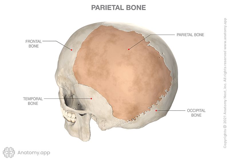
Parietal bone
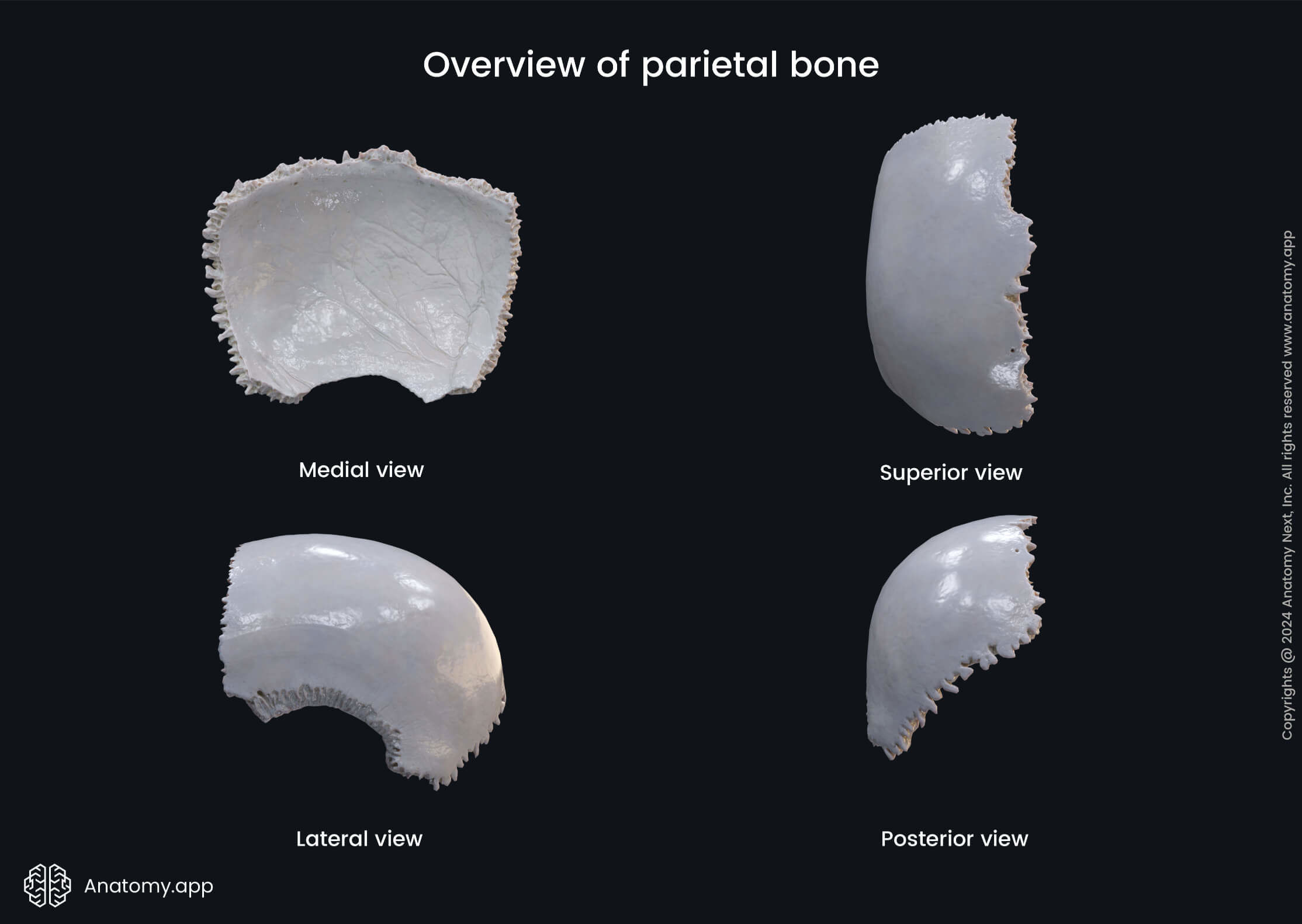
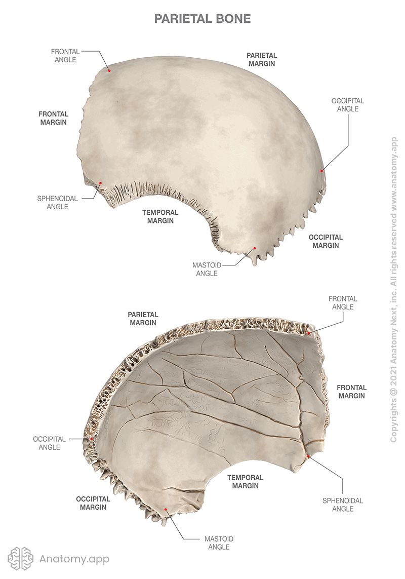
The parietal bone (Latin: os parietale) is located on each side of the skull right behind the frontal bone. Both parietal bones together form most of the calvaria. Each bone takes an irregular quadrilateral shape and has four angles, four margins, and two surfaces.
Angles of parietal bone
The four angles of the parietal bone are called:
- Frontal angle
- Sphenoidal angle
- Occipital angle
- Mastoid angle
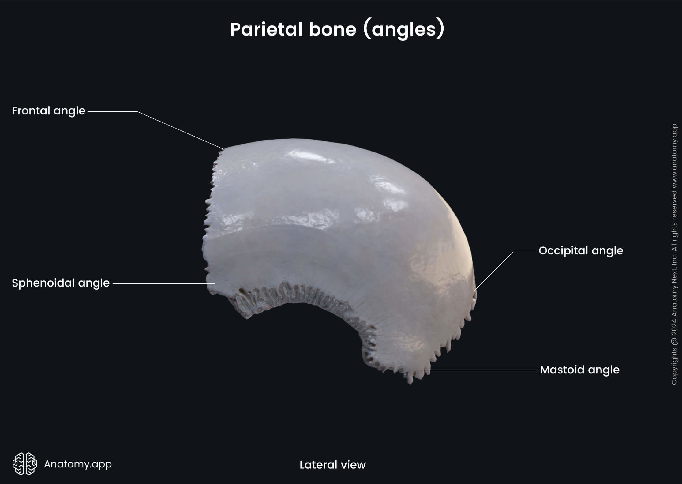
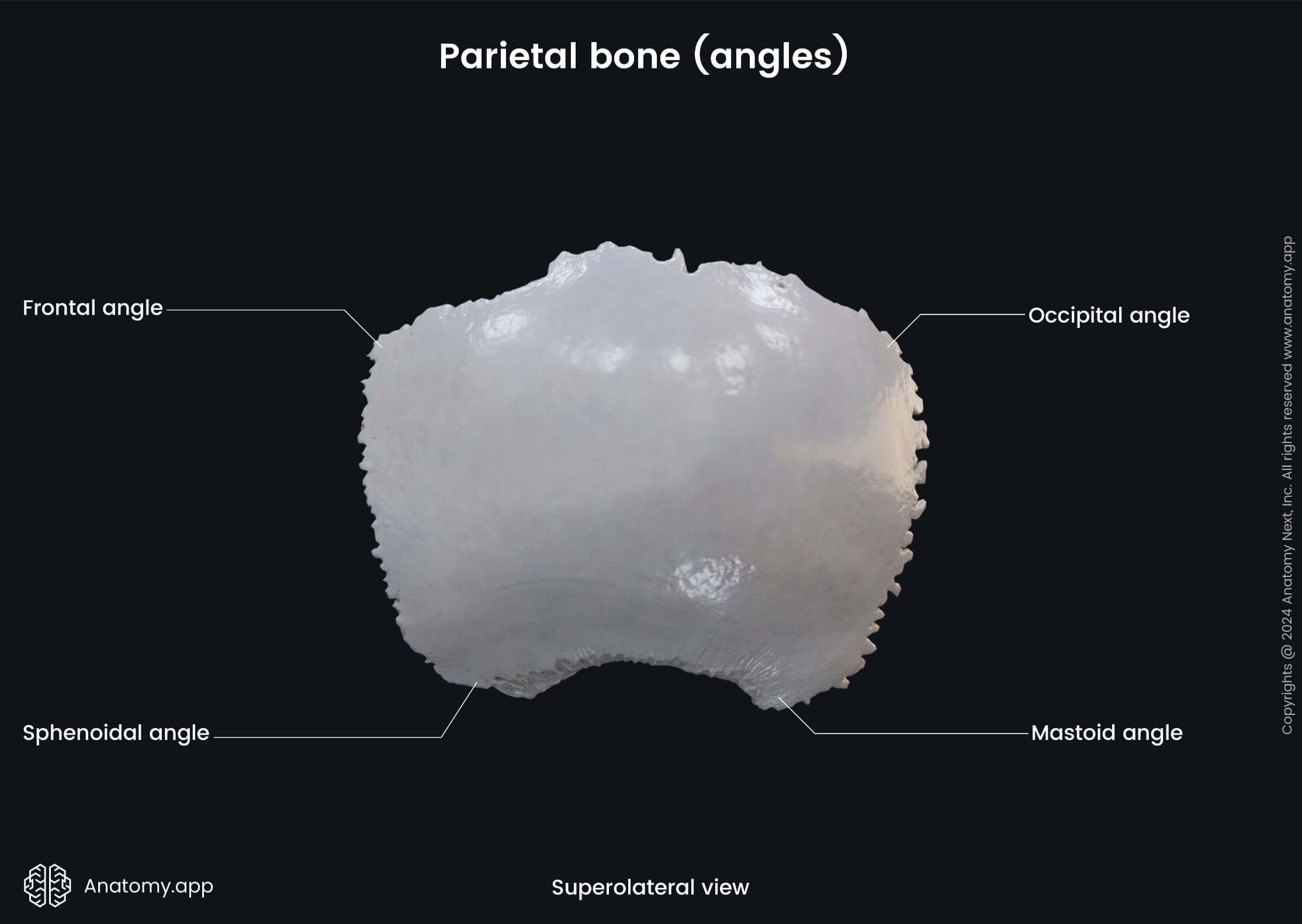
The frontal angle of the parietal bone is its anterior superior angle. The sphenoidal angle is the anterior inferior angle, and the most sharpest angle of the parietal bone. The occipital angle is the posterior superior angle. And the mastoid angle of the parietal bone is its posterior inferior angle.
Margins of parietal bone
The four margins or borders of the parietal bone are the following:
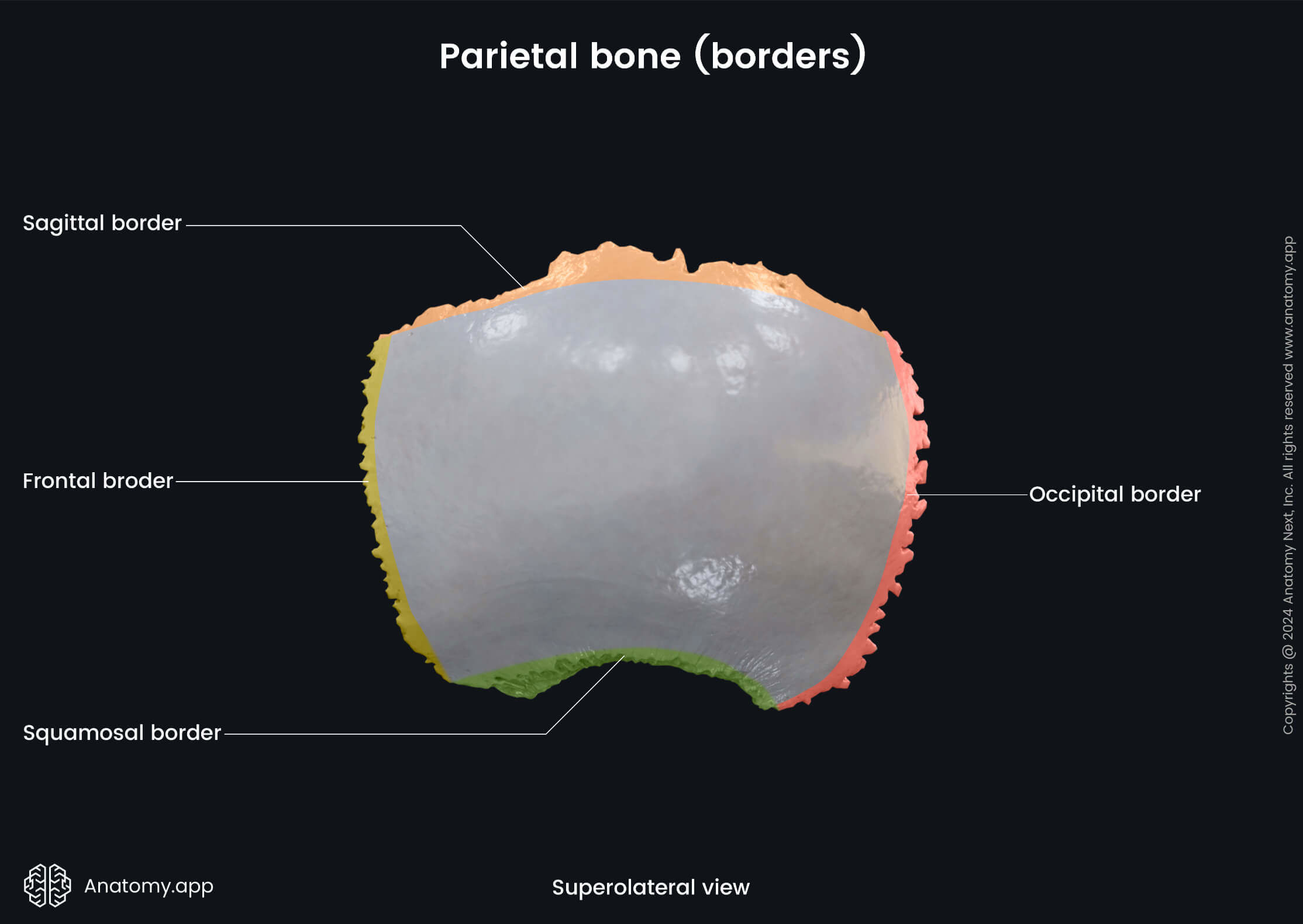
The frontal border of the parietal bone forms its anterior border. The occipital border forms the posterior border. The squamous border is the inferior border of the parietal bone. And the sagittal border of the parietal bone is its upper, medial border.
Surfaces and landmarks of parietal bone
External surface
The external surface of the parietal bone features the following anatomical landmarks:
- Parietal tuber
- Superior temporal line
- Inferior temporal line
- Parietal foramen
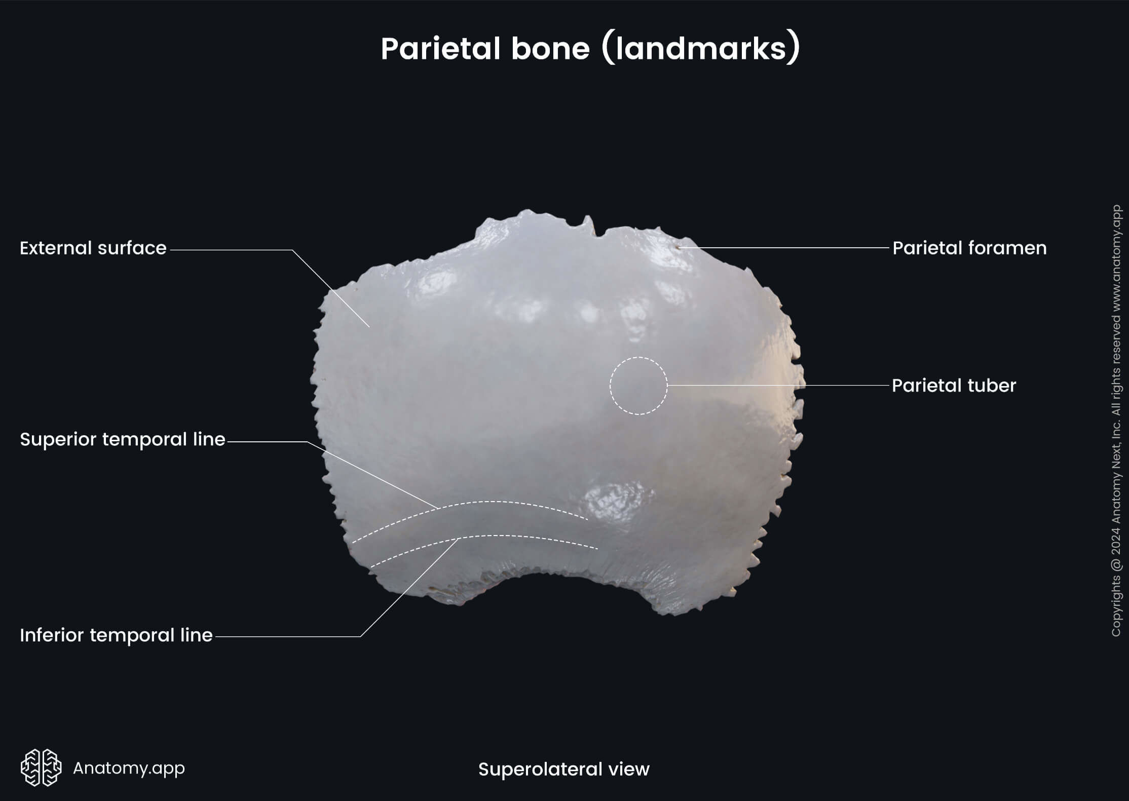
The parietal tuber (or parietal tuberosity, parietal eminence) is a smooth prominence located near the middle of the external surface of the parietal bone.
The superior temporal line is a curved line seen on the external surface of the parietal bone. It serves for attachment of the temporal fascia. The superior temporal line also represents the upper margin of the temporal plane.
The inferior temporal line is another curved line on the parietal bone bellow the superior temporal line. It serves for attachment of the temporalis muscle. This line marks the upper limit of the origin site of the mentioned muscle.
The parietal foramen is an opening occasionally present at the back part of the parietal bone. It serves as a passage for the parietal emissary vein, which drains into the superior sagittal sinus.
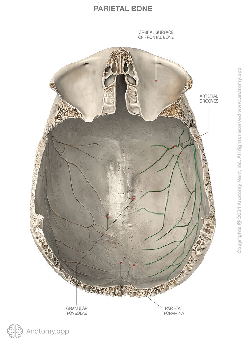
Internal surface
The internal surface of the parietal bone features the following landmarks:
- Groove for superior sagittal sinus
- Groove for sigmoid sinus
- Granular foveolae
- Impressions of cerebral gyri
- Arterial grooves
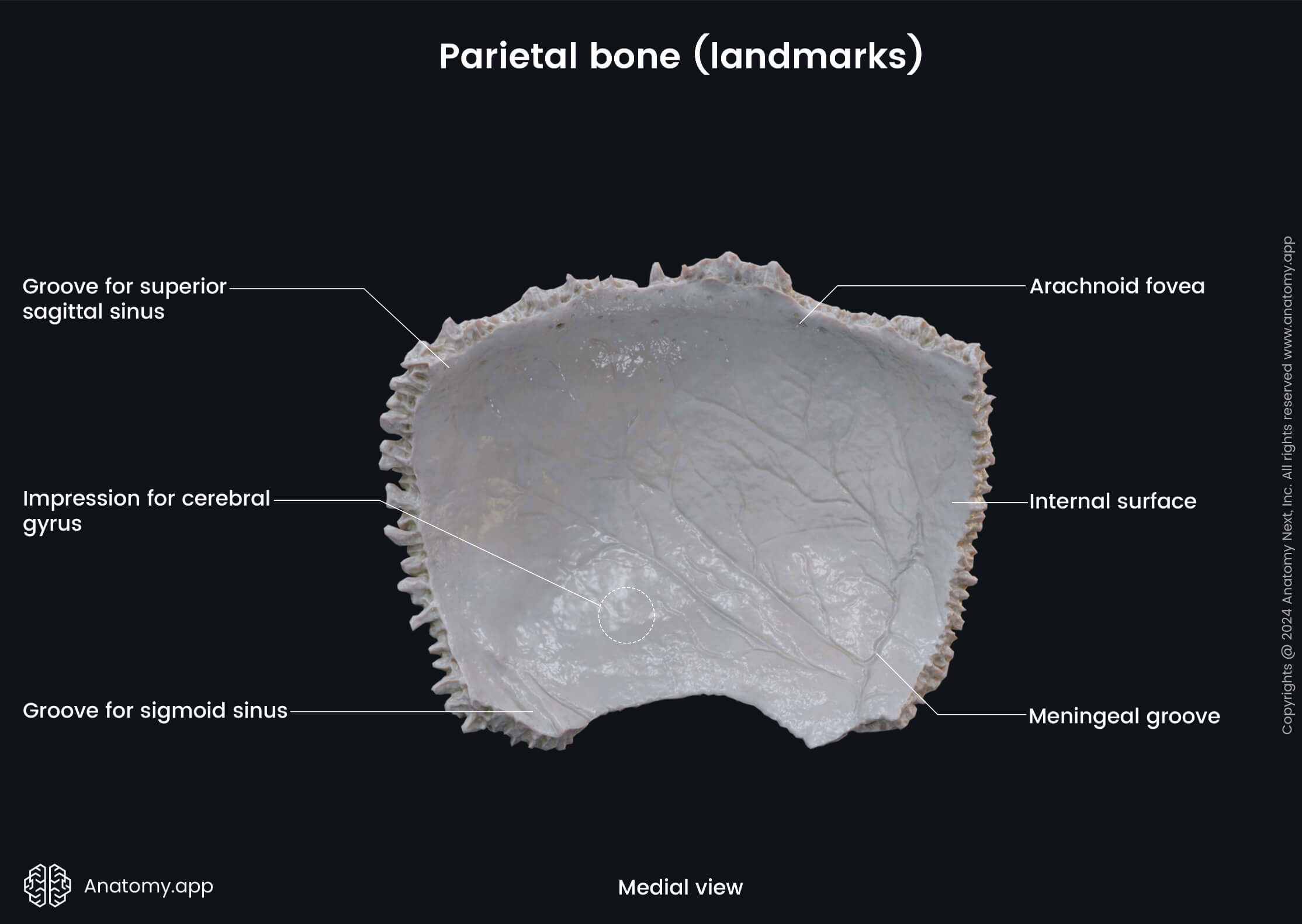
The groove for the superior sagittal sinus is a shallow depression that can be seen on the parietal, frontal, and occipital bones. It forms a channel for the superior sagittal sinus. The margins of this groove come together as it passes downward and become continuous with the frontal crest.
The groove for the sigmoid sinus can be seen in the posterior cranial fossa, found on three bones: occipital, temporal, parietal. It begins from the lateral part of the occipital bone, curves around the jugular process on the mastoid part of the temporal bone. Finally, turns sharply on the inner surface of the parietal bone continuing as the transverse groove.
The granular foveolae are small pits located on the internal surface of the parietal bone along the groove for the superior sagittal sinus. These pits are created by penetration of the arachnoid sheath (one of the meninges that cover the brain) in the bone.
The impressions of cerebral gyri (also called the digital impressions) are flat indentations on the inner surface of the skull that correspond to the cerebral gyri (folds of the cerebral cortex) which produce them.
The arterial grooves are many grooves found on the inner surface of the skull. They are produced by pressure from arteries, primarily the middle meningeal artery and its branches.
Anatomy.app
Contact information
- For questions regarding business inquiries. Please contact:
- info@anatomy.app