- Anatomical terminology
- Skeletal system
- Joints
- Muscles
- Heart
- Blood vessels
- Lymphatic system
- Nervous system
- Respiratory system
- Digestive system
- Urinary system
- Female reproductive system
- Male reproductive system
- Endocrine glands
- Eye
- Ear
Cervical vertebrae
The cervical vertebrae (Latin: vertebrae cervicales) are seven vertically aligned small bones that are positioned in the neck. At the same time, they form the cervical part of the spine, extending between the skull and the first rib of the thorax.
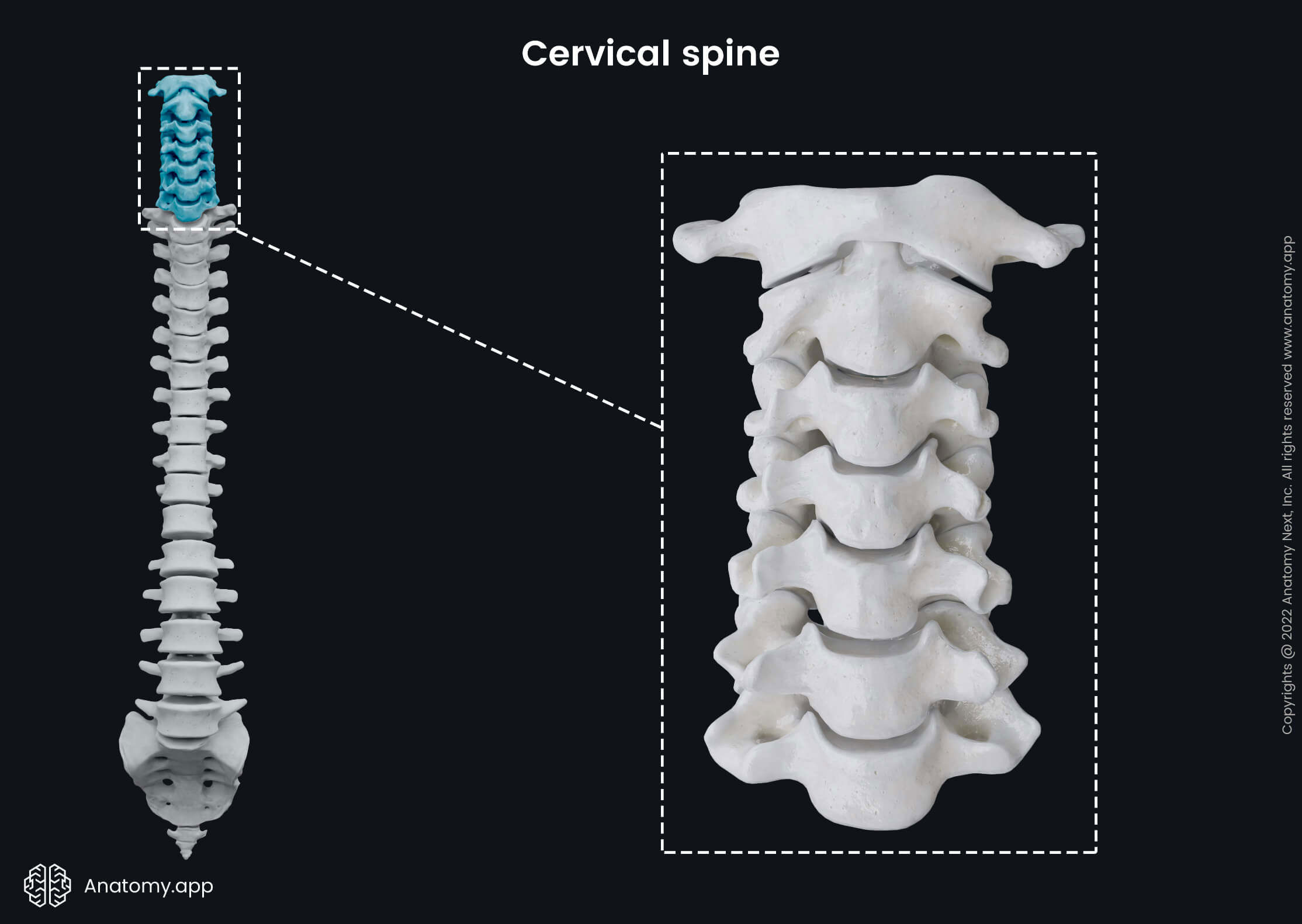
The cervical vertebrae are denoted C1 to C7, and they form the most mobile part of the spine. The spinous processes of the cervical vertebrae are covered by a thick ligament called the nuchal ligament. The most easily palpable cervical vertebra is the seventh cervical vertebra (C7) - it has a long spinous process that protrudes toward the skin of the posterior neck.
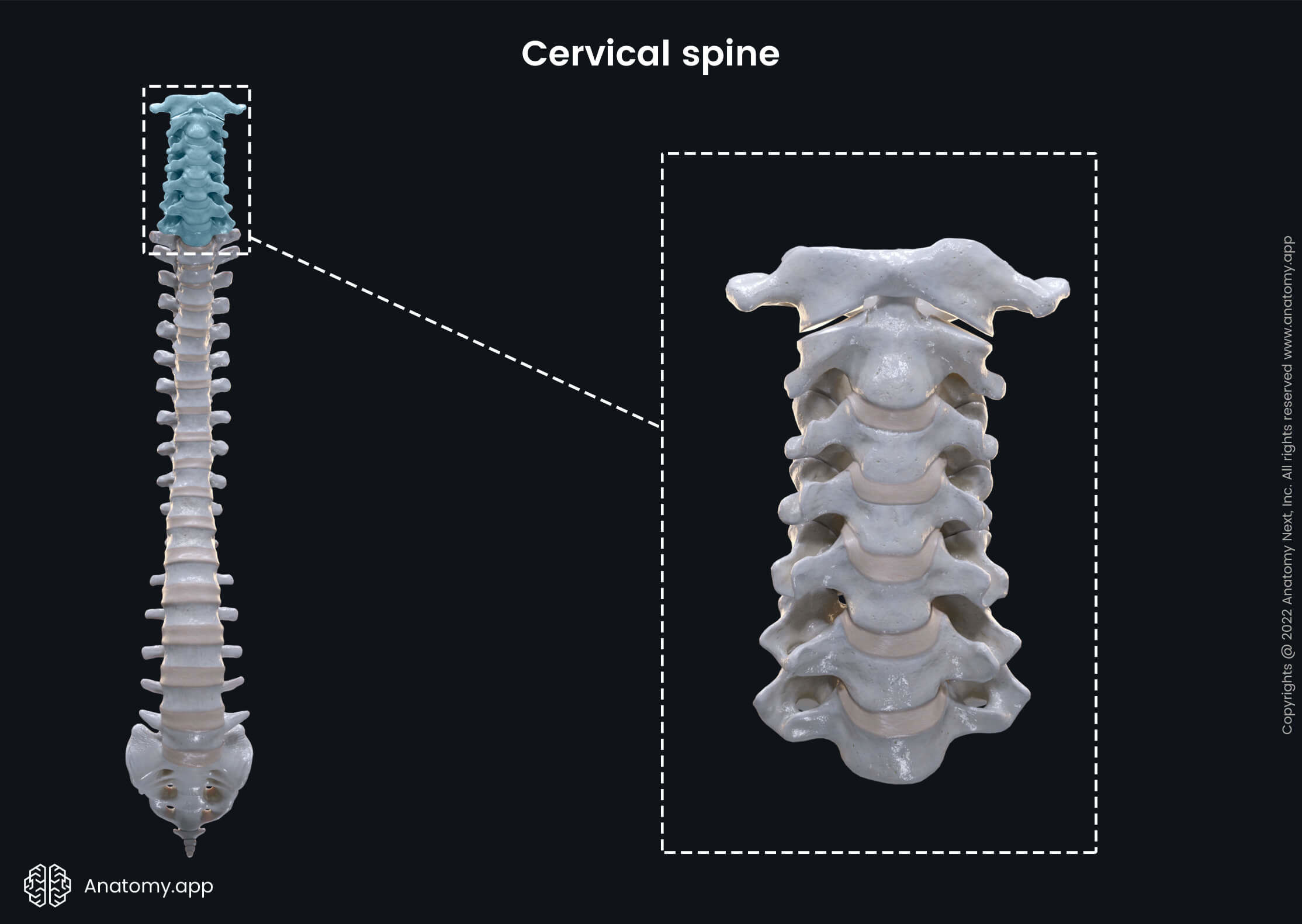
Each part of the spine has typical features that distinguish one type of vertebrae from the other. The cervical vertebrae can be easily recognized by the presence of the openings - transverse foramina - in their transverse processes. These openings are formed by the fusion of the rib rudiments and transverse processes.
Classification of cervical vertebrae
Each cervical vertebra can either be classified as typical or atypical.
- The typical cervical vertebrae are characterized mainly by their small size, presence of the transverse foramina and short and bifid spinous processes. This group includes the third through fifth cervical vertebrae (C3 - C5).
- Every atypical cervical vertebra has distinct characteristic features distinguishing them from the other vertebrae. This group includes four vertebrae - the first (C1), second (C2), sixth (C6) and seventh (C7) cervical vertebrae.
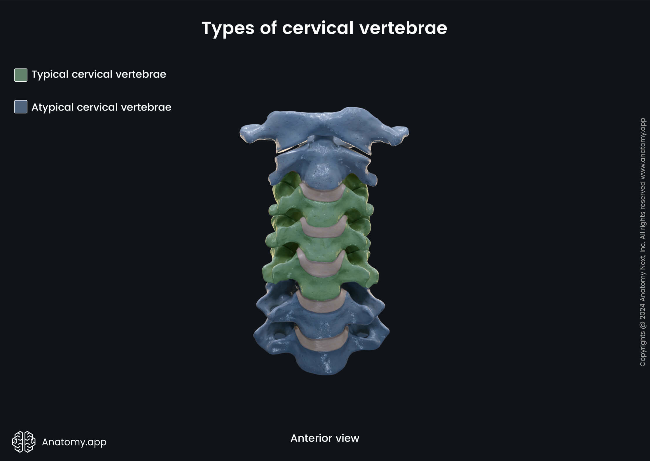
Note: Some authors do not consider the sixth cervical vertebra (C6) as an atypical cervical vertebra because its distinctive feature is not as significant as the anatomical features of the other atypical cervical vertebrae. However, we will classify and review this vertebra as an atypical vertebra.
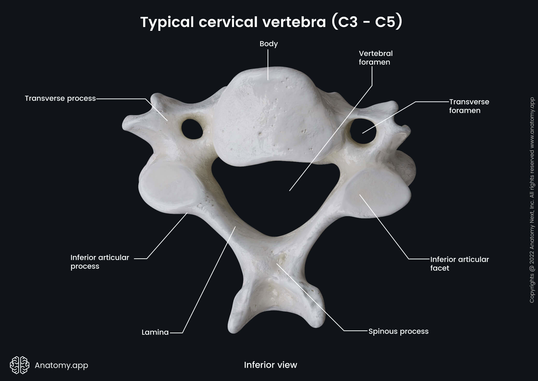
Landmarks of typical cervical vertebrae
As mentioned, the typical cervical vertebrae are the third through fifth cervical vertebrae (C3 - C5). All these vertebrae present with the following characteristic features:
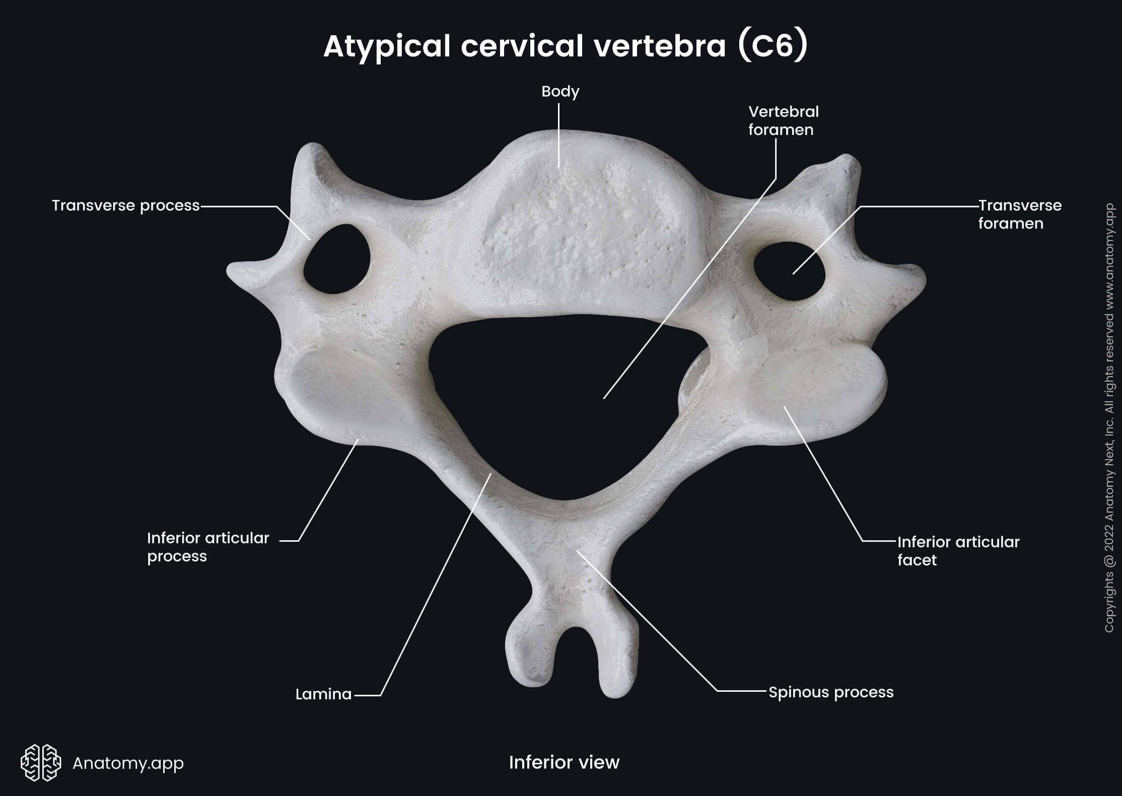
- Vertebral body (1) - large cylindrical-shaped anterior part of a vertebra that serves as the weight-bearing structure; two adjacent vertebral bodies are separated by a fibrocartilaginous intervertebral disc;
- Vertebral arch (1) - forms the lateral and posterior portions of a vertebra; it is composed of two parts:
- Pedicle (2) - bony structure that anchors the vertebral arch to the posterior surface of the vertebral body;
- Lamina (2) - bony plate found posterior to the pedicle; both laminae merge in the midline of the arch connecting the transverse and spinous processes;
- Spinous process (1) - a single process that projects posteriorly and inferiorly from the central aspect of the vertebral arch;
- Superior articular process (2) - extends upward from the vertebral arch and contains an articular facet:
- Superior articular facet (2) - surface that articulates with the inferior articular facet of a vertebra above;
- Inferior articular process (2) - extends downward from the vertebral arch and has an articular facet:
- Inferior articular facet (2) - surface that articulates with the superior articular facet of a vertebra below;
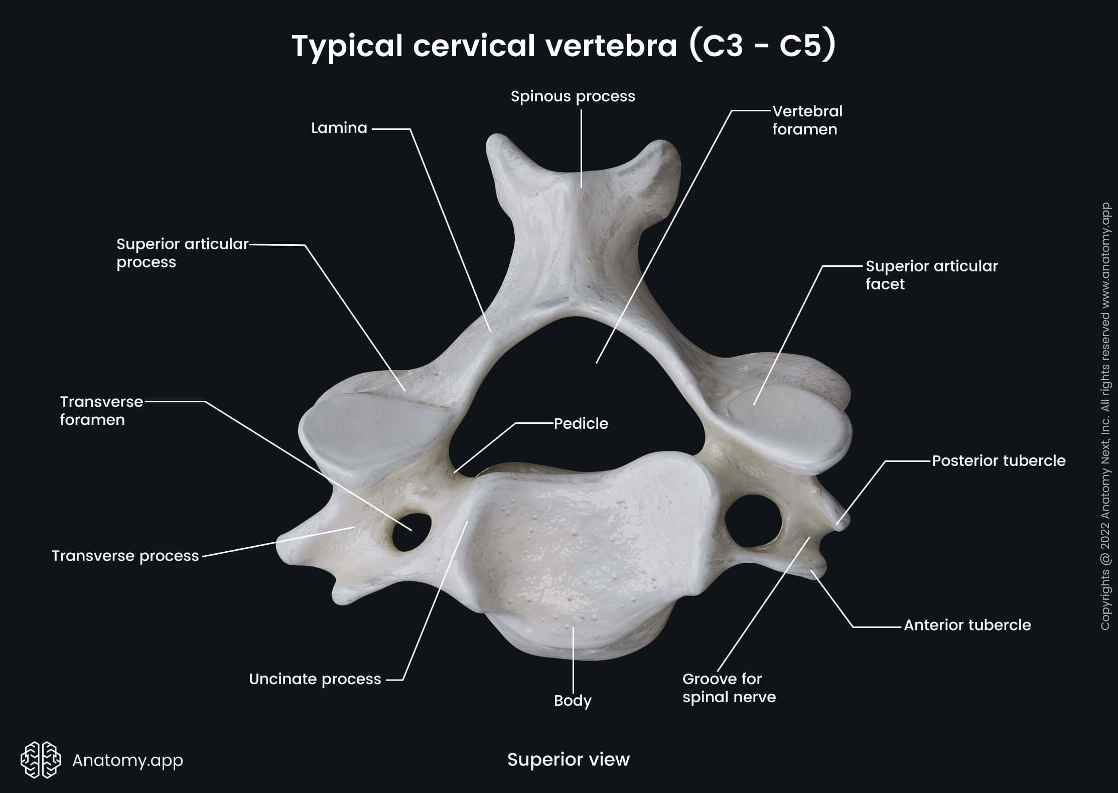
- Uncinate process (2) - bony projection found on the superior aspect of the lateral side of the vertebral body; it forms the medial aspect of the intervertebral foramen and uncovertebral joint (Luschka joint) with the body of the vertebra above;
- Transverse process (2) - projects from the posterolateral aspect of the vertebral arch; it has several landmarks:
- Transverse foramen (2) - a small opening formed by the fusion of the transverse process and rudimentary rib; transmits the vertebral artery, vertebral vein and sympathetic fibers from the lower cervical ganglion of the sympathetic trunk;
- Groove for spinal nerve (2) - a shallow bony channel for the passage of the spinal nerve; found on the superior surface of the transverse process;
- Anterior tubercle (2) - round eminence found anterior to the previous groove; serves as an attachment site for muscles, such as the longus capitis;
- Posterior tubercle (2) - round eminence found posterior to the groove for spinal nerve; serves as an attachment site for muscles, such as the levator scapulae.
The superior and inferior aspects of the pedicles contain superior (2) and inferior(2) vertebral notches. The inferior vertebral notch is larger than the superior one. Notches of adjacent vertebrae align and form intervertebral foramina (2) that transmit the spinal nerves.
And finally, the vertebral body and arch form an enclosed opening called the vertebral foramen (1). All vertebral foramina create the vertebral canal, through which the spinal cord passes.
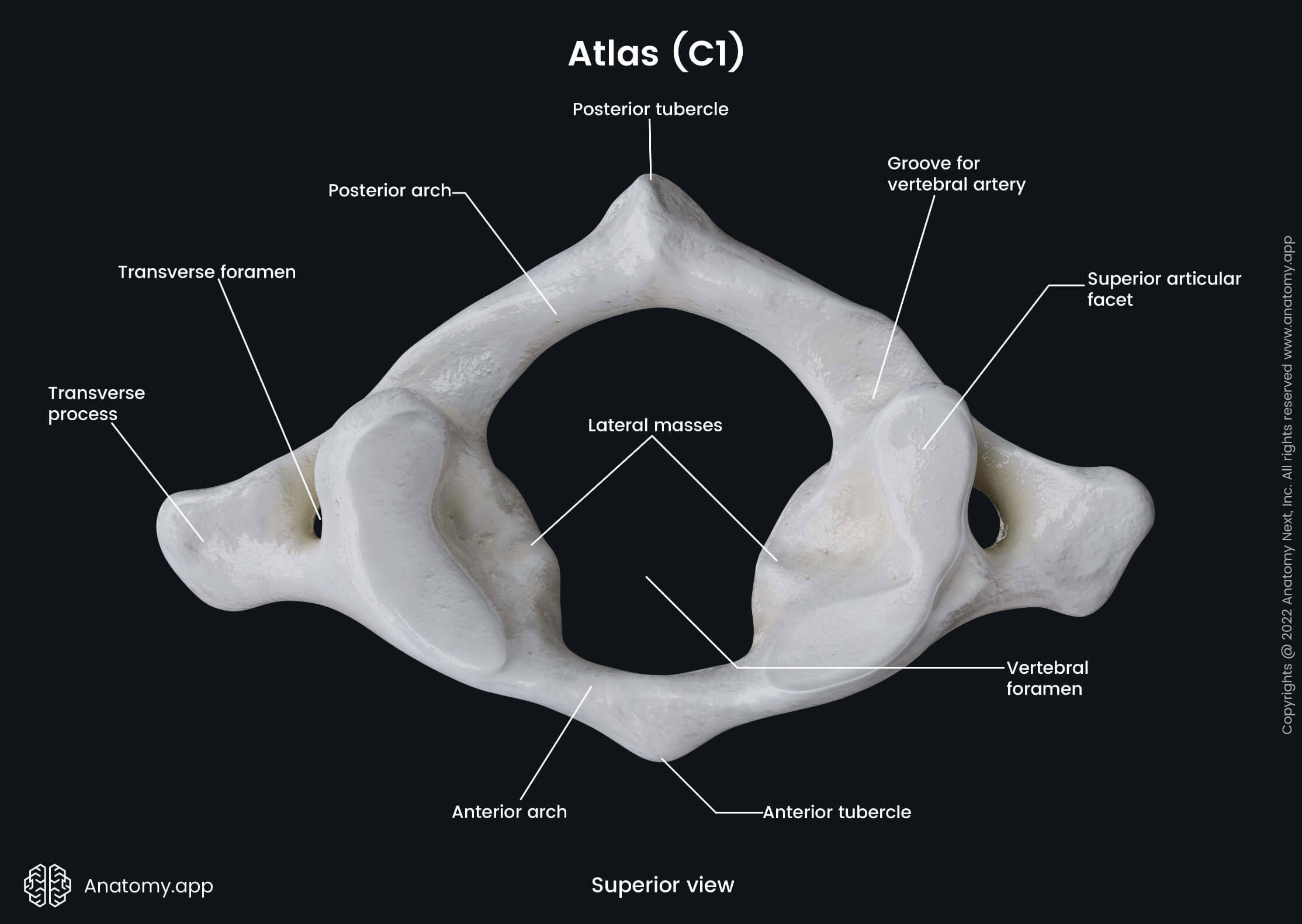
Atlas (C1)
The first cervical vertebra is also known as the atlas (C1). It articulates with the occipital bone of the skull at the atlanto-occipital joint and with the second cervical vertebra (C2) at the lateral and median atlanto-axial joints. The major distinguishing feature of the atlas is the lack of a vertebral body and a spinous process. Instead, it has two arches - anterior and posterior arches. Both are interconnected by two lateral masses.
Each lateral mass is formed by articular processes and presents with two articular surfaces - the superior and inferior articular facets. The superior facet articulates with the occipital bone of the skull at the atlanto-occipital joint. In contrast, the inferior articular facet articulates with the axis (C2), forming the atlanto-axial joint.
The structural features of the first two cervical vertebrae allow performing a great degree of neck and head movements. The atlanto-occipital joint is responsible for head shaking (neck flexion) when saying “yes.”
The inner or posterior aspect of the anterior arch has an impression for the dens of the axis (C2). It is called the fovea for dens. Moreover, this impression contains a facet for dens. The superior surface of the posterior arch posterior to the lateral masses has a groove for vertebral artery.
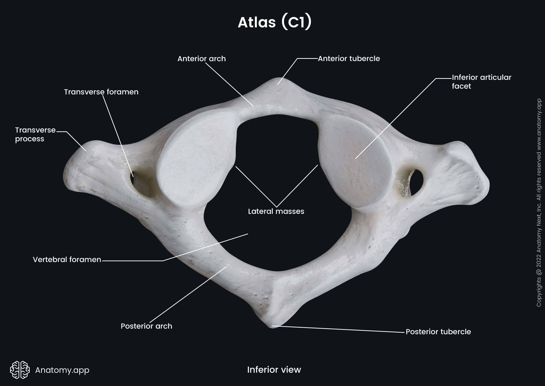
The central aspects of both arches present with anterior and posterior protrusions known as the anterior and posterior tubercles. And finally, the two transverse processes of the atlas are large and project further laterally than those of other cervical vertebrae. They extend from the lateral masses and act as levers for muscle action.
Axis (C2)
The axis (C2) is the second cervical vertebra characterized by all landmarks found on the typical vertebra. However, its body has a large tooth-like projection called the dens (odontoid process). It extends superiorly from the vertebral body toward the foramen magnum. The dens articulates with the atlas (C1) at the atlanto-axial joint. The movements provided by the joint include rotation of the head. Therefore, the dens is involved in head shaking when saying "no."
The highest point of the dens is called the apex. It serves as the attachment site for the apical ligament of dens. The anterior aspect of the dens has an articular surface - anterior articular facet - that articulates with the facet for dens of the atlas (C1). The posterior aspect of the dens has one more articular facet - posterior articular facet.
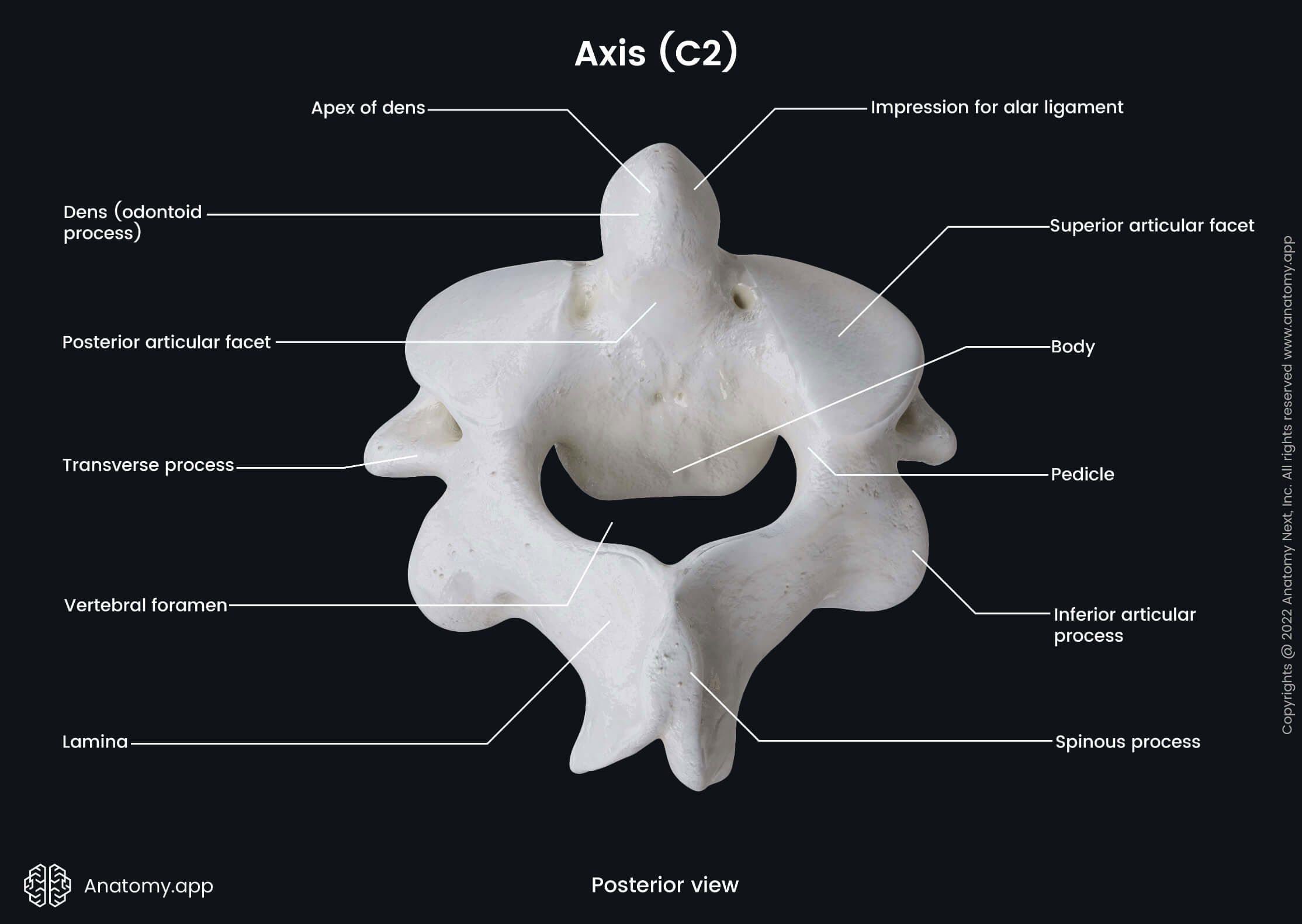
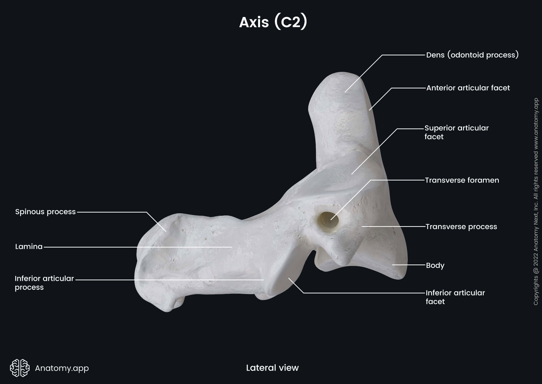
Besides the mentioned features, the axis (C2) also has two impressions for alar ligaments. They are shallow grooves located on each upper aspect of the lateral side of the dens. Both serve as an attachment site for the alar ligament.
Sixth cervical vertebra (C6)
The sixth cervical vertebra (C6) is also considered an atypical vertebra. Some authors do not classify the sixth cervical vertebra (C6) as an atypical cervical vertebra because its distinctive feature is not as significant as the anatomical features of the other atypical cervical vertebrae.
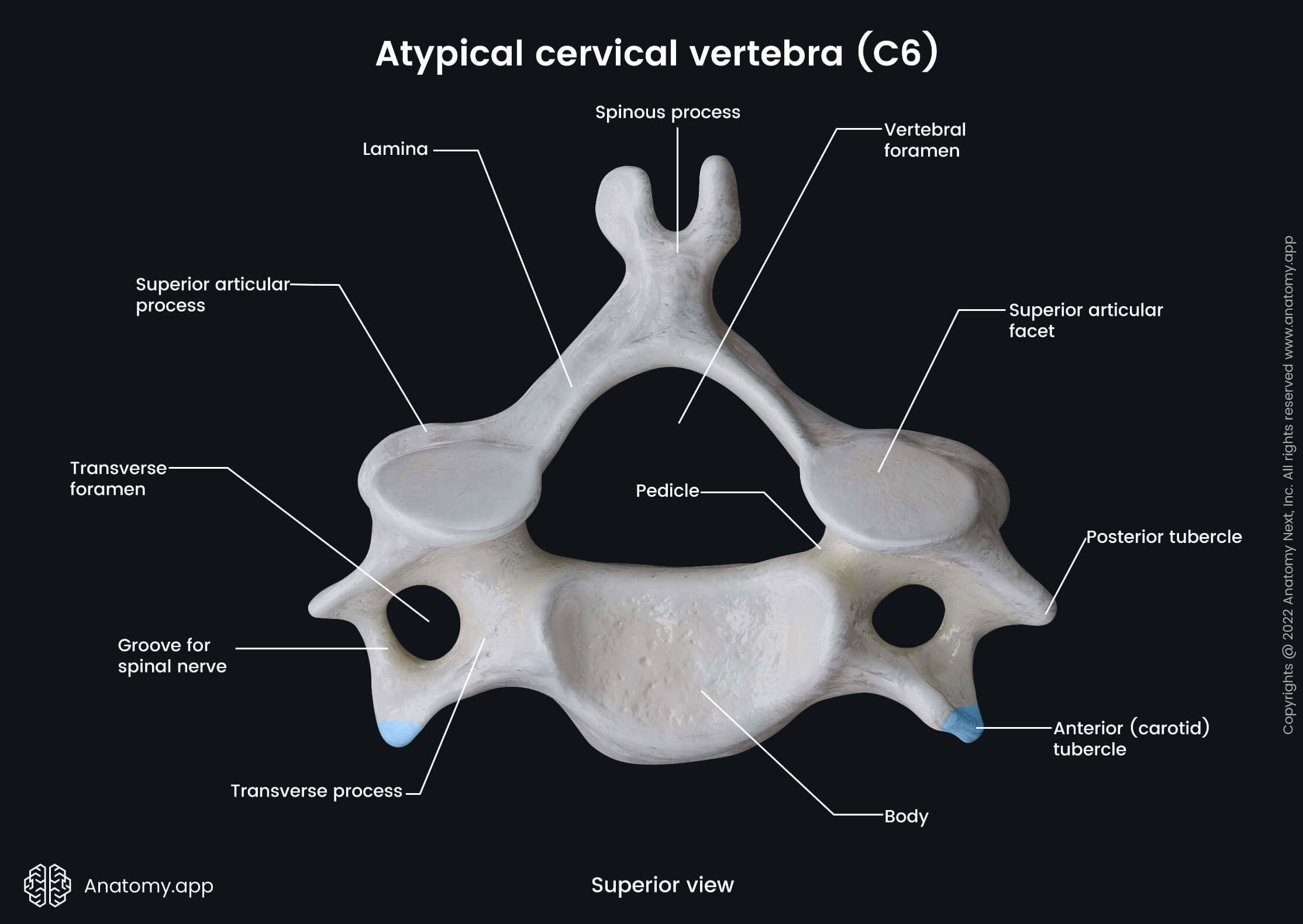
Besides all the anatomical landmarks of a typical cervical vertebra, the sixth cervical vertebra (C6) presents a larger anterior tubercle. This tubercle is also known as the carotid tubercle or the tubercle of Chassaignac. Anterior to the tubercle goes the common carotid artery, and it can be compressed by fingers against the tubercle. This landmark is helpful in carotid massage and serves as a site to palpate the carotid pulse.
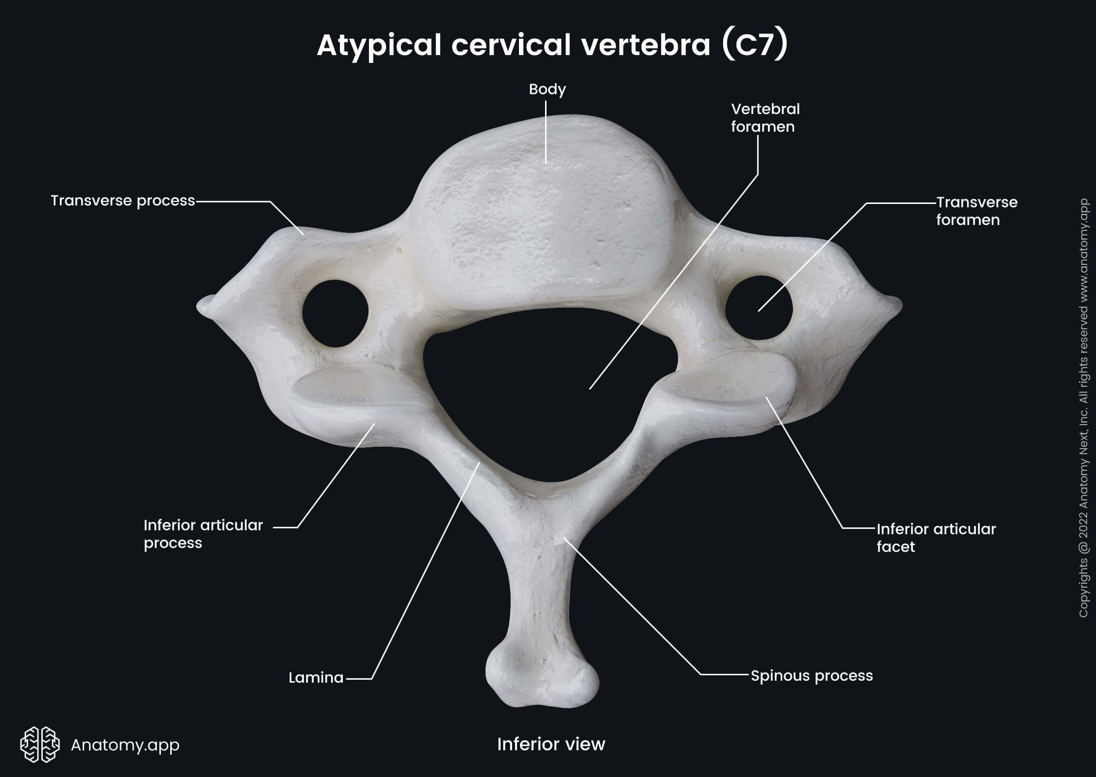
Vertebra prominens (C7)
The seventh cervical vertebra is also called the vertebra prominens (C7). It is very similar to a typical vertebra, but it has some distinct features. Thus, the vertebra prominens is considered an atypical vertebra.
The main distinguishing feature of the seventh cervical vertebra (C7) is its spinous process - it is the longest among all cervical vertebrae. Also, it is not bifid but ends with a rounded tubercle. Moreover, the spinous process of the vertebra prominens is easily palpable as it protrudes posteriorly towards the skin at the back of the neck.
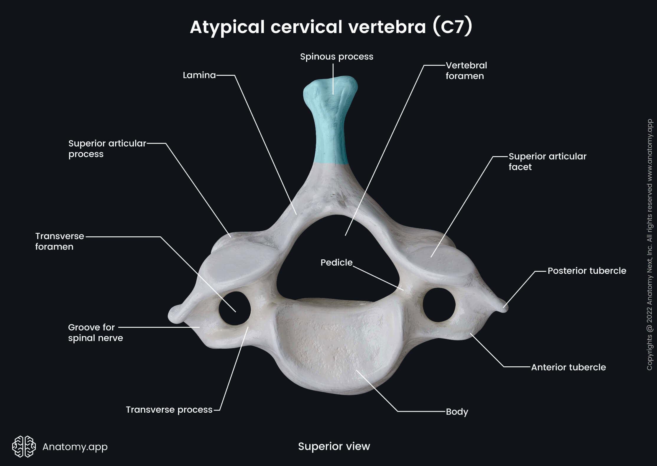
Besides the distinct features related to the spinous process, the seventh cervical vertebra (C7) also has a relatively small transverse foramen. Moreover, it does not transmit the vertebral artery.
Anatomy.app
Contact information
- For questions regarding business inquiries. Please contact:
- info@anatomy.app