- Anatomical terminology
- Skeletal system
- Joints
- Muscles
- Heart
- Blood vessels
- Lymphatic system
- Nervous system
- Respiratory system
- Digestive system
- Urinary system
- Female reproductive system
- Male reproductive system
- Endocrine glands
- Eye
- Ear
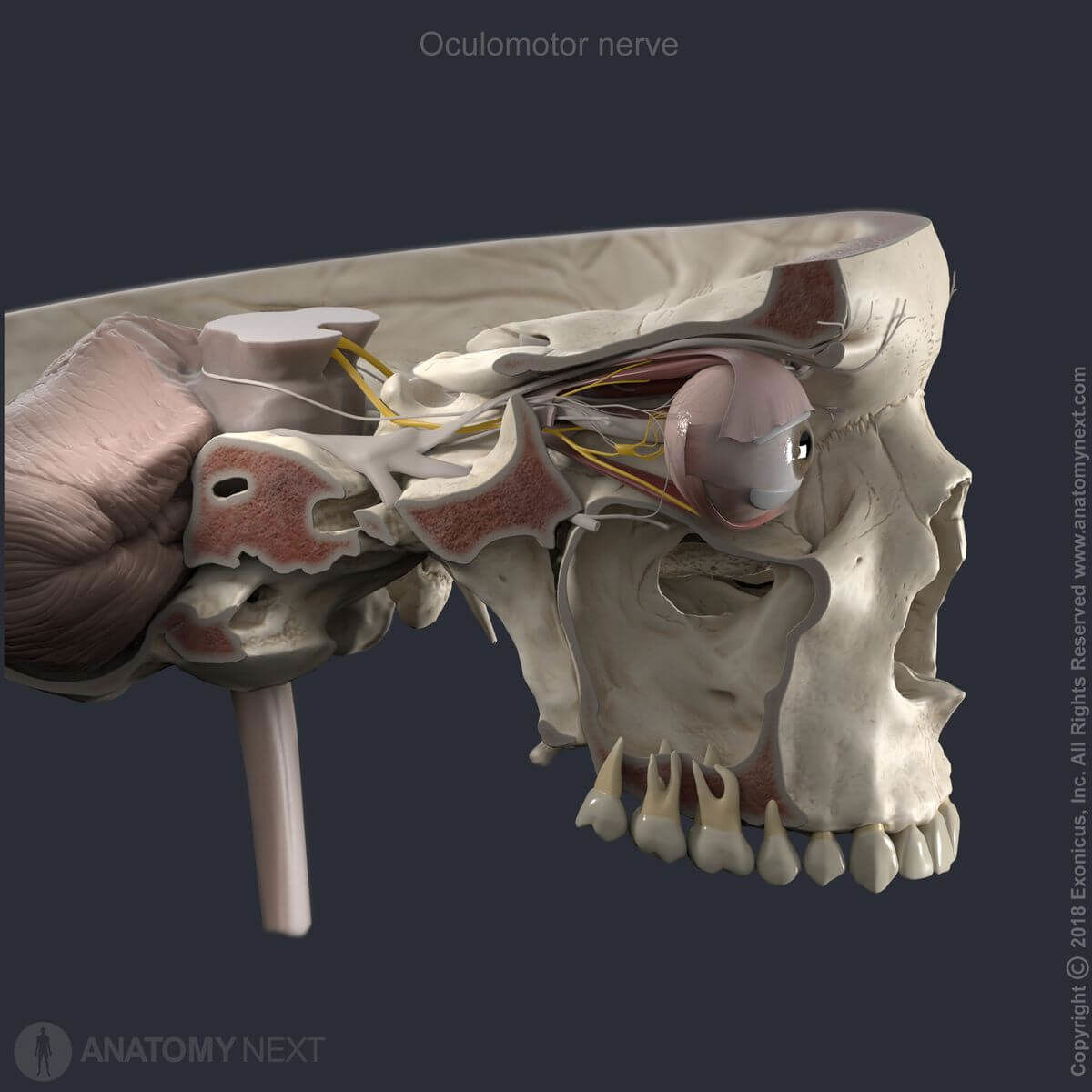
Oculomotor nerve (CN III)
The oculomotor nerve (Latin: nervus oculomotorius), the third cranial nerve (CN III) is a mixed peripheral nerve containing motor and parasympathetic fibers. The motor fibers of the oculomotor nerve supply most of the extrinsic muscles of the eye. The parasympathetic fibers carried by the third cranial nerve participate in controlling the intraocular muscles that provide pupillary constriction and accommodation.
Oculomotor nerve nuclei
The oculomotor nerve fibers are axons that originate from neurons in two nuclei located in the posteromedial aspect of the midbrain at the level of the superior colliculus:
- Oculomotor nucleus - a somatic motor nucleus which provides the oculomotor nerve with general somatic efferent fibers to most of the extraocular muscles, including the levator palpebrae superioris, medial, superior and inferior recti muscles, and the inferior oblique muscle.
- Accessory nuclei of the oculomotor nerve - parasympathetic nuclei, among which the clinically most important one is the Edinger-Westphal nucleus; the accessory nuclei give rise to preganglionic parasympathetic (general visceral efferent) fibers of the oculomotor nerve innervating intrinsic muscles of the eye.
Oculomotor nerve course
The oculomotor nerve appears outside on the anterior aspect of the midbrain, emerging on the oculomotor sulcus passing along the interpeduncular fossa between the cerebral peduncles. The third cranial nerve then travels forward below the posterior cerebral artery and above the superior cerebellar artery.
In the subarachnoid space, the oculomotor nerve travels medial to the trigeminal nerve (CN V). Then it pierces the dura mater and continues in ventral direction inside the lateral wall of the cavernous sinus. The oculomotor nerve leaves the cranial cavity and enters the orbit through the superior orbital fissure dividing into two - superior and inferior - divisions that run beneath the trochlear (CN IV) and ophthalmic (CN V1) nerves.
Oculomotor nerve branches
The superior division of the oculomotor nerve contains only motor fibers. This division passes above the optic nerve (CN II) to supply the superior rectus muscle, also giving off a branch that innervates the levator palpebrae superioris muscle.
The inferior division contains motor and parasympathetic fibers. This division splits into medial, central and lateral branches. The medial branch enters the ocular surface of the medial rectus innervating it. The central branch enters the ocular surface of the inferior rectus muscle.
Finally, the lateral branch of the inferior division of the oculomotor nerve enters the orbital surface of the inferior oblique muscle and communicates with the ciliary ganglion to distribute parasympathetic fibers to the sphincter pupillae (providing constriction of the pupil) and to the ciliary muscle (accommodation of the lens).
Anatomy.app
Contact information
- For questions regarding business inquiries. Please contact:
- info@anatomy.app