- Anatomical terminology
- Skeletal system
- Joints
- Muscles
- Heart
- Blood vessels
- Lymphatic system
-
Nervous system
- Central nervous system
-
Peripheral nervous system
- Cranial nerves
- Spinal nerves
- Respiratory system
- Digestive system
- Urinary system
- Female reproductive system
- Male reproductive system
- Endocrine glands
- Eye
- Ear
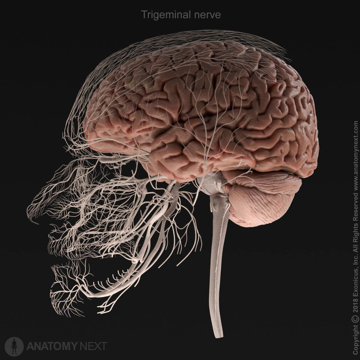
Trigeminal nerve (CN V)
The trigeminal nerve (Latin: nervus trigeminus), the fifth cranial nerve (CN V), is a mixed nerve containing sensory and motor fibers. It provides sensory innervation for the mucous membrane of the oral and nasal cavities, orbit, ear, conjunctiva, and the skin of the face and scalp. The motor fibers of the trigeminal nerve innervate the muscles of the first branchial arch, which include all masticatory muscles.
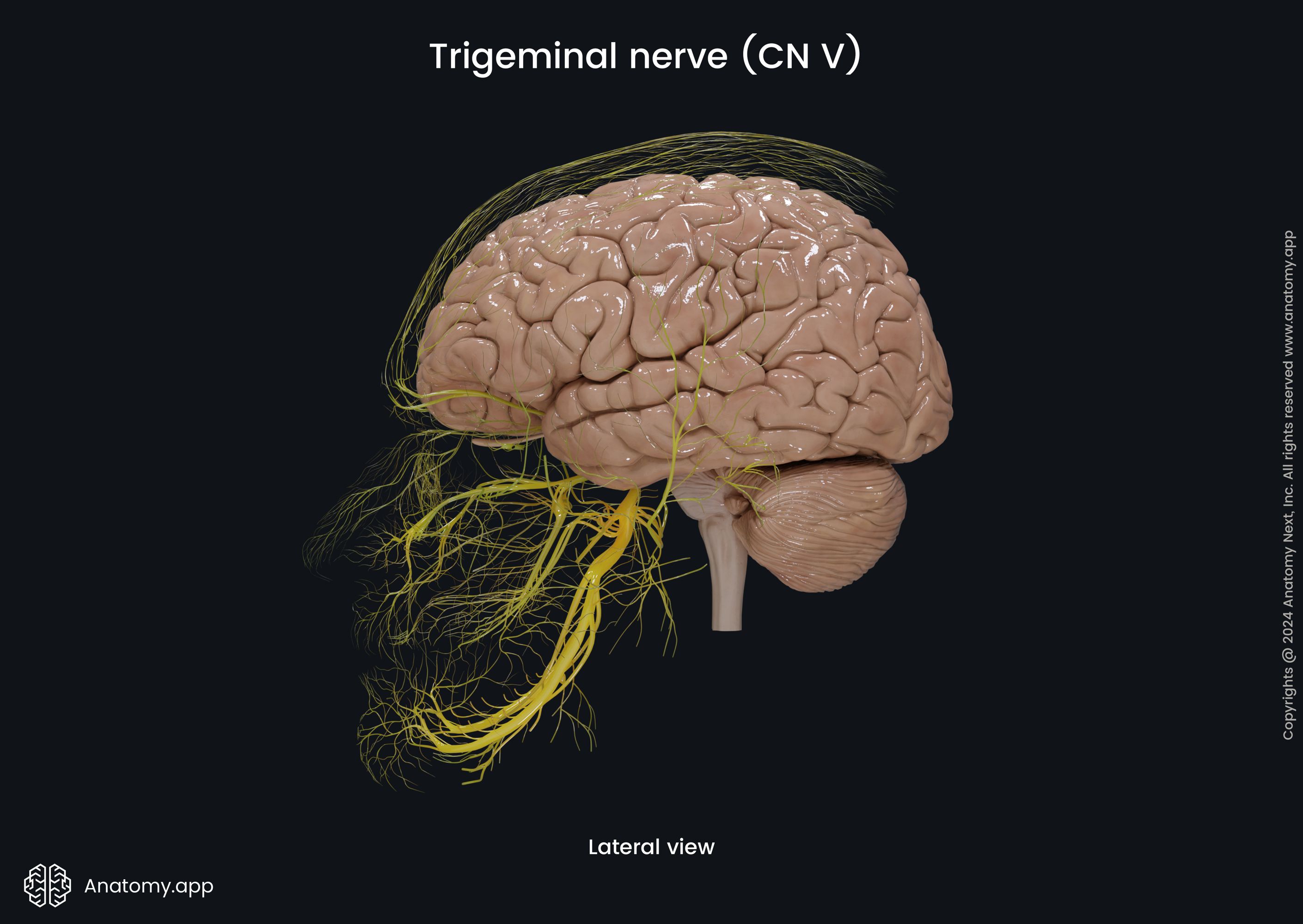
The trigeminal nerve is the largest of the cranial nerves and it develops from the mesoderm of the first branchial arch. It emerges from the brainstem between the pons and the middle cerebral peduncles with two roots: the sensory and the motor root of the trigeminal nerve. The sensory root is larger than the motor one.
Trigeminal nerve nuclei
The trigeminal nerve is connected with four nuclei in the central nervous system located in the superior aspect of the rhomboid fossa. These nuclei provide the nerve with fibers of different modalities, and they include the following:
- Trigeminal motor nucleus - the only somatic motor (special visceral efferent) nucleus of the trigeminal nerve;
- Principal sensory nucleus - general sensory afferent nucleus located in the pons;
- Spinal trigeminal nucleus - general sensory afferent nucleus located in the medulla oblongata;
- Mesencephalic trigeminal nucleus - another general sensory afferent nucleus located in the midbrain.
Trigeminal nerve roots and trigeminal ganglion
The sensory root of the trigeminal nerve has an enlargement - the trigeminal ganglion (Gasserian ganglion), which is formed by the cell bodies (perikaryons) of pseudounipolar neurons providing sensory fibers for the trigeminal nerve. This ganglion is located in the slit-like cavity or pouch of the dura mater called the trigeminal (Meckel's) cave and is lying on the trigeminal impression of the petrous part of the temporal bone. The motor root is located below the trigeminal ganglion.
Trigeminal nerve branches
Beyond the trigeminal ganglion, the trigeminal nerve divides into three major branches or divisions:
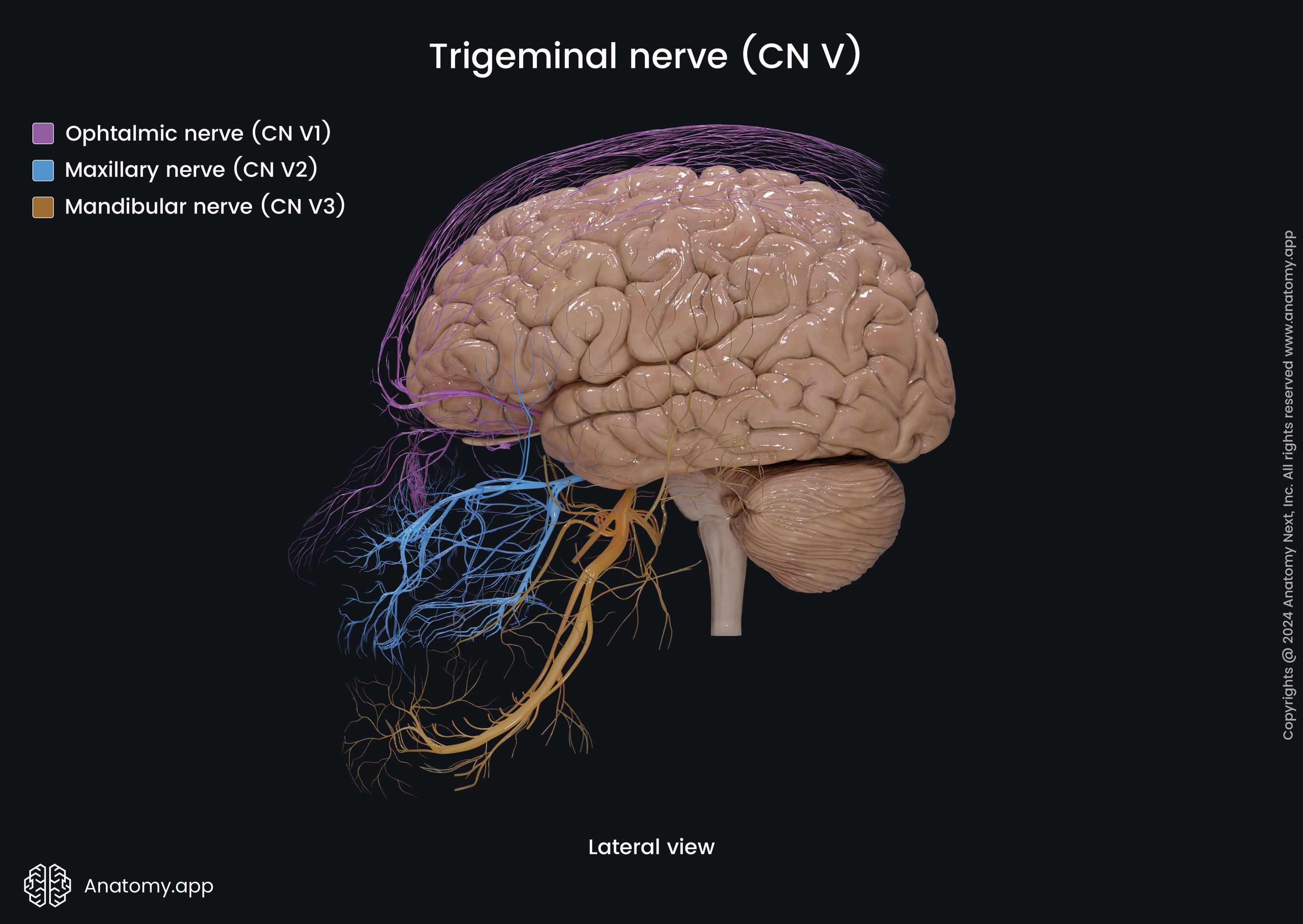
The first branch of the trigeminal nerve is the ophthalmic nerve (CN V1) - a sensory nerve that carries information to the central nervous system (CNS) from structures of the upper part of the face and scalp - eyeball (for example, cornea and conjunctiva) and orbit, lacrimal glands, skin of the upper face (forehead) and anterior scalp, lining of the upper part of the nasal cavity and ethmoid air cells, as well as frontal sinus and the meninges of the anterior cranial fossa.
The maxillary nerve is the second division of the trigeminal nerve. This nerve, with its branches, carries sensory information to the CNS from the middle part of the face - upper gingiva, upper jaw and upper teeth, skin of the middle part of the face (zygomatic area), mucosa of the palate, and nasal cavity, upper lip, and cheek, as well as the dura mater of the middle cranial fossa.
The third branch of the trigeminal nerve is known as the mandibular nerve. It is a mixed nerve, and its branches innervate the lower parts of the face. Its sensory branches innervate several skin regions of the face (mandibular area, lower lip), as well as the mucosa of the oral cavity, lower teeth, and lower gingiva, while the motor branches of the mandibular nerve supply muscles of the first branchial arch, including all muscles of mastication.
Anatomy.app
Contact information
- For questions regarding business inquiries. Please contact:
- info@anatomy.app