- Anatomical terminology
- Skeletal system
- Joints
- Muscles
- Heart
- Blood vessels
- Lymphatic system
- Nervous system
- Respiratory system
- Digestive system
- Urinary system
- Female reproductive system
- Male reproductive system
- Endocrine glands
- Eye
- Ear
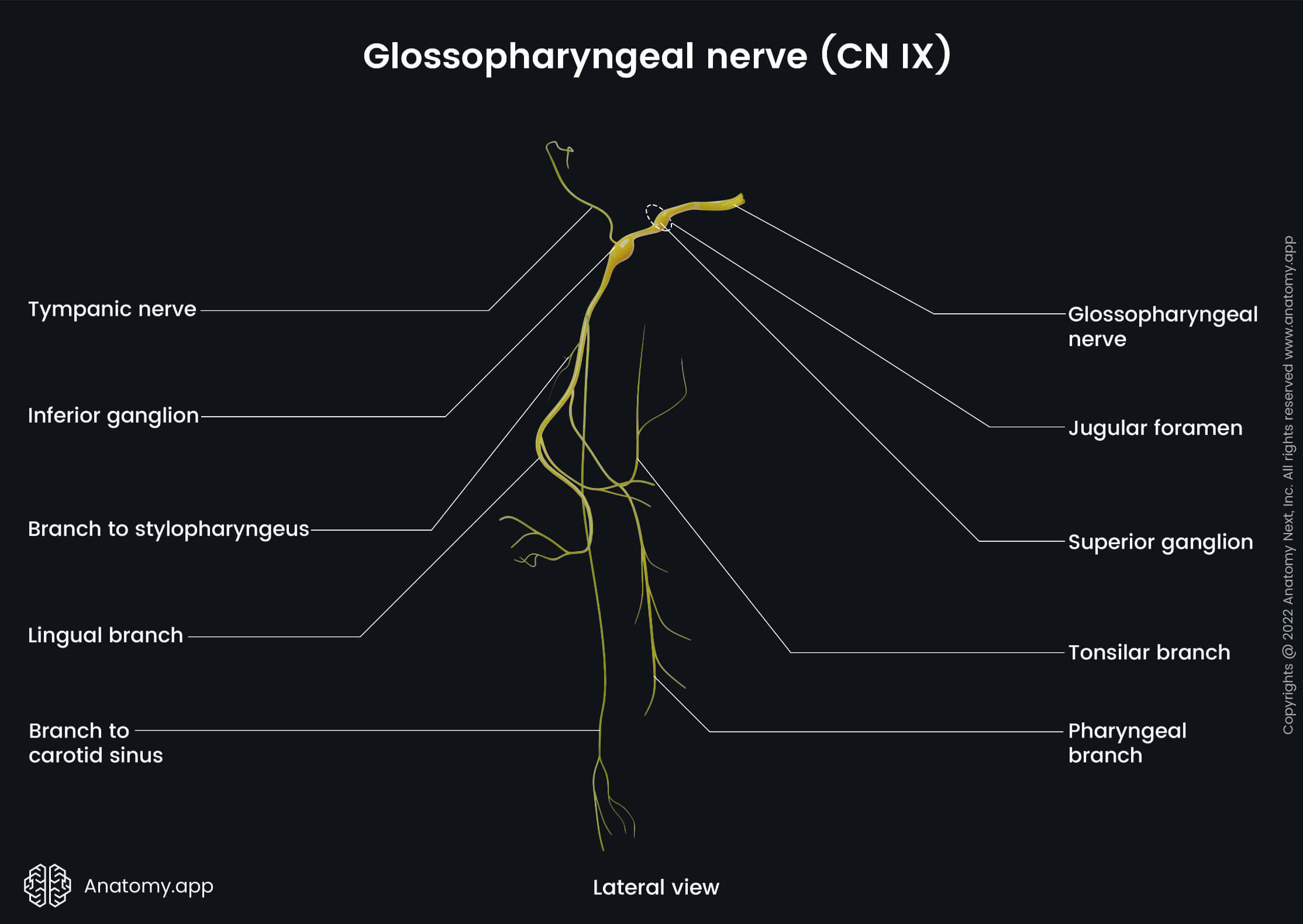
Glossopharyngeal nerve (CN IX)
The glossopharyngeal nerve (Latin: nervus glossopharyngeus), the ninth cranial nerve (CN IX), is a mixed cranial nerve, which provides motor innervation to the stylopharyngeus muscle and the superior pharyngeal constrictor muscle. Its sensory fibers supply the root of the tongue (including the vallate papillae), palatine tonsil and oropharynx, as well as the mucosa of the tympanic cavity, the auditory tube, and the mastoid cells. The CN IX also controls the secretion of the parotid gland via its parasympathetic fibers.
Glossopharyngeal nerve nuclei
The glossopharyngeal nerve has three nuclei located in the lower aspect of the rhomboid fossa:
- Nucleus ambiguous (motor) - shared with the vagus (CN X) and accessory nerve (CN XI); provides innervation for the stylopharyngeus and the superior pharyngeal constrictor muscles;
- Solitary tract nucleus (sensory) - shared with the vagus (CN X) and intermediate portion of the facial nerve (CN VII); receives fibers from the posterior one-third of the tongue;
- Inferior salivatory nucleus (parasympathetic) - supplies the parotid gland.
Glossopharyngeal nerve course and branches
The ninth cranial nerve emerges from the medulla oblongata on the posterolateral sulcus together with the vagus nerve (CN X) and accessory nerve (CN XI). The glossopharyngeal nerve, along with the two other mentioned cranial nerves, goes forward and laterally and leaves the skull through the jugular foramen.
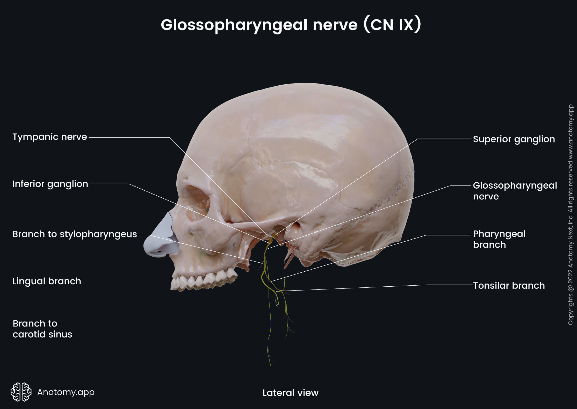
The CN IX forms two enlargements in the area of the jugular foramen: the superior ganglion in the jugular foramen and the inferior ganglion of the glossopharyngeal nerve below the jugular foramen at the level of petrosal fossula. Both ganglia contain cell bodies of sensory neurons - pseudounipolar neurons - with dendrites receiving information in the periphery and traveling to the central nervous system (CNS) via the glossopharyngeal nerve and axons traveling to the sensory nuclei in the brainstem.
After emerging on the external surface of the skull, the glossopharyngeal nerve descends along with the stylopharyngeus muscle, at first, behind the internal carotid artery, and then between the same artery and the internal jugular vein. Then it wraps around the stylopharyngeus muscle and passes between the superior and middle pharyngeal constrictor muscles, enters the oral part of the pharynx, reaching the root of the tongue, where it diverges into lingual branches.
The lingual branches of the glossopharyngeal nerve innervate the mucosa of the posterior one-third of the tongue between the terminal sulcus and the epiglottis with general visceral afferent fibers, vallate papillae with special visceral afferent (taste) fibers, and lingual glands with general visceral efferent (parasympathetic) fibers. On its course, the glossopharyngeal nerve gives off several side branches, including the following:
- Branch to stylopharyngeus (motor)
- Pharyngeal branches (sensory)
- Lingual branches (sensory and parasympathetic)
- Tonsillar branches (sensory)
- Branch to carotid sinus (sensory)
- Tympanic nerve (sensory and parasympathetic)
Branch to stylopharyngeus muscle
The branch to the stylopharyngeus muscle is a small motor branch of the glossopharyngeal nerve supplying the stylopharyngeus muscle.
Pharyngeal branches
The pharyngeal branches of the glossopharyngeal nerve are 2 to 3 side branches of the CN IX that mainly carry sensory fibers. Together with branches of the vagus nerve (CN X) and laryngopharyngeal nerve of the cervical sympathetic trunk, these branches participate in forming the pharyngeal nerve plexus on the posterior wall of the pharynx. This plexus eventually innervates the mucosa lining the upper part of the pharynx.
Tonsillar branches
The tonsillar branches of the glossopharyngeal nerve are also 2 - 3 side branches carrying sensory fibers. These branches supply the mucosal lining of the palatoglossal arch and the palatopharyngeal arch, as well as the palatine tonsils.
Carotid branch
The carotid branch of the glossopharyngeal nerve (carotid sinus nerve) is a side branch of the ninth cranial nerve that arises from it right below the nerve emerges on the external cranial base. It descends in the neck region, going along with the internal carotid artery, and running anterior to it. When reaching the bifurcation of the common carotid artery, the nerve divides and innervates the carotid sinus and the carotid body.
The general visceral afferent fibers carried by the carotid branch of the glossopharyngeal nerve transmit information from the baroreceptors (in the carotid sinus) and chemoreceptors (in the carotid body), thus participating in the reflector regulation of blood pressure and forming the afferent pathway of the cardiac reflexes.
Tympanic nerve
The tympanic nerve is a branch of the CN IX that arises from the inferior ganglion and carries sensory and preganglionic parasympathetic fibers to the tympanic cavity. It arises right below the jugular foramen and then travels forward and laterally. This nerve enters the tympanic cavity via the tympanic canaliculi.
In the tympanic cavity, the nerve forms the tympanic plexus on its medial wall providing sensory innervation for the mucosa of the tympanic cavity, auditory tube, and the mastoid cells. The parasympathetic preganglionic fibers separate from the nerve and leave the middle ear as the lesser petrosal nerve.
Lesser petrosal nerve
The lesser petrosal nerve is a branch of the tympanic nerve that carries parasympathetic fibers originating from the glossopharyngeal nerve (CN IX) that ultimately innervate the parotid gland. These parasympathetic fibers make up the majority of the nerve, however, two other cranial nerves contribute to the lesser petrosal nerve, and these are: the intermediate portion of the facial nerve (CN VII), and the auricular branch of the vagus nerve (CN X).
The lesser petrosal nerve via the hiatus for lesser petrosal nerve leaves the tympanic cavity, reaching the anterior surface of the petrous part of the temporal bone. Then the nerve penetrates the cartilage of the foramen lacerum and emerges on the external cranial base. The nerve goes into the otic ganglion, forming its parasympathetic root, and finally, the parasympathetic postganglionic fibers reach the parotid gland and innervate it.
Anatomy.app
Contact information
- For questions regarding business inquiries. Please contact:
- info@anatomy.app