- Anatomical terminology
- Skeletal system
- Joints
- Muscles
- Heart
- Blood vessels
- Lymphatic system
- Nervous system
- Respiratory system
- Digestive system
- Urinary system
- Female reproductive system
- Male reproductive system
- Endocrine glands
- Eye
- Ear
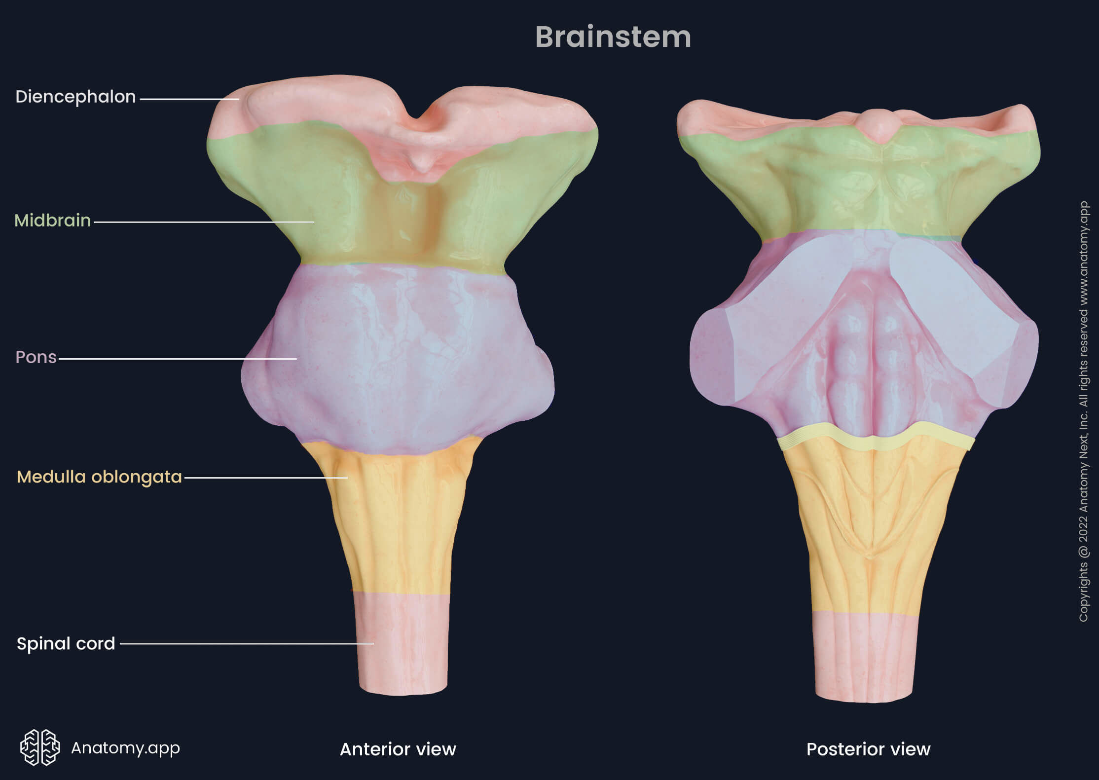
Brainstem
The brainstem (Latin: truncus encephali) is the distal part of the brain situated in the posterior cranial fossa. It is connected with other parts of the central nervous system, including the spinal cord, cerebellum, diencephalon, and cerebral hemispheres. The brainstem is involved in regulating such essential functions as heart rate, blood pressure, and respiration.
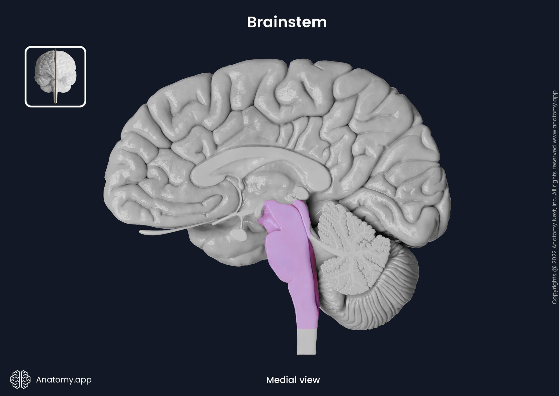
The length of the brainstem is about 7 - 7.5 cm, and it is approximately 3 cm wide. Anteriorly, it faces the clivus of the occipital bone and dorsum sellae of the sphenoid bone. Posteriorly to the brainstem are the following structures: cisterna magna, fourth ventricle and cerebellum. Superiorly, it is continuous with the thalamus of the diencephalon, while inferiorly it continues as the spinal cord. The brainstem is made up of three parts: medulla oblongata, pons, and midbrain.
The brainstem is the place of origin for ten of the twelve cranial nerves (CN). Also, it contains many nuclei (collections of neural cell bodies), including most of the cranial nerve nuclei, and many tracts (neural pathways). An additional network of interconnected neurons called the reticular formation is situated between the tracts and cranial nerve nuclei of the brainstem.
During embryogenesis, the brainstem originates from two of the three primary brain vesicles: mesencephalon and rhombencephalon. Eventually, from the primary vesicles, the secondary brain vesicles develop and give rise to the mature parts of the brain. The mesencephalon becomes the midbrain, and the rhombencephalon differentiates into the metencephalon (pons and cerebellum) and the myelencephalon (medulla oblongata).
Medulla oblongata
The medulla oblongata is the lowest part of the brainstem. Inferiorly it transitions and continues as the spinal cord, while superiorly connects to the pons. Also, it communicates with the cerebellum via the inferior cerebellar peduncles. The upper rear part of the medulla oblongata forms the lower portion of the fourth ventricle.
Besides being a conduit for fibers between the spinal cord and higher brain regions, the medulla oblongata also contains control centers for involuntary functions such as regulation of blood pressure and breathing. Four pairs of cranial nerves emerge on the ventral surface of the medulla oblongata:
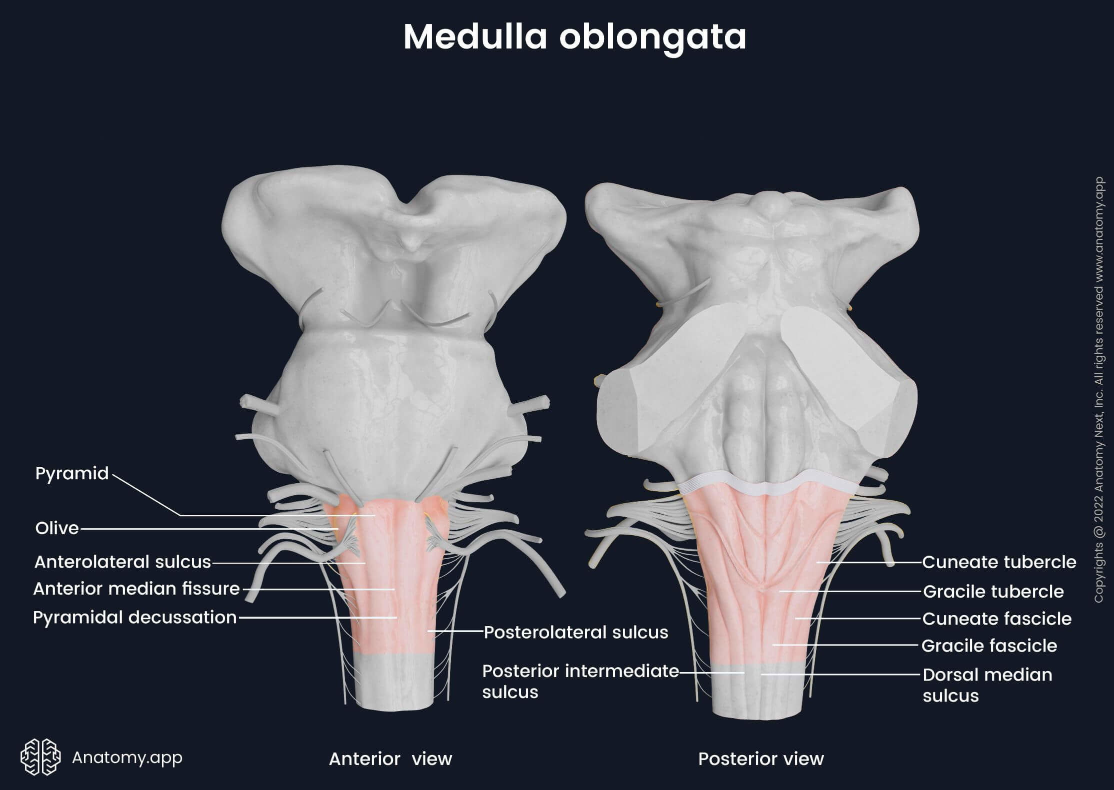
Like other brainstem parts, the medulla oblongata has two external surfaces: ventral and dorsal. Along the ventral surface, five grooves can be distinguished. The anterior median fissure runs between the two eminences called pyramids along the midline of the ventral surface. On each lateral side of the pyramids is a groove called the anterolateral sulcus. It separates the pyramid from the olive - another eminence of the ventral surface. Further laterally to each olive is an additional groove - the posterolateral sulcus.
The pyramids are paired prominences that contain the corticospinal tract fibers. They are located on the ventral surface between the anterior median fissure and the anterolateral sulcus. Near the junction of the medulla oblongata and the spinal cord, the motor fibers of the corticospinal tracts cross in the midline, forming the pyramidal decussation. Superiorly, on the ventral surface of the medulla oblongata is a pair of eminences called the olives, containing the superior and inferior olivary nuclei.
Along the midline of the dorsal surface descends a shallow groove called the dorsal median sulcus (or posterior median sulcus). It continues as the posterior median sulcus of the spinal cord. Lateral to the dorsal median sulcus is a paired eminence known as the gracile fascicle, and a bit more laterally to it is one more eminence - the cuneate fascicle. Superiorly, both fascicles form two prominences: the gracile tubercle medially and the cuneate tubercle laterally. Inferiorly, the posterior intermediate sulcus separates the cuneate and gracile fascicles.
The internal anatomy of the medulla oblongata is very complex, and it contains a multitude of nuclei and tracts. To better understand these structures and their relations, the medulla oblongata is typically examined at three cross-section levels: pyramidal decussation level, sensory decussation level and level of the olives.
Pons
The pons forms the middle part of the brainstem. It is located in the posterior cranial fossa beneath the midbrain, anterior to the cerebellum and above the medulla oblongata. The pons contains a multitude of nuclei and is the site of passage and relay of many neural tracts. It regulates respiration, sleep, equilibrium and other vital functions of the body. Four paired cranial nerves arise from the pons:
- Trigeminal nerve (CN V)
- Abducens nerve (CN VI)
- Facial nerve (CN VII)
- Vestibulocochlear nerve (CN VIII)
The pons can be subdivided into two portions known as the ventral (basilar) and dorsal (pontine tegmentum) parts. Like the medulla oblongata and midbrain, it also has two surfaces - ventral and dorsal. The ventral surface of the pons appears convex and faces the clivus of the occipital bone. A central groove, called the basilar sulcus, runs along the anterior midline of the pons. The basilar artery is located within this sulcus. Laterally, the pons narrows and transitions into the middle cerebellar peduncles connecting the pons with the cerebellum. Inferiorly, a transverse groove called the bulbopontine sulcus separates the pons from the pyramids of the medulla oblongata.
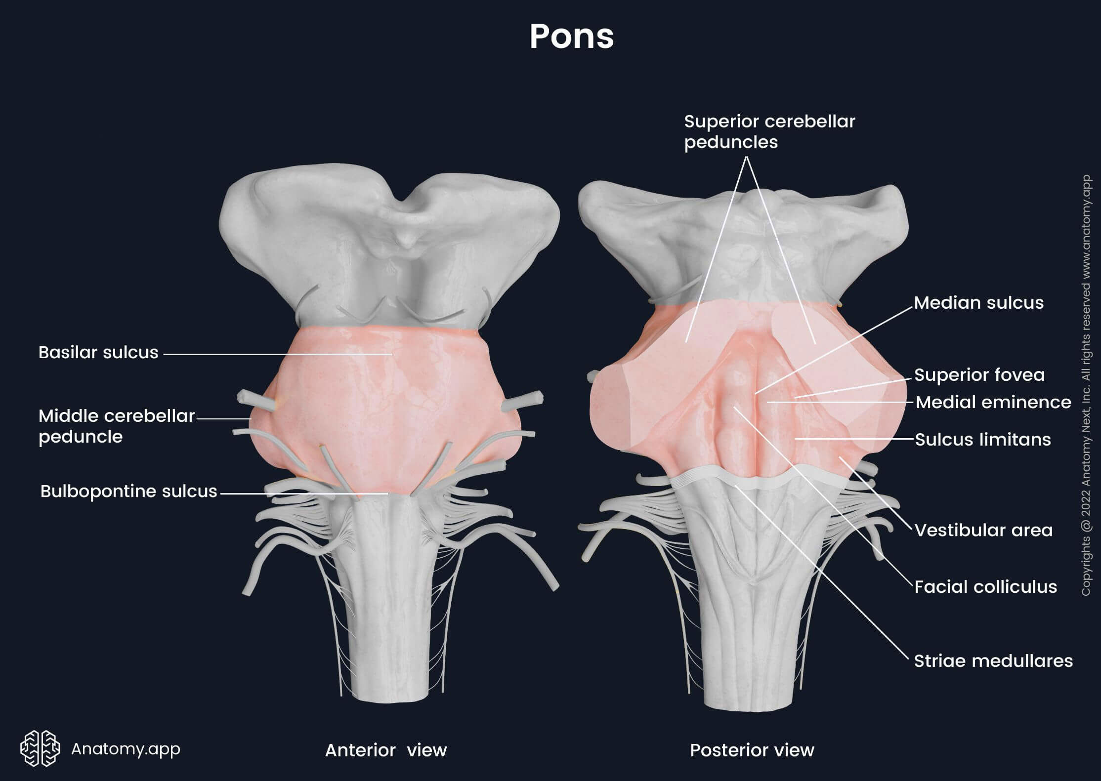
The dorsal surface of the pons is covered by the cerebellum, and it participates in forming the floor of the fourth ventricle. Moreover, the dorsal surface constitutes the upper half of the rhomboid fossa. Superiorly and laterally, it is limited by the superior cerebellar peduncles. The striae medullares - collections of axons originating from the arcuate nucleus - form the inferior border of the dorsal surface and separate the pons from the medulla oblongata. Vertically, the median sulcus runs along the midline of the dorsal pons. It separates a pair of structures called the medial eminences.
The upper aspect of the medial eminence is slightly more noticeable than the lower one, and it is known as the facial colliculus. Another slightly elevated region is seen on each lateral side of the dorsal pons (in each corner of the rhomboid fossa), and it is the vestibular area. Lateral to each medial eminence is the sulcus limitans, which widens at the level of the facial colliculus into a flattened depression - the superior fovea.
The internal anatomy of the pons looks at its deeper-located structures such as the nuclei and tracts, and it is best inspected in transverse cross-sections. The transverse sections can be done at different levels of the pons. Accordingly, the pons is subdivided into three parts: the caudal pons, midpons and rostral pons.
Midbrain
The midbrain, also called the mesencephalon, is the uppermost part of the brainstem. It lies above the pons and below the diencephalon in the posterior cranial fossa. The midbrain is involved in regulating many essential functions, including eye movements, auditory processes, movement planning, motivation, excitation, temperature, and others. It is also the site of origin for two pairs of cranial nerves - the oculomotor (CN III) and trochlear (CN IV) nerves.
The midbrain presents with two surfaces - a ventral and a dorsal one. On the ventral surface paired cerebral peduncles are situated. Both are separated by a deep depression called the interpeduncular fossa. On the dorsal surface of the midbrain four rounded formations known as the colliculi are seen. They include two superior and two inferior colliculi.
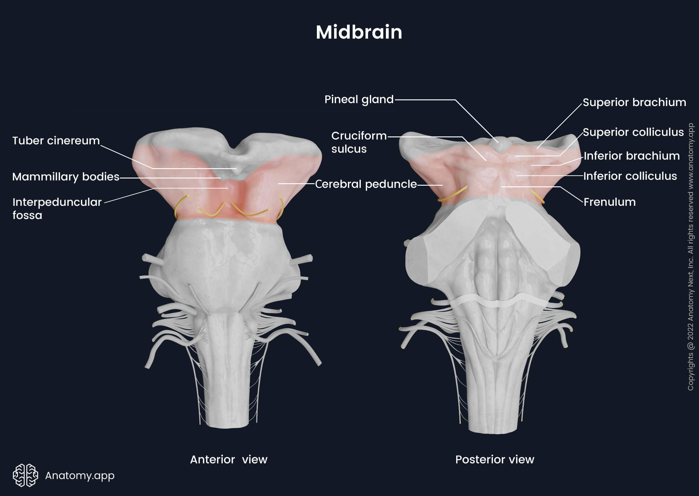
The midbrain has two parts separated by the cerebral aqueduct of Sylvius, and these are the anterior part and posterior part. The internal anatomy of the midbrain can be better understood when looked at from the sagittal and transverse planes. In the sagittal cross-section, the midbrain is further divided into crus cerebri, tegmentum and tectum. The midbrain is the point of origin for the oculomotor (CN III) and trochlear (CN IV) cranial nerves.
Reticular formation
The reticular formation is an interconnected system of nuclei and neurons located within the brainstem. The word “reticular” derives from Latin reticulum, meaning “small net.” The reticular formation extends from the upper segments of the spinal cord to the thalamus. Therefore, it interacts with the medulla oblongata, pons, midbrain and diencephalon. It is the site of integration and relay of vital information from the cerebrum, cerebellum and spinal cord.
There are more than 100 nuclei that form the reticular formation located throughout the brainstem. These nuclei are bilaterally situated on each half of the brainstem. Anatomically, they are subdivided into three groups: lateral, medial, and median (raphe). The nuclei of the reticular formation can be best studied at different brainstem levels in transverse cross-sections.
The reticular formation includes the ascending and descending tracts. The ascending reticular activating system (ARAS) is the major ascending pathway that sends information to the cortex. In contrast, the major descending tract is the reticulospinal tract that sends information to the spinal cord. Overall, the reticular formation is responsible for the modulation of pain, sleep and consciousness cycle (circadian rhythm), arousal state, somatic motor control, habituation, and cardiovascular control.
Rhomboid fossa
The rhomboid fossa (floor of the fourth ventricle) is a shallow and diamond-shaped depression situated on the dorsal aspects of the medulla oblongata and pons. It is so named because of its rhomboid shape. The rhomboid fossa contains four margins and four corners. Two superolateral margins are formed by the superior and inferior cerebellar peduncles. The inferolateral margins of the rhomboid fossa are formed by bands of white matter known as taeniae of the fourth ventricle.
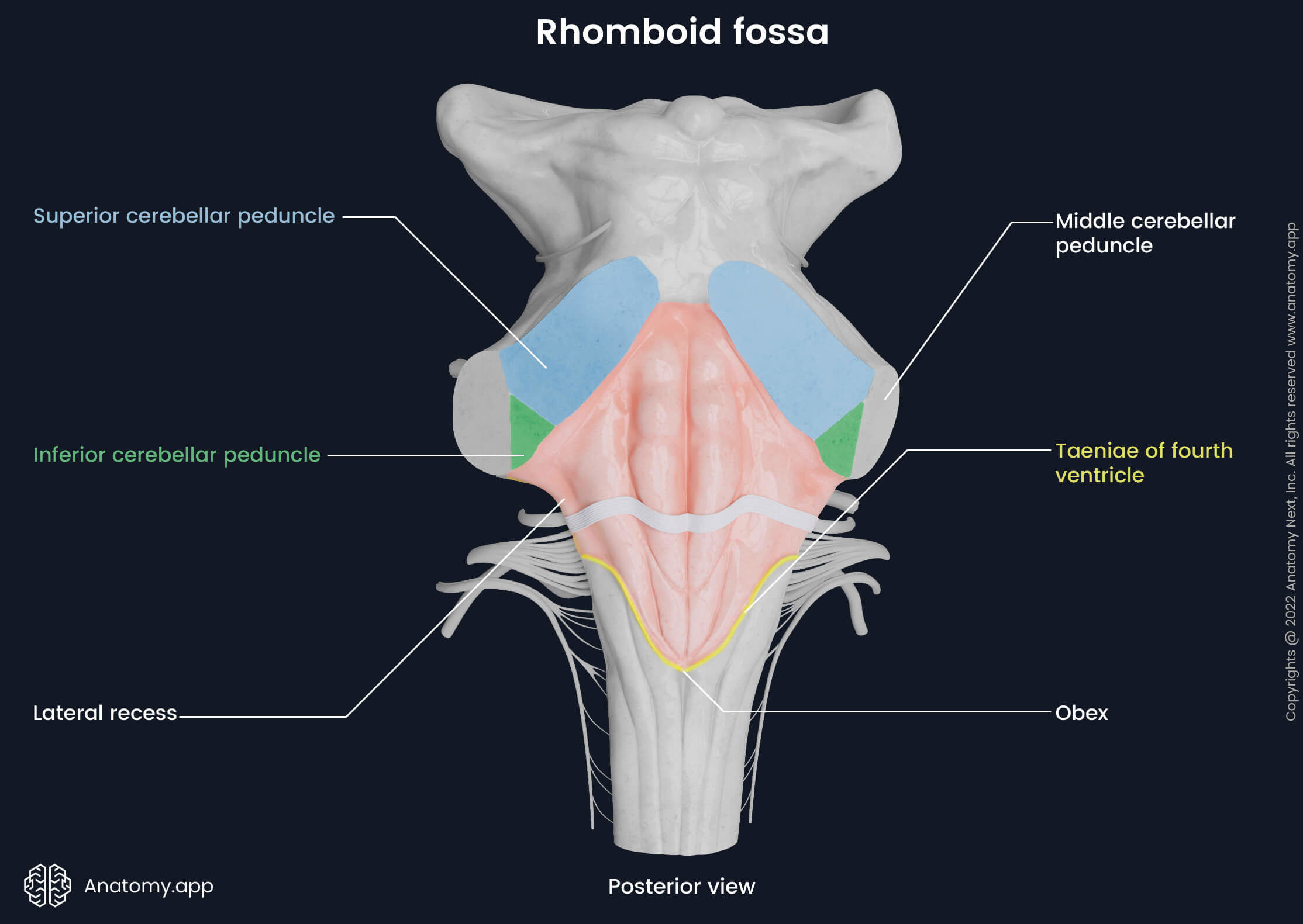
The upper corner of the fossa is continuous with the cerebral aqueduct (aqueduct of Sylvius) connecting the fourth ventricle to the third one. The lower corner contains the most caudal part of the fourth ventricle called the obex, which narrows and connects with the central canal of the spinal cord. The lateral corners of the rhomboid fossa are formed by the lateral recesses - lateral extensions of the inferior cerebellar peduncles. At the same time, lateral recesses are also sites where the medulla and pons merge.
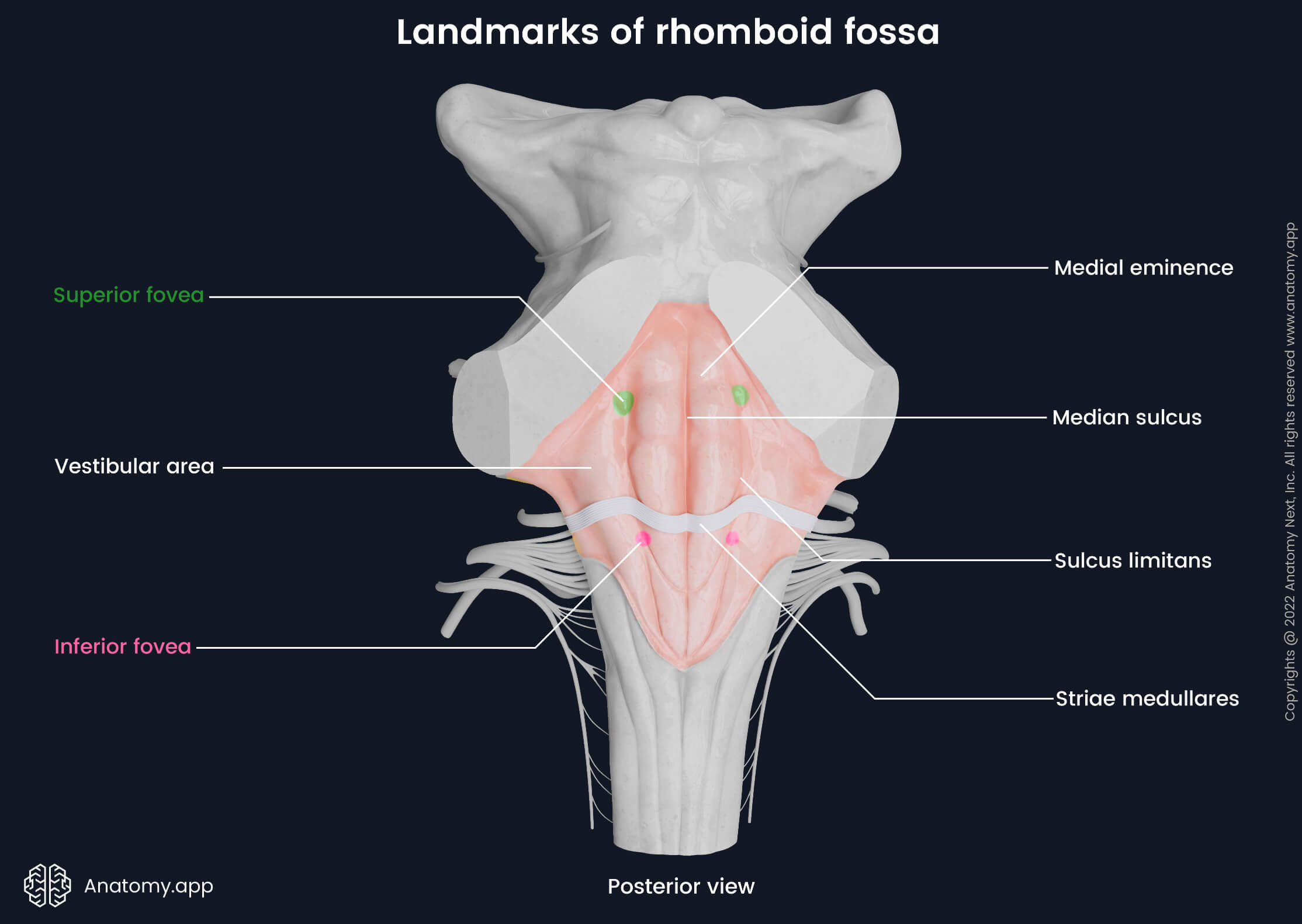
Parts and landmarks of rhomboid fossa
The surface of the rhomboid fossa is not smooth, but it contains several grooves, elevations and depressions. In the midline of the rhomboid fossa from the upper to the lower corner goes a longitudinal groove called the median sulcus. It divides the fossa into the right and left halves.
In transverse directions from both lateral corners to the midline, bundles of fibers known as the striae medullares pass. They divide the fossa into upper and lower triangular-shaped parts - superior triangle and inferior triangle. At the same time, the striae medullares represent a border between the dorsal surfaces of the pons and medulla oblongata.
The superior triangle of the rhomboid fossa is formed by the pons. The inferior triangle - by the medulla oblongata and a small portion of the lower aspect of the dorsal pons. The rhomboid fossa can also be divided into the following three parts:
- Upper part - formed by the superior triangle;
- Intermediate part - represented by the striae medullares;
- Lower part - formed by the inferior triangle.
Parallel to the median sulcus and on each lateral side of it, another slightly convex groove known as the sulcus limitans goes. This sulcus contains two slight depressions at both ends: superior fovea - located above the striae medullares, and inferior fovea - situated below the striae at the lower aspect of the sulcus limitans.
A longitudinal elevation known as the medial eminence is located between the sulcus limitans and median sulcus along the entire length of the rhomboid fossa. The surfaces of the lateral aspects of the rhomboid fossa laterally to the sulcus limitans are a bit elevated. These elevated sites are known as vestibular areas - they house the vestibular nuclei.
Superior triangle of rhomboid fossa
The triangle-shaped part of the rhomboid fossa above the striae medullares is known as the superior triangle. This part of the fossa is formed by the pons.
Just above the striae medullares on the medial eminence is located a swelling of the facial nerve (CN VII) called the facial colliculus. It represents the location of the abducens motor nucleus and internal genu of the facial nerve. Lateral to the facial colliculus is another landmark - the superior fovea.
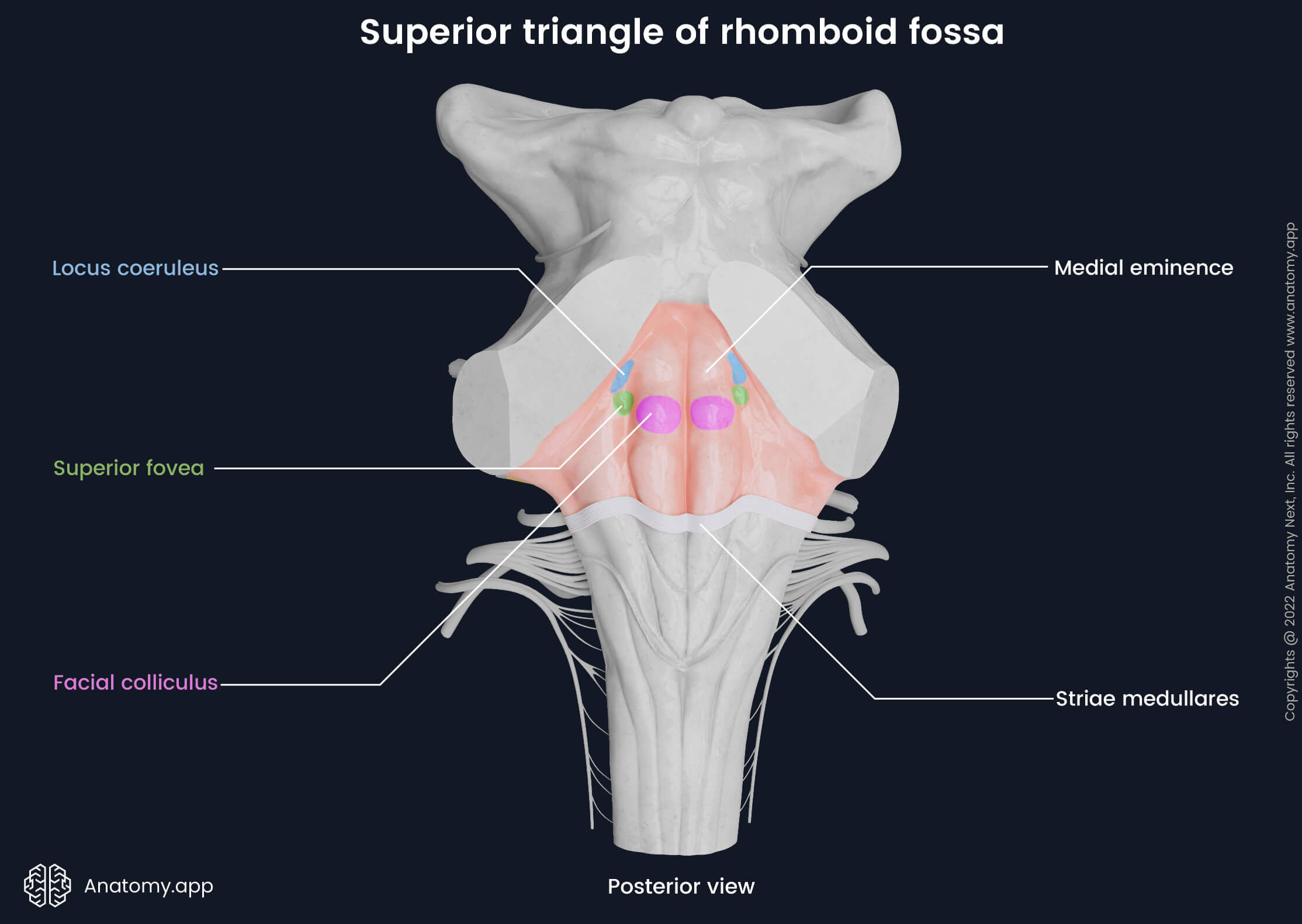
At the uppermost aspect of the sulcus limitans, an area known as the locus coeruleus (blue spot) is situated. It contains a similarly named nucleus. The nucleus coeruleus is the main source of norepinephrine in the brain and therefore has a high concentration of this neurotransmitter which gives a blue appearance to this region.
Inferior triangle of rhomboid fossa
The inferior triangle of the rhomboid fossa is the part of the rhomboid fossa below the striae medullares. It is formed by the medulla oblongata and a small portion of the pons. Inferior to the striae medullares, the fossa presents with two bilateral triangles (trigones):
- Hypoglossal trigone (or triangle) - an elevated and triangular-shaped narrowing of the medial eminence; it houses the hypoglossal nuclei;
- Vagal trigone (or triangle) - located laterally and downward from the hypoglossal triangle; it contains the vagal nuclei.
Laterally to both mentioned triangles is the inferior fovea. Each inferolateral margin of the rhomboid fossa contains narrow and small elevations or ridges called taeniae (also known as taeniae of the fourth ventricle). They extend from the striae medullares to the obex where the taeniae from the left and right sides merge.
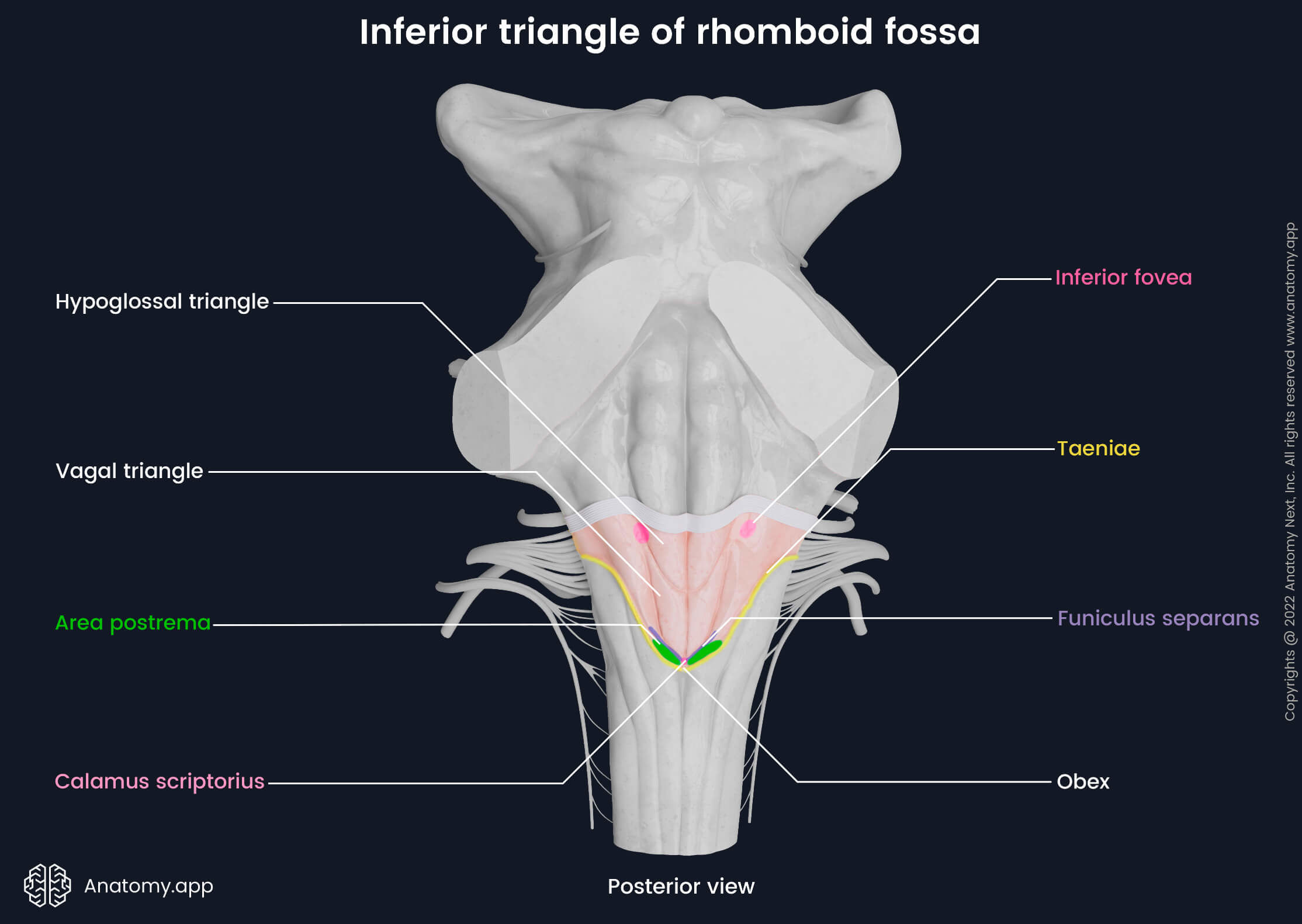
Just before the obex below the vagal trigone, is a small protuberance known as the area postrema. It is thought to be the vomiting control center, which induces vomiting in response to potential toxins. The area postrema is separated from the vagal trigone by a thin ridge named funiculus separans. The lowest part of the rhomboid fossa or its caudal ending is also known as calamus scriptorius.
Cranial nerve nuclei
The brainstem is a site of origin for ten of the twelve cranial nerves (CN), therefore, it contains most of the cranial nerve nuclei. The brainstem houses nuclei of the third through twelfth cranial nerves (CN III - XII). Most of them are located in the rhomboid fossa (CN V - XII); however, some are situated in the midbrain (CN III - IV). All cranial nerve nuclei are paired structures and can be found on either side of the rhomboid fossa and midbrain.
NOTE: The accessory nerve (CN XI) has two motor nuclei. One is located in the rhomboid fossa, and it is known as the ambiguus nucleus. The other is called the spinal accessory nucleus, and it forms an elongated column within the upper cervical spinal cord levels. Although the spinal accessory nucleus is not located in the brainstem, it is included in this article as it extends right below the ambiguus nucleus and some authors consider it to be the spinal continuation of the ambiguus nucleus.
The cranial nerve nuclei can be either sensory, motor or parasympathetic. Nerve fibers of the sensory nuclei participate in the formation of ascending tracts that send sensory impulses from the periphery of the body and lower parts of the brain to the higher regions of it. In contrast, the descending tracts are motor pathways that convey information in the opposite direction - from the higher brain regions to lower areas, ending in the motor nuclei of the cranial nerves or spinal cord. The parasympathetic nuclei are general visceral motor nuclei, and as the motor nuclei, they are components of the descending pathways.
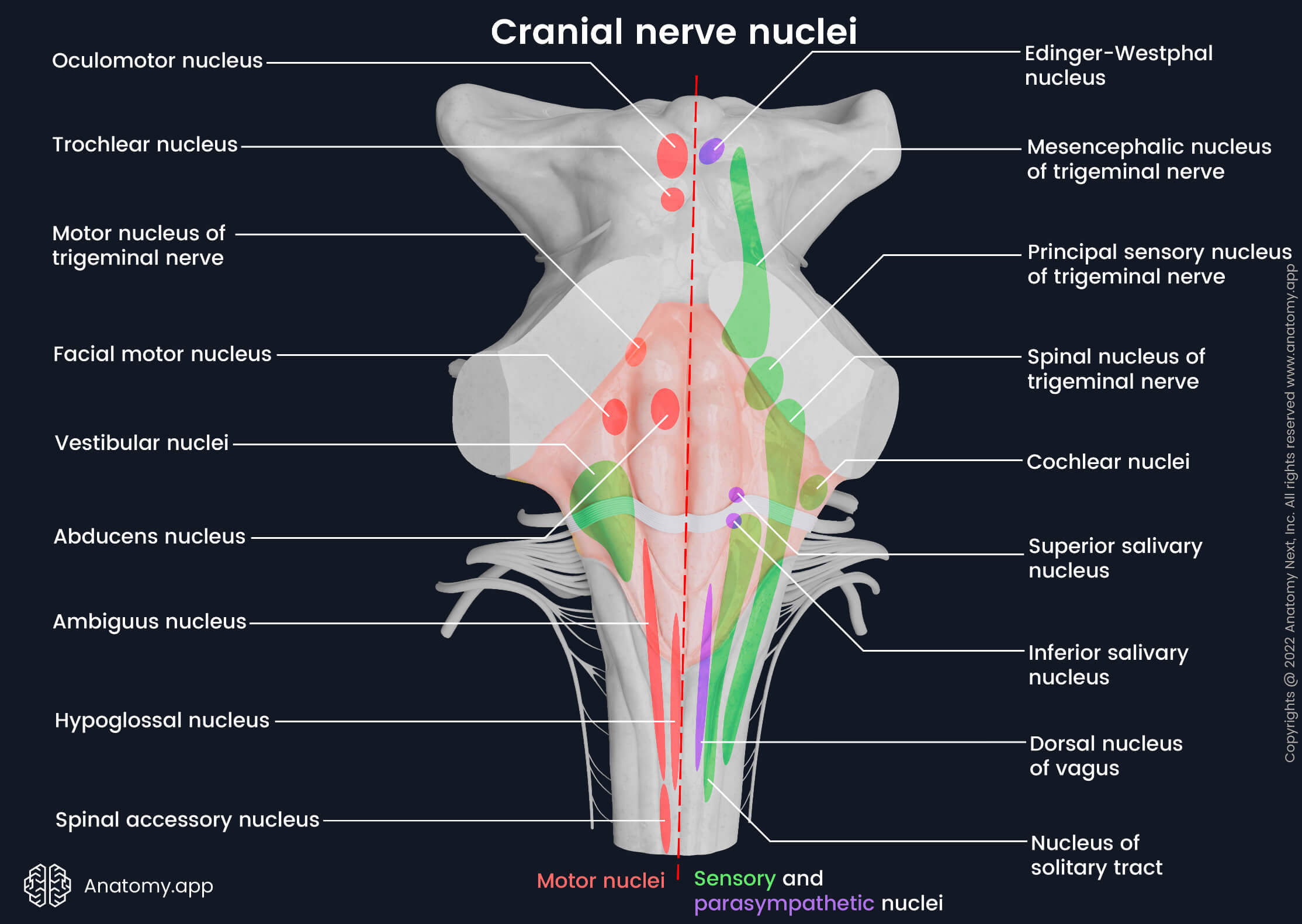
The motor nuclei of the cranial nerves are positioned within the midbrain, rhomboid fossa and spinal cord, and they include the following nuclei:
- Oculomotor nucleus - a general somatic motor nucleus of the oculomotor nerve (CN III); located in the midbrain at the level of the superior colliculus; sends fibers to most of the extraocular muscles - superior rectus, inferior rectus, medial rectus, inferior oblique and levator palpebrae superioris;
- Trochlear nucleus - a general somatic motor nucleus of the trochlear nerve (CN IV); situated in the midbrain at the level of the inferior colliculus; it contains nerve fibers that supply the superior oblique extraocular muscle;
- Motor nucleus of the trigeminal nerve - a special visceral motor nucleus of the trigeminal nerve (CN V); positioned close to the locus coeruleus in the upper aspect of the rhomboid fossa (superior triangle); axons of this nucleus innervate mastication muscles (temporalis, masseter, pterygoids), mylohyoid, anterior belly of the digastric, tensor tympani and tensor veli palatini muscles;
- Abducens nucleus - a general somatic motor nucleus of the abducens nerve (CN VI); located in the superior triangle of the rhomboid fossa near the median sulcus and deep within the facial colliculus; it sends fibers to the lateral rectus extraocular muscle;
- Facial motor nucleus - a special visceral motor nucleus of the facial nerve (CN VII); situated laterally and deeper from the facial colliculus, close to the lateral angle of the fossa at the level of the middle cerebellar peduncle; axons of the nucleus supply muscles of the second embryological branchial arch - facial muscles, some muscles of the neck (stylohyoid and posterior belly of the digastric) and stapedius;
- Ambiguus nucleus - a special visceral motor nucleus of the glossopharyngeal (CN IX), vagus (CN X) and accessory (CN XI) nerves; positioned right below the striae medullares; cells of this nucleus form an elongated column within the medulla oblongata that is situated laterally from the hypoglossal trigone and dorsal nucleus of the vagus nerve; it contains fibers that innervate striated muscles of the soft palate, larynx, pharynx and upper esophagus;
- Spinal accessory nucleus - a general somatic motor nucleus of the accessory nerve (CN XI); neurons of this nucleus form an elongated column that is located in the anterior horn of the spinal cord at the C1 - C5 levels (rarely ends at the C6 - C7 levels); the cell column extends inferior to the ambiguus nucleus; axons of this nucleus form the spinal accessory nerve that innervates trapezius and sternocleidomastoid muscles;
- Hypoglossal nucleus - a general somatic motor nucleus of the hypoglossal nerve (CN XII); a column formed by this nucleus extends downward from the hypoglossal trigone and is situated in the floor of the rhomboid fossa close to the median sulcus and medial to the dorsal nucleus of the vagus nerve; axons of this nucleus supply all the intrinsic and three extrinsic (genioglossus, styloglossus, hyoglossus) tongue muscles and geniohyoid.
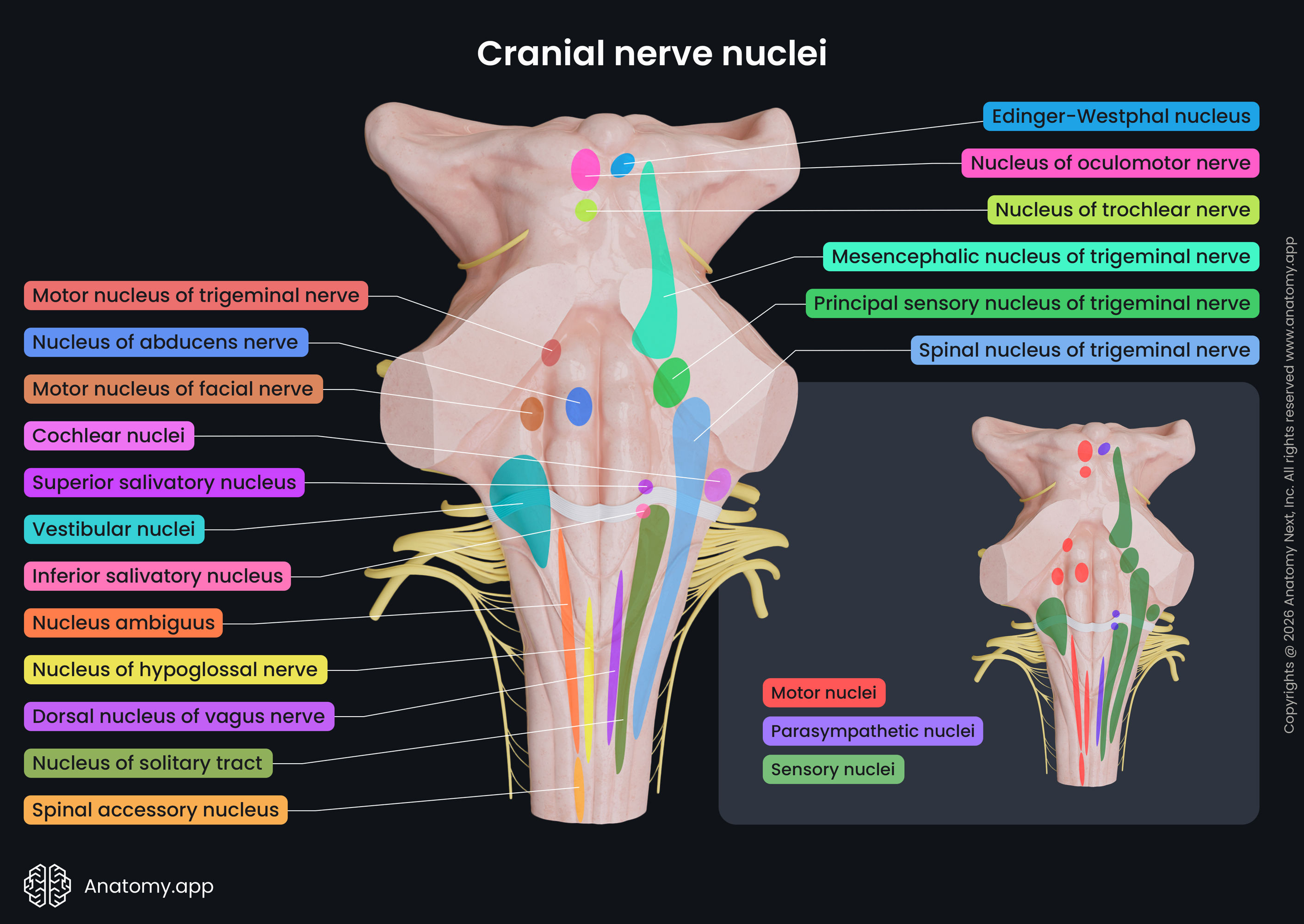
The rhomboid fossa contains the following sensory nuclei:
- Principal (main or chief) sensory nucleus of the trigeminal nerve - a general somatic sensory nucleus of the trigeminal nerve (CN V); located lateral to the motor nucleus of the trigeminal nerve and medial to the middle cerebellar peduncle within the dorsal aspect of the pons; receives information about discriminative and light touch sensation from the face and conscious proprioception from the jaws and temporomandibular joint (proprioception is an ability to sense the position of the body, movements, and actions of various body parts perceived in conscious and unconscious levels);
- Mesencephalic nucleus of the trigeminal nerve - a general somatic sensory nucleus of the trigeminal nerve (CN V); cells of this nucleus form a column that goes upward from the upper aspect of the principal sensory nucleus of the trigeminal nerve and ends in the tegmentum of the midbrain; it contains cell bodies of afferent proprioceptors and is responsible for processing proprioceptive impulses coming from the teeth, mastication muscles, jaws and temporomandibular joint;
- Spinal nucleus of the trigeminal nerve - a general somatic sensory nucleus of the trigeminal nerve (CN V), it also incorporates and relays sensory information from the facial (CN VII), glossopharyngeal (CN IX) and vagus (CN X) nerves; it receives information about pain, deep touch and temperature sensation from the face, external auditory canal, tympanic membrane, external ear, larynx, pharynx, soft palate, meninges and dura of the posterior cranial fossa; cells of this nucleus form a column that goes downward from the caudal end of the principal sensory nucleus; it extends throughout the entire length of the medulla oblongata and ends in the upper two to three cervical segments of the spinal cord; this nucleus has three parts (subnuclei) - pars caudalis, pars interpolaris and pars oralis;
- Nucleus of the solitary tract (nucleus tractus solitarius) - a general and special visceral sensory nucleus of the intermediate (part of the facial nerve (CN VII)), glossopharyngeal (CN IX) and vagus nerves (CN X); positioned below the striae medullares; cells of this nucleus form a column that goes downward along the sides of the hypoglossal and vagal trigones; this nucleus has two parts - the upper is the rostral part with gustatory nuclei containing taste fibers, while the lower is the caudal part with visceral or cardiorespiratory nuclei for reflex control of respiratory and cardiovascular functions; overall, it receives cardiovascular, respiratory, visceral, taste and orotactile sensory information;
- Vestibular nuclei - special somatic sensory nuclei of the vestibular part of the vestibulocochlear nerve (CN VIII); located in the vestibular area more in the medulla oblongata; they relay information coming from the semicircular canals, utricle and saccule of the inner ear about head position, motion, spatial orientation, balance and posture to the vestibular areas of the brain; the vestibular nuclei complex contains four nuclei - lateral, medial, superior and inferior vestibular nuclei;
- Cochlear nuclei - special somatic sensory nuclei of the cochlear part of the vestibulocochlear nerve (CN VIII); they are also situated in the vestibular area, but more laterally - closer to the lateral angles - and more in the pons; they are part of the auditory pathway and transmit auditory information; they integrate, process and relay auditory input from the cochlea of the inner ear to the auditory areas of the brain; the cochlear nuclei complex is composed of two nuclei - posterior (dorsal) cochlear nucleus and anterior (ventral) cochlear nucleus.
From the midbrain to the lower parts of the fossa, the parasympathetic nuclei of the cranial nerves are located in the following order:
- Edinger-Westphal nucleus (accessory oculomotor nucleus) - a general visceral motor (parasympathetic) nucleus of the oculomotor nerve (CN III); situated in the midbrain superior to the superior colliculus and almost at the midline; sends fibers to the sphincter pupillae (iris sphincter muscle) and ciliary muscles;
- Superior salivatory nucleus - a general visceral motor (parasympathetic) nucleus of the intermediate nerve (part of the facial nerve (CN VII)); positioned above the striae medullares and ambiguus nucleus and medial to the nucleus of the solitary tract; axons of this nucleus supply the submandibular, sublingual and lacrimal glands;
- Inferior salivatory nucleus - a general visceral motor (parasympathetic) nucleus of the glossopharyngeal nerve (CN IX); located right below the striae medullares and superior salivary nucleus; its fibers innervate the parotid gland;
- Dorsal (motor) nucleus of the vagus nerve - a general visceral motor (parasympathetic) nucleus of the vagus nerve (CN X); this nucleus extends downward from the vagal trigone and presents as a column that is located medial to the ambiguus nucleus; it sends fibers to the internal organs of the thorax and abdomen, such as the heart, lungs, bronchi, esophagus, stomach, liver, pancreas, spleen, small intestines and proximal colon.
Knowing how these nuclei are arranged makes it easier to understand what kind of fibers they contain. Motor and parasympathetic nuclei are situated in the medial aspect and close to the midline of the fossa or midbrain, while more lateral are the sensory nuclei.
References:
- Crossman, A. R., & Neary, D. (2019). Neuroanatomy: An Illustrated Colour Text (6th ed.). Elsevier.
- Vanderah, T. W., & Gould, D. J. (2020). Nolte’s The Human Brain (8th ed.). Elsevier.
- Mangold, S. A., & Das, J. M. (2021). Neuroanatomy, Reticular Formation. NCBI. https://www.ncbi.nlm.nih.gov/books/NBK556102/
Anatomy.app
Contact information
- For questions regarding business inquiries. Please contact:
- info@anatomy.app