- Anatomical terminology
- Skeletal system
- Joints
- Muscles
- Heart
- Blood vessels
- Lymphatic system
- Nervous system
- Respiratory system
- Digestive system
- Urinary system
- Female reproductive system
- Male reproductive system
- Endocrine glands
- Eye
- Ear
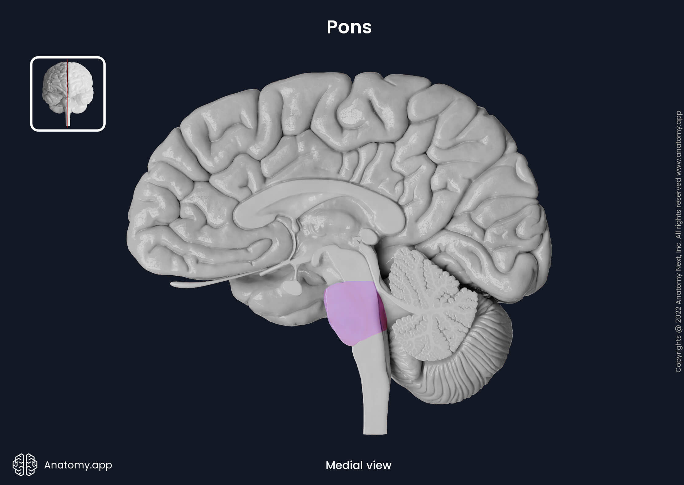
Pons
The pons (Latin: pons) forms the middle portion of the brainstem and is a part of the metencephalon. The word pons comes from Latin, meaning "bridge." It is situated beneath the midbrain and above the medulla oblongata in the posterior cranial fossa. Hence, the pons serves as a bridge, connecting the lower-lying medulla oblongata to the cerebellum and midbrain.
Like other brainstem structures, the pons contains many nuclei and is a passage and relay site of numerous neural tracts. The pons regulates respiration, sleep cycle, balance and other vital functions of the body. Moreover, the pons sends signals to the cerebellum. It also allows communication between the right and left hemispheres of the cerebellum.
Anatomically, the pons is divided into external and internal structures. The external anatomy includes the superficial aspects and features of the pons. At the same time, the pons may be divided into two parts: ventral (basilar) and dorsal (pontine tegmentum). The exterior of the ventral part is known as the ventral surface, while the exterior of the dorsal part is the dorsal surface.
Four cranial nerves arise from the pons. On the ventral surface of the pons, at the border with the middle cerebellar peduncles, the trigeminal nerve (CN V) emerges. Lower, at the border between the pons and medulla oblongata, three more cranial nerves appear: abducens (CN VI), facial (CN VII), and vestibulocochlear (CN VIII) nerves. The nuclei of these cranial nerves are found in the dorsal part of the pons.
Several neural pathways pass through the pons, such as the corticospinal, corticobulbar and spinothalamic tracts. The pons also contains some nuclei (collections of neural cell bodies) and other important structures internally. The ventral part of the pons contains the pontine nuclei. Tracts originating from the pontine nuclei combine and form the cerebellar peduncles. The dorsal part of the pons includes structures of the reticular formation.
Pons external anatomy
The pons is the middle part of the brainstem and is approximately 2.5 cm long. Anteriorly, it rests against the clivus of the occipital bone, and posteriorly it is covered by the cerebellum. The pons is separated from the medulla oblongata by the inferior pontine sulcus that marks the pontomedullary junction. Superiorly, the superior pontine sulcus separates the pons from the midbrain. The pons has two surfaces: ventral and dorsal.
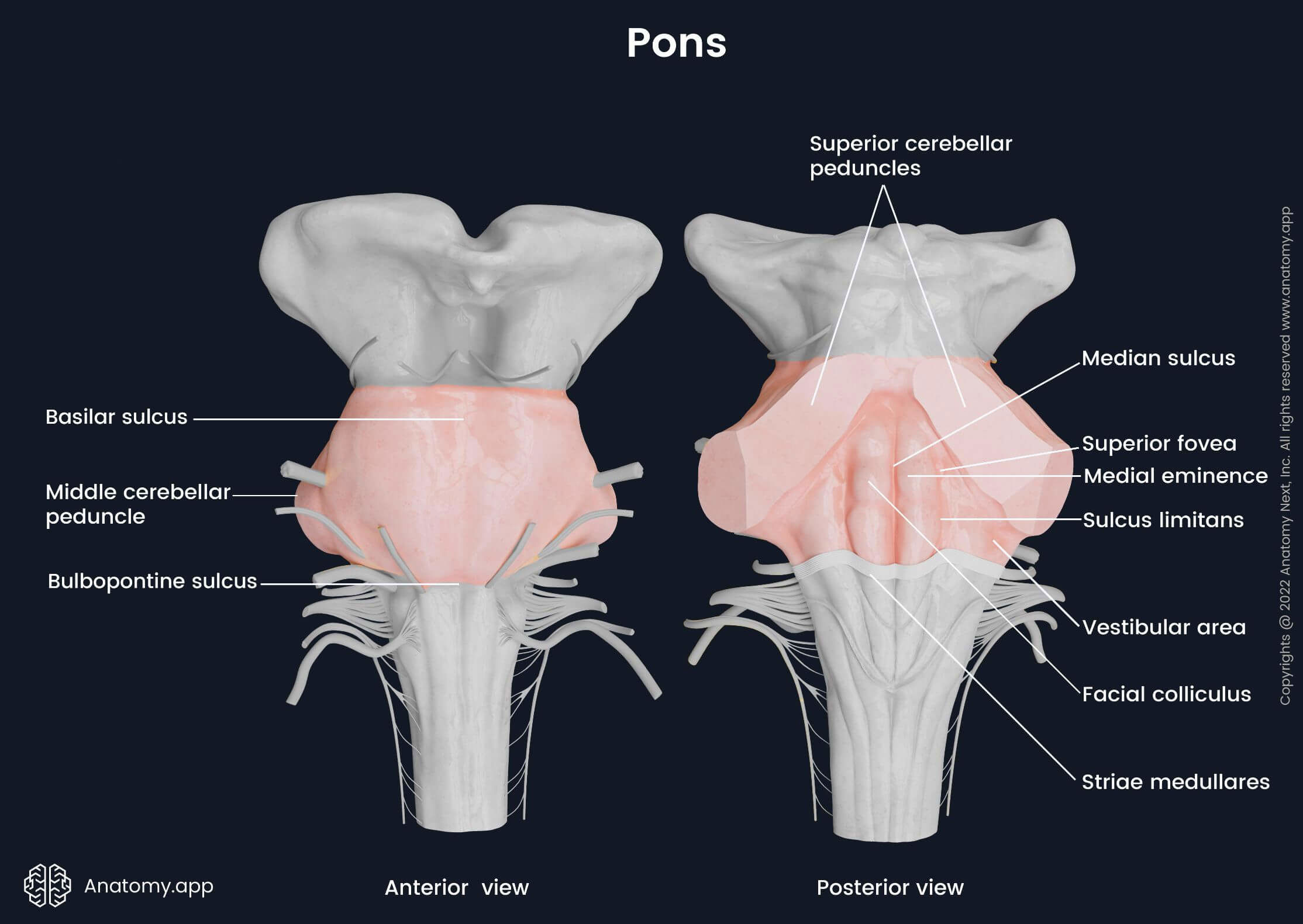
Ventral surface of pons
The ventral surface of the pons is the outer aspect of its ventral (basilar) part. It appears convex and faces the clivus of the occipital bone. In the inferior direction, the ventral part of the pons becomes slightly narrower until it reaches the pontomedullary junction. Laterally, the pons also narrows and transitions into the middle cerebellar peduncles - paired structures containing nerve tracts that connect the pons to the posteriorly lying cerebellum.
On the anterior surface of the ventral part, horizontal lines known as the transverse pontine fibers can be seen. A central groove, called the basilar sulcus, runs along the midline of the pons. Anteriorly is the pontine (prepontine) cistern - an unpaired subarachnoid space located between the ventral surface of the pons and the clivus. The basilar artery ascends in the pontine cistern along the basilar sulcus.
On each lateral side of the ventral part of the pons is an additional cistern - the cerebellopontine angle cistern. This cistern is a paired subarachnoid space formed between the anterolateral aspect of the cerebellum and the lateral side of the pons. The cerebellopontine angle cisterns contain the trigeminal (CN V), facial (CN VII) and vestibulocochlear (CN VIII) nerves, superior cerebellar and anterior inferior cerebellar arteries, and numerous veins.
Four pairs of cranial nerves arise from the ventral surface of the pons. The trigeminal nerve (CN V) emerges between the pons and the middle cerebellar peduncles. The three other cranial nerves arise in the pontomedullary junction between the lower border of the pons and the superior border of the medulla oblongata.
The abducens nerve (CN VI) is the most medially positioned of all four cranial nerves emerging from the ventral surface of the pons. On each side, the nerve exits on the bulbopontine sulcus between the pyramid of the medulla oblongata and the pons. The facial (CN VII) and vestibulocochlear (CN VIII) nerves emerge from the area called the cerebellopontine angle that is located on each side between the olive of the medulla oblongata and middle cerebellar peduncle.
Dorsal surface of pons
The dorsal surface is the posterior aspect of the pons, and the cerebellum covers it. Therefore, it can only be seen when the cerebellum is removed. This surface forms the upper part of the fourth ventricle's floor and also constitutes the upper half of the rhomboid fossa. The dorsal surface is bounded laterally by the superior cerebellar peduncles in the upper part and the inferior cerebellar peduncles in the lower part.
If the cerebellum is resected, the dorsal surface of the pons reveals prominent peduncles, called the middle cerebellar peduncles. They connect the pons to the cerebellum and contain pontocerebellar fibers. The middle peduncles are the largest of all three cerebellar peduncles. They house notable amounts of afferent fibers and send information from the pons to the contralateral side of the cerebellum.
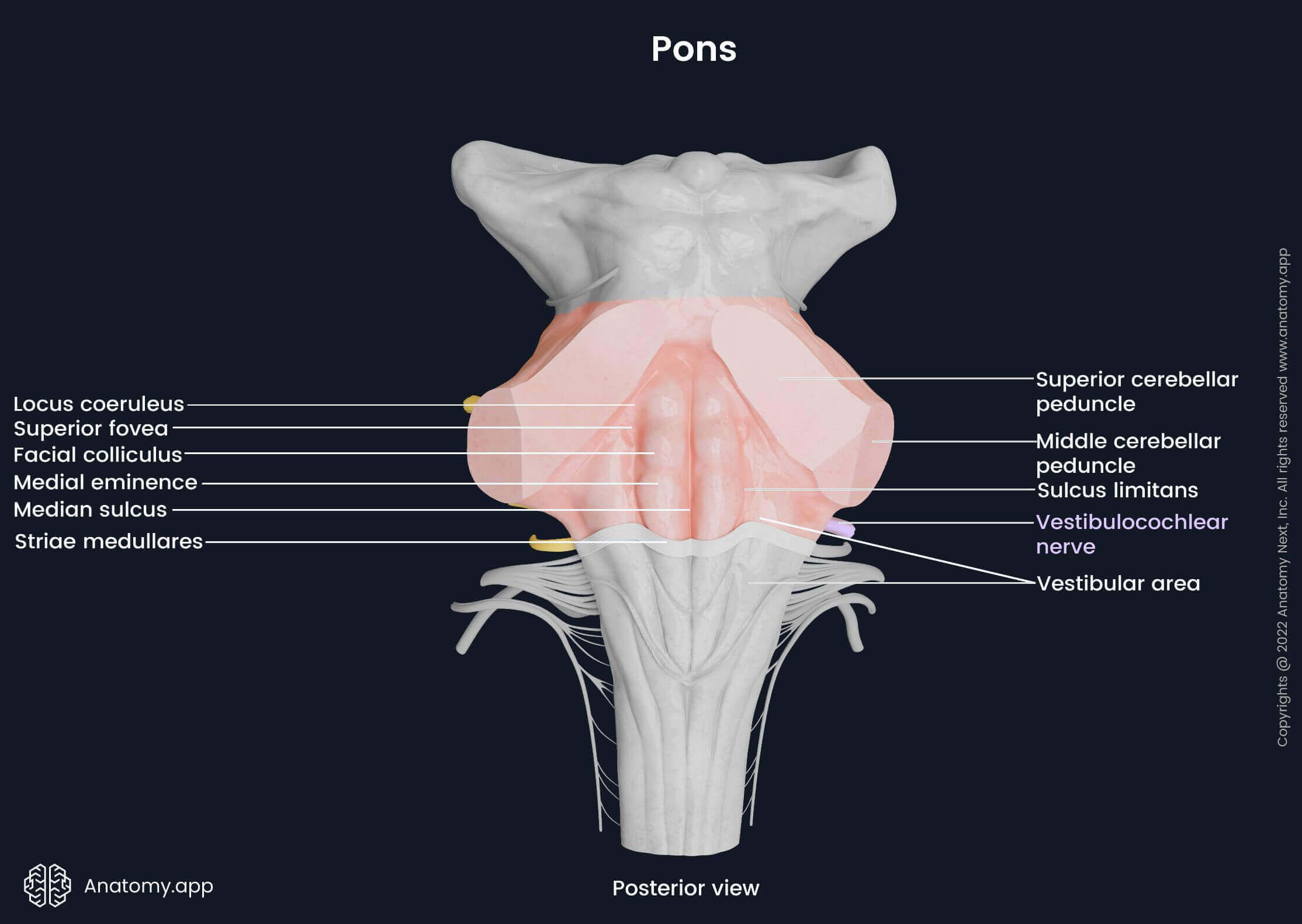
The dorsal surface of the pons forms the upper half of the rhomboid fossa, namely, the superior triangle. Superiorly and laterally, it is limited by the superior cerebellar peduncles. Thin transparent lamina (containing white matter) between the superior cerebellar peduncles known as the superior medullary velum forms the upper border of the dorsal surface, and the striae medullares - the inferior. The striae medullares are collections of axons originating from the arcuate nucleus.
Vertically, the median sulcus runs along the midline of the dorsal pons. It separates a pair of structures called the medial eminences. Lateral to both medial eminences is the sulcus limitans, which widens at the level of the facial colliculus into a flattened depression - the superior fovea. Above it is the bilaterally located locus coeruleus, which contains the nucleus coeruleus.
The upper aspect of the medial eminence is slightly more noticeable than the lower one, and it is known as the facial colliculus. The facial colliculus is formed by the internal genu of the facial nerve motor fibers and the abducens nerve nucleus. The fibers of the facial nerve posteriorly wrap around the abducens nerve nucleus. Another slightly elevated region is seen on both lower lateral sides of the dorsal pons (in each corner of the rhomboid fossa) - the vestibular area.
Pons internal anatomy
The internal anatomy of the pons looks at its deeper-located structures. The pons can be divided into the ventral (basilar) and dorsal (pontine tegmentum) parts. Moreover, it can be looked at in the transverse sections. The transverse sections can be done at different levels of the pons. Accordingly, it is then subdivided into three parts: the caudal pons, midpons, and rostral pons. This cross-section allows us to better describe and understand the structures seen in the pons and the level at which they are situated.
The pons is made up of white and gray matter. Gray matter consists mostly of nuclei, which are mainly situated in the dorsal part of the pons and are billateral. The gray matter structures include the following:
- Pontine nuclei
- Trapezoid body nuclei
- Gray matter of reticular formation with the caudal pontine reticular nucleus
- Nuclei of the trigeminal nerve (CN V)
- Nucleus of the abducens nerve (CN VI)
- Nuclei of the facial nerve (CN VII)
- Nuclei of the vestibulocochlear nerve (CN VIII)
The white matter mostly consists of axons - extensions of neurons. Collections of axons create pathways or tracts. The white matter of pons contain both ascending and descending tracts. These tracts are listed below.
- Ascending tracts:
- Medial lemniscus
- Spinothalamic fibers (spinal lemniscus)
- Anterior spinocerebellar tract
- Trigeminal lemniscus
- Trapezoid body
- Lateral lemniscus
- Descending tracts:
- Corticospinal tract
- Corticobulbar tract
- Cortico-pontine tract
- Pontocerebellar tract
- Tectospinal tract
- Rubrospinal tract
- Reticulospinal tract
- Medial longitudinal fasciculus
The ventral part is the anterior portion of the pons, also known as the ventral pons. If looked at from the sagittal plane, it appears oval-shaped. The ventral part of the pons is an extension of the midbrain's crus cerebri (superior) and medulla oblongata's pyramids (inferior). The ventral pons is comprised of the pontine nuclei and several descending tracts.
The dorsal pons, also known as the tegmentum of the pons, is the dorsal or posterior portion of the pons. It is a continuation of the midbrain's tegmentum (superior) and the tegmentum of the medulla oblongata (inferior). The dorsal pons contains the pontine reticular formation, most nuclei of the pons (including the nuclei of CN V, CN VI, CN VII, CN VIII), and numerous ascending and descending tracts.
Ventral pons
Pontine nuclei
The pontine nuclei are a group of nuclei located in the ventral part of the pons. The nuclei receive information from the primary motor, premotor and somatosensory areas of the cerebral cortex and adjacent parts of the parietal lobe, as well as from the limbic, visual and association areas via the corticopontine fibers that terminate in the pontine nuclei. Further, from the pontine nuclei, transverse pontine (pontocerebellar) fibers arise and cross the midline of the pons, forming the middle cerebellar peduncles. Thus, the pontocerebellar fibers advance to the contralateral cerebellum. These nuclei and their associated fibers are involved in motor function and movement coordination.
Descending tracts of ventral pons
The descending tracts in the pons are neural pathways that pass downward from the higher neural regions, such as the cerebral cortex, to the lower-lying parts, including the cerebellum and spinal cord. The tracts descending through the ventral part of the pons are the following:
- Corticospinal and corticobulbar fibers of the pyramidal tract
- Cortico-cerebellar system composed of:
- Corticopontine fibers
- Pontocerebellar fibers
The corticospinal fibers connect the cerebral cortex to the spinal cord. They regulate voluntary movements of the limbs and trunk via the interneurons and lower motor neurons of the spinal cord. The corticobulbar (corticonuclear) fibers also originate from the cerebral cortex and terminate in the pyramids of the medulla oblongata. A part of these fibers engage with the cranial nerve motor nuclei and, through them, regulate voluntary movements of the face, head and neck.
The corticopontine fibers descend from the cerebral cortex to the pontine nuclei. The continuing pontocerebellar fibers originate from the pontine nuclei and project through the contralateral middle cerebellar peduncles to the opposite side of the cerebellum. Both fiber groups act together as the cortico-cerebellar system. They ensure communication between the cerebrum and the cerebellum and coordinate movements of the whole body.
Dorsal pons
The dorsal pons, also known as the pontine tegmentum, contains many ascending and descending tracts and nuclei. Most of the nuclei are cranial nerve nuclei of the trigeminal (CN V), abducens (CN VI), facial (CN VII), and vestibulocochlear nerves (CN VIII). However, there are a few exemptions, namely, the nuclei of the trapezoid body and the caudal pontine reticular nucleus.
The nuclei of the trapezoid body are situated in the caudal pons between the fibers of the trapezoid body that is located between the pontine nuclei and the medial lemniscus. They are a part of the auditory pathway. The caudal pontine reticular nucleus, as the name suggests, is located in the caudal pons posterior to the trapezoid body. It is a part of the reticular formation, ensuring the acoustic startle responses, muscular activities produced as a response to a sudden loud sound.
Cranial nerve nuclei
Located in the dorsal part of the pons are numerous cranial nerve nuclei, which are paired and thus found on both sides of the midline. They are either sensory or motor nuclei. All nuclei located in the dorsal pons are part of either one of the following cranial nerves: the trigeminal nerve (CN V), the abducens nerve (CN VI), the facial nerve (CN VII) or the vestibulocochlear nerve (CN VIII). The cranial nerve nuclei of the dorsal pons include the following:
- Trigeminal nerve nuclei:
- Abducens nerve nucleus
- Facial nerve nuclei:
- Vestibulocochlear nerve nuclei:
The trigeminal nerve (CN V) nuclei are the largest group extending throughout the entire length of the brainstem. There are two nuclei of the trigeminal nerve located only in the pons. And two other stretch from the pons to the upper or lower parts of the brainstem. The mesencephalic nucleus extends from the pons into the midbrain. The spinal nucleus extends from the pons into the medulla oblongata and upper two to three cervical segments of the spinal cord. Both are direct extensions of the principal sensory nucleus located in the pons.
The trigeminal nerve nuclei located within the dorsal part of the pons include the principal sensory nucleus of the trigeminal nerve and the motor nucleus of the trigeminal nerve. The principal sensory nucleus of the trigeminal nerve, also called the 'chief' or 'main' sensory nucleus of the trigeminal nerve, is situated on both sides of the pons, lateral to the motor nucleus of the trigeminal nerve.
The principal sensory nucleus receives afferent information about touch, proprioceptive impulses and pressure from the face and other head regions. This information is further sent upwards to the thalamus. The motor nucleus of the trigeminal nerve receives motor input from the motor cortex of the cerebrum, and its efferent fibers further innervate the muscles of mastication, mylohyoid, tensor tympani, tensor palati muscles and the anterior belly of the digastric muscle.
The abducens nerve nucleus is a paired motor nucleus responsible for the conjugate horizontal eye movements (simultaneous movements of both eyes in the same direction - horizontal, in this case). The nucleus is located in the caudal pons beneath the respective facial colliculus in the most posterior portion of the dorsal pons. The abducens nerve nucleus consists of motor, internuclear and interneurons. The efferent axons arising from this nucleus form the abducens nerve (CN VI) and innervate the lateral and medial recti muscles of the eyes.
The facial motor and superior salivatory nuclei are both nuclei of the facial nerve (CN VII) located in the pons. The facial motor nucleus is located in the caudal part of the dorsal pons, anterior and lateral to the abducens nucleus. It innervates the muscles responsible for facial expression, stapedius, stylohyoid, platysma, auricular muscles and the posterior belly of the digastric muscle.
The superior salivatory nucleus is bilaterally located within the dorsal part of the caudal pons, superior to the pontomedullary junction. The superior salivatory nucleus is a parasympathetic nucleus. Nerve fibers that originate from it travel over branches of the facial nerve to the pterygopalatine (greater petrosal nerve) and submandibular ganglia (chorda tympani). The chorda tympani provides general visceral efferent fibers that further innervate the submandibular and sublingual salivary glands and regulate their secretion, while the greater petrosal nerve provides supply to the lacrimal and nasal glands.
The vestibular and cochlear nuclei receive sensory input from the vestibulocochlear nerve (CN VIII). There are ventral and dorsal cochlear nuclei and a group of four vestibular nuclei. The nuclei are paired and located bilaterally in the posterior part of the caudal pons and the medulla oblongata.
There are four paired vestibular nuclei: medial vestibular nucleus, lateral vestibular nucleus, inferior vestibular nucleus and superior vestibular nucleus. The medial, lateral and inferior vestibular nuclei are located in the medulla oblongata. In contrast, the superior vestibular nucleus is situated in the pons.
In the pons, the vestibular nuclei are located more medially than the cochlear nuclei. In the cochlear nuclei, the cochlear nerve fibers synapse. Axons mostly from the ventral cochlear nuclei form horizontal fibers called the trapezoid body. In the vestibular nuclei, the vestibular axons synapse with the vestibular nuclei.
The cochlear nuclei contribute to the cochlear nuclear complex and are part of the auditory pathway. They integrate, process and relay auditory input from the cochlea to further auditory areas of the brain. In turn, the vestibular nuclei maintain head position, motion, spatial orientation, equilibrium, posture, and clear vision with movement.
Ascending tracts of dorsal pons
There are multiple ascending pathways in the white matter of the dorsal pons. They are usually paired and symmetrically located on both lateral sides of the midline. The ascending tracts passing through the dorsal part of the pons include the following:
- Medial lemniscus
- Trigeminal lemniscus
- Spinothalamic fibers (spinal lemniscus)
- Lateral lemniscus
- Trapezoid body
- Anterior spinocerebellar tract
The medial, spinal, trigeminal and lateral lemnisci are bilaterally situated pathways in the anterior portion of the dorsal part of the pons. The four lemnisci are located next to one another in a chain-like formation. The medial lemniscus is located near the midline of the pons, the middle portion is medially formed by the trigeminal lemniscus and laterally by the spinal lemniscus, and posterolaterally is the lateral lemniscus.
The medial lemniscus is a second-order neuron of the dorsal column - medial lemniscus pathway. The dorsal column-medial lemniscus tract transports sensory information from the spinal cord to the medulla oblongata and further from the medulla to the thalamus and cerebral cortex. The medial lemniscus starts from the nucleus gracilis and nucleus cuneatus in the medulla oblongata. It carries sensory information about vibration, fine touch, pressure and proprioception to the thalamus and postcentral gyrus.
Laterally to the medial lemniscus is the trigeminal lemniscus, also called the trigeminothalamic tract. It is formed by the axons of the trigeminal nerve's (CN V) sensory nuclei. The trigeminal lemniscus is composed of the ventral and dorsal trigeminal lemnisci. It transmits pain, temperature, and deep sensations from the facial regions to the contralateral ventral posteromedial nucleus of the thalamus. Fibers decussate in the pons.
The spinal lemniscus is made of the anterior and lateral spinothalamic tracts. It conveys sensory information about crude pain, touch, pressure, and temperature. The lateral lemniscus is the most posterolaterally situated pathway of all lemnisci. It originates from the cochlear nucleus, passes to the superior olivary nucleus and advances to the contralateral inferior colliculus of the midbrain. The lateral lemniscus is a part of the ascending auditory pathway.
The trapezoid body is a part of the auditory pathway. It is mostly composed of axons of the ventral cochlear nuclei. A portion of the fibers decussate and continue on the contralateral side. These axons ascend, creating the lateral lemniscus. The anterior spinocerebellar tract transmits unconscious proprioceptive information from the lower limbs to the cerebellum via the superior cerebellar peduncles.
Descending tracts of dorsal pons
The descending tracts found in the white matter of the dorsal pons are as follows:
- Tectospinal tract
- Medial longitudinal fasciculus
- Rubrospinal tract
- Medial (pontine) reticulospinal tract
The tectospinal tract is a motor descending pathway and part of the extrapyramidal system. It originates from the superior colliculus of the midbrain and descends in the posterior portion of the dorsal pons, anterior to the medial longitudinal fascicle. The tract transmits impulses that coordinate reflector head and eye movements as a protective reaction to unexpected visual or auditory stimuli. This pathway connects the midbrain with the upper cervical spinal cord segments.
The medial longitudinal fasciculus is located posterior to the tectospinal tract in the posterior portion of the dorsal pons. The fasciculus connects the motor nuclei of the cranial nerves CN III, CN IV, CN VI, and CN VIII with each other. The medial longitudinal fasciculus descends as the medial vestibulospinal tract from the medial vestibular nuclei of the medulla oblongata to the motor nuclei in the spinal cord's cervical and upper thoracic segments. The medial longitudinal fasciculus coordinates eye movements and therewith associated head and neck movements.
The bilateral rubrospinal tract is located centrally close to the midline of the pons and posterolateral to the medial lemniscus. It originates from the red nucleus in the midbrain and further decussates and descends. The rubrospinal tract terminates in the spinal cord. The pathway connects the red nucleus with the motor nuclei of the spinal cord. It controls the muscle tone of the upper limb flexors.
The medial (pontine) reticulospinal tract originates from the medial pontine reticular formations: the oral and caudal pontine reticular nuclei. This tract extends to all levels of the spinal cord and communicates with the spinal reticular formations and motor nuclei of the spinal cord. The pontine reticulospinal pathway regulates and enables postural control and proximal stability, inhibits flexors of the limbs and stimulates extensors.
Cross-section of pons
When looked at in the transverse cross-section, the pons can be divided into three levels: the caudal pons, midpons, and rostral pons. These divisions allow us to describe better and understand the structures seen in the pons and their location. All mentioned parts can be further subdivided into the ventral (basilar) and dorsal (tegmental) segments.
Apart from a few minor changes in location, many structures, especially tracts, are present at all three levels. These are the medial, spinal, trigeminal and lateral lemnisci, the medial longitudinal fasciculus, rubrospinal and tectospinal tracts, corticospinal fibers, and corticobulbar fibers.
Rostral pons
The rostral pons is the uppermost part of the pons, and it connects to the midbrain superiorly. Its posterior aspect forms the floor of the fourth ventricle. Also, some periaqueductal gray remains in the posterior rostral pons from the midbrain. The trigeminal nerve's mesencephalic nuclei are found in the dorsal part of the rostral pons, stretching upwards into the midbrain.
The medial lemniscus and the adjacently situated spinal and trigeminal lemnisci in the rostral pons are all positioned centrally. The spinothalamic tracts (spinal lemniscus) are found adjacent and lateral to the medial lemniscus. The lateral lemniscus lies posterolaterally to the spinothalamic tracts, but the rubrospinal tract is posterolateral to the lateral lemniscus.
The corticospinal, corticobulbar, and corticopontine fibers travel down through the basilar part of the rostral pons. The corticospinal and corticobulbar tracts within the basilar part are in the form of many bundles. Bundles of the corticospinal and corticobulbar fibers are located anterior and more in the center of this part, while the corticopontine fibers are situated posterolaterally from the previous ones. All three tracts enter the pons from the crus cerebri of the midbrain and split into fascicles separated by the pontine nuclei and transverse pontine fibers.
The corticospinal fibers travel across the pons to the medullary pyramids without disruptions. In the medullary pyramids, the fibers join together and create compact tracts. The corticobulbar fibers end in the cranial nerve nuclei, but the corticopontine fibers – in the pontine nuclei.
Midpons
The main structures seen at the level of the midpons are the middle cerebellar peduncles and the exit point of the trigeminal nerve. Other prominent structures of the midpons include the motor, principal sensory and mesencephalic nuclei of the trigeminal nerve, pontocerebellar fibers, and the pontine nuclei.
As the corticobulbar tract connects the primary motor cortex with the nuclei of the cranial nerves, some of the descending corticobulbar fibers end in the motor nucleus of the trigeminal nerve. The motor nucleus of the trigeminal nerve lies in the lateral aspect of the dorsal midpons, medial to the principal sensory nucleus of the trigeminal nerve.
The principal sensory nucleus of the trigeminal nerve is a prominent nucleus situated lateral to the previously mentioned motor nucleus. The axons of the principal sensory nucleus go to the contralateral anterior trigeminothalamic tract and ipsilateral posterior trigeminothalamic tract. Both of these tracts terminate in the thalamus. The mesencephalic nucleus of the trigeminal nerve is found posterolateral to the pontine reticular formation.
The pontine nuclei are scattered all over the basilar pons at all its levels. The pontine nuclei receive corticopontine fibers from the cerebral cortex. The axons of the pontine nuclei form the pontocerebellar tract that sends the major afferent fibers to the cerebellum.
Upon exiting the pontine nuclei, the pontocerebellar fibers from both sides of the pons decussate in the midline and enter the contralateral cerebellum through the paired middle cerebellar peduncles. The middle cerebellar peduncles attach the cerebellum to the dorsal part of the pons. These peduncles consist only of the pontocerebellar fibers.
Caudal pons
The caudal pons houses the corticobulbar tract, facial colliculus, medial longitudinal fasciculus, pontine nuclei, corticospinal tract, spinal trigeminal tract, spinal nucleus of the trigeminal nerve, and nuclei and tracts associated with the abducens (CN VI), facial (CN VII), and vestibulocochlear (CN VIII) nerves. Moreover, from the caudal pons emerge all previously mentioned cranial nerves.
The ventral part of the caudal pons houses the corticospinal, corticobulbar, and corticopontine fibers, as well as the pontine nuclei and pontocerebellar fibers. These are also seen in the midpons and rostral pons. The rest of the mentioned and described structures are located within the dorsal part of the caudal pons, and a few of the structures are right on the border with the medulla oblongata.
The spinal trigeminal tract and spinal nucleus of the trigeminal nerve are located next to each other, with the nucleus being medial to the tract. They descend into the medulla oblongata from the caudal end of the principal sensory nucleus. The axons of the spinal nucleus form the ventral trigeminothalamic tract going to the contralateral thalamus.
The abducens nerve nucleus is situated very close to the dorsal surface of the caudal pons, near the floor of the fourth ventricle and beneath the respective facial colliculus. Part of the corticobulbar tract fibers ends in the motor nucleus of the facial nerve. The facial colliculus is formed by the facial nerve fibers known as the internal genu of the facial nerve that wraps around the abducens nerve nucleus.
After forming the internal genu, the fibers of the facial nerve continue moving in the ventral and lateral direction and finally exit the brainstem at the cerebellopontine angle. These fibers become the motor root of the facial nerve, which exits the pons next to the sensory root of the mentioned nerve. The superior salivatory nucleus is another nucleus of the facial nerve (CN VII), which lies anterolateral to the nucleus of the abducens nerve.
The medial longitudinal fasciculus is located medial to the abducens nerve nucleus, and it is also seen in the rostral pons and midpons. The tegmental part of the caudal pons also houses the vestibular nuclei. The superior vestibular nucleus is located entirely in the caudal pons.
The cochlear root is the constituent of the vestibulocochlear nerve (CN VIII) and has two nuclei: the dorsal and ventral cochlear nuclei. Both nuclei are located posterolaterally in the dorsal part of the caudal pons.
Vasculature of pons
The pons receives oxygenated blood from the vertebrobasilar system. It is supplied by the pontine arteries - branches of the basilar artery. Small portions of the pons are also supplied by the anterior inferior cerebellar artery and the superior cerebellar artery. Both are also branches of the basilar artery.
The venous drainage is ensured through the anterior pontomesencephalic vein collecting blood from the smaller pontine and mesencephalic veins. Superiorly, the anterior pontomesencephalic vein carries blood to the basal vein, which further links up with and drains into the deep cerebral veins. Inferiorly, the blood is drained into the inferior petrosal sinus, which further carries it to the internal jugular veins.
References:
- Crossman, A. R., & Neary, D. (2019). Neuroanatomy: an Illustrated Colour Text (6th ed.). Elsevier.
- Goetz, C. (2007). Textbook of Clinical Neurology (3rd edition). Saunders.
- Gupta, D. (2017). Neuroanatomy. In Essentials of Neuroanesthesia (pp. 3–40). Elsevier.
- Hernandez, E., & Da, J. (2020). Neuroanatomy, Nucleus Vestibular. NCBI. https://www.ncbi.nlm.nih.gov/books/NBK562261/
- Rahman, M., & Tadi, P. (2021). Neuroanatomy, Pons. NCBI. https://www.ncbi.nlm.nih.gov/books/NBK560589/
- Vanderah, T. W., & Gould, D. J. (2020). Nolte's The Human Brain (8th ed.). Elsevier.
Anatomy.app
Contact information
- For questions regarding business inquiries. Please contact:
- info@anatomy.app