- Anatomical terminology
- Skeletal system
- Joints
- Muscles
- Heart
- Blood vessels
- Lymphatic system
- Nervous system
- Respiratory system
- Digestive system
- Urinary system
- Female reproductive system
- Male reproductive system
- Endocrine glands
- Eye
- Ear
Midbrain
The midbrain (Latin: mesencephalon), also called the mesencephalon, is the uppermost part of the brainstem. The name mesencephalon comes from the Greek word mesos, meaning "middle," and enkephalos, meaning "brain". The midbrain is located beneath the thalamus and above the pons in the posterior cranial fossa. It connects the brainstem and cerebellum with the cerebrum.
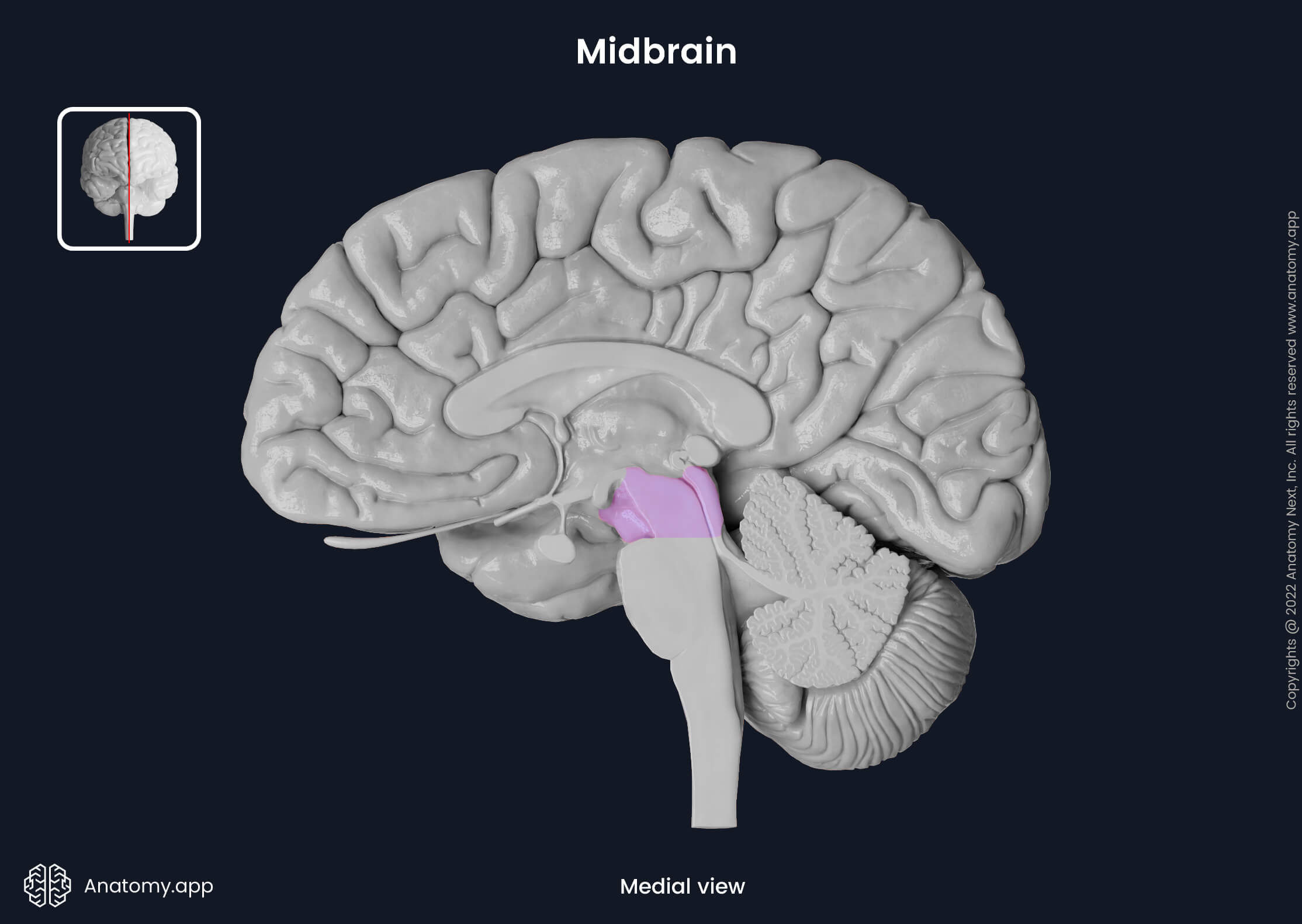
The midbrain is the most rostral segment of the brainstem, and it is also the shortest part, measuring less than 2 cm in length. Superiorly, the midbrain meets the thalamus of the diencephalon. Inferiorly it connects to the pons. The parahippocampal gyri surround its lateral surfaces. The midbrain passes through the tentorial notch between the free anterior margin of the tentorium cerebelli and the clivus of the occipital bone.
Like all other structures of the brainstem, the midbrain contains a multitude of nuclei, and it is the site of passage and relay of numerous neural pathways. It is involved in a wide array of vital functions, including regulating eye movement, movement planning, auditory processes, motivation, excitation, temperature, and others. It is also the site of origin of two cranial nerves - oculomotor (CN III) and trochlear (CN IV), and contains four pairs of cranial nerve nuclei.
Anatomically, the midbrain can be further divided into external and internal components. The external anatomy of the midbrain looks at its superficial structures and features. In contrast, the internal anatomy looks at the deeper-located parts of the midbrain and is very complex as it contains numerous nuclei and tracts.
Midbrain external anatomy
The external anatomy of the midbrain includes its superficial structures and features. Externally, the midbrain has two surfaces: ventral (anterior) and dorsal (posterior). The midbrain may also be separated into anterior and posterior parts by a passageway known as the cerebral aqueduct of Sylvius. The main feature of the anterior aspect of the midbrain is the pair of cerebral peduncles. The posterior part of the midbrain is formed by the tectum.
Ventral surface
The ventral or anterior surface is the external aspect of the anterior part of the midbrain. The main features of this surface are the landmarks of the cerebral peduncles. The cerebral peduncles resemble two stalks and, as they descend, they slightly converge towards each other. The cerebral peduncles contain white matter tracts.
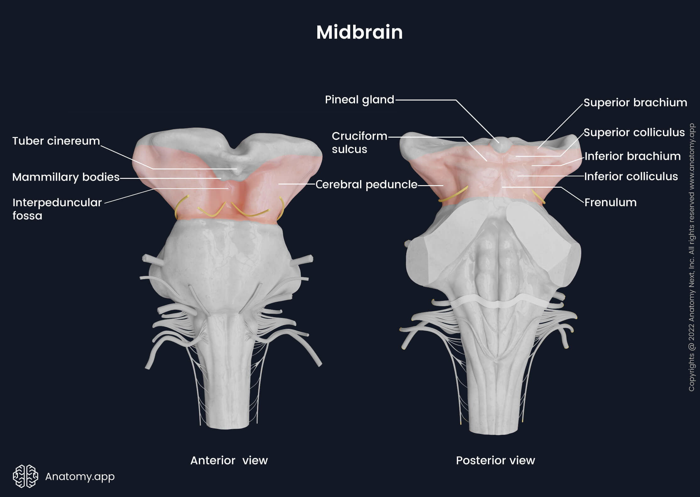
Superiorly above the ventral surface of the midbrain are the mammillary bodies and tuber cinereum. Both structures are parts of the diencephalon. The optic tract leaves the optic chiasm located anterior to the tuber cinereum. At the superior aspect of the midbrain, the optic tract horizontally crosses the cerebral peduncles and passes posteriorly.
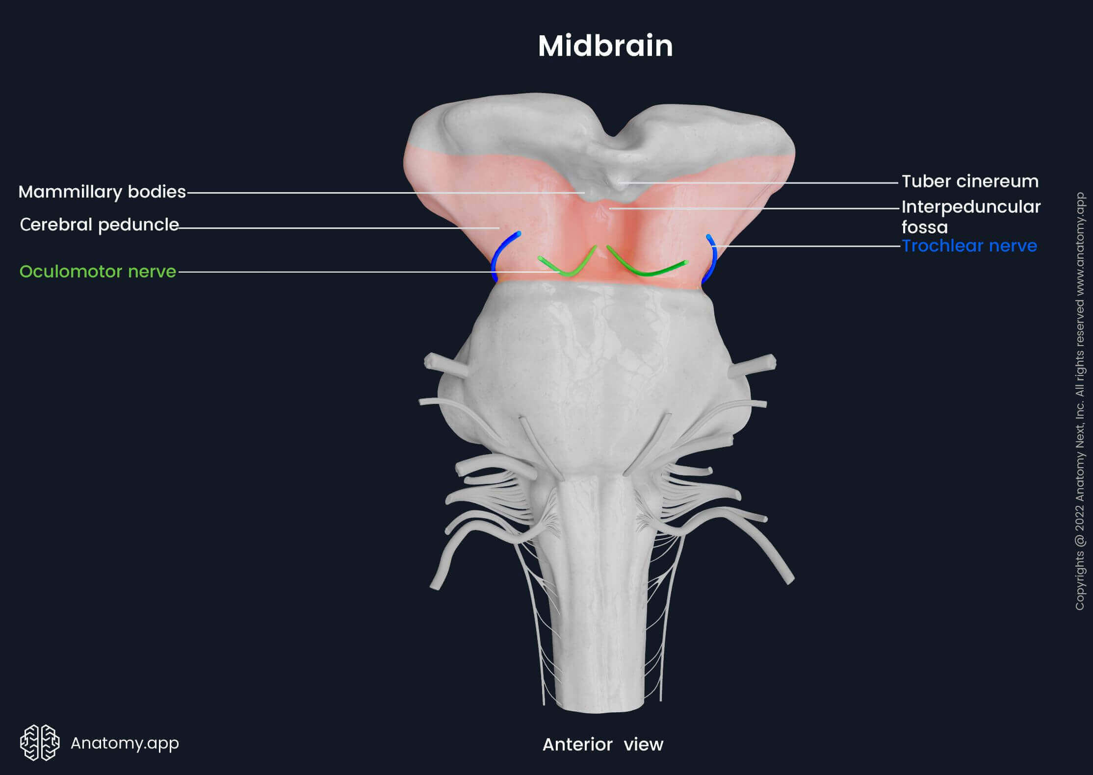
In the lower part of the anterior surface, between the two cerebral peduncles, is a depression known as the interpeduncular fossa, and it consists of gray matter. The floor of the fossa is composed of the posterior perforated substance, which is pierced by the branches of the posterior cerebral artery. By perforating the base of the interpeduncular fossa, the branches of the posterior cerebral artery supply the midbrain with oxygenated blood.
Moreover, the interpeduncular fossa is the site of terminal bifurcation of the basilar artery. The basilar artery travels upwards from the pons, running along the basilar sulcus. It reaches the midbrain and divides into the right and left posterior cerebral arteries. The posterior communicating arteries that originate from the internal carotid arteries join the posterior cerebral arteries and so link them with the internal carotid arteries.
Two oculomotor sulci are located in the lower anterior part on either side of the interpeduncular fossa. Each oculomotor nerve (CN III) emerges from the oculomotor sulcus between the posterior cerebral artery and the superior cerebellar artery. The nerve emerges close to (or inferior to) the corresponding posterior cerebral artery and travels further above the superior cerebellar artery.
The space in front of the interpeduncular fossa is a part of the interpeduncular cistern. The interpeduncular cistern is a cavity formed by the widening of the subarachnoid space between the arachnoid mater and pia mater. Within it are located the previously mentioned arteries and oculomotor nerves. Anteriorly, the interpeduncular cistern communicates with the suprasellar (or chiasmatic) cistern and, inferiorly, with the prepontine (or pontine) cistern.
Dorsal surface
The dorsal surface, also called the posterior surface, is the rear part of the midbrain. The tectum is the posterior part of the midbrain, separated from the anterior part by the aqueduct of Sylvius. The dorsal surface is the superficial part of the tectum. On the dorsal surface are four round formations known as the superior and inferior colliculi. The cerebral peduncles mark the lateral sides of the posterior surface.
Superior to the dorsal surface are structures of the diencephalon: the pineal gland and the two thalami with their associated structures. Just beneath the diencephalon are the two upper tubercles of the midbrain - the right and left superior colliculi. Beneath them are the two lower tubercles – the right and left inferior colliculi. The superior colliculi are more prominent and darker than the inferior colliculi.
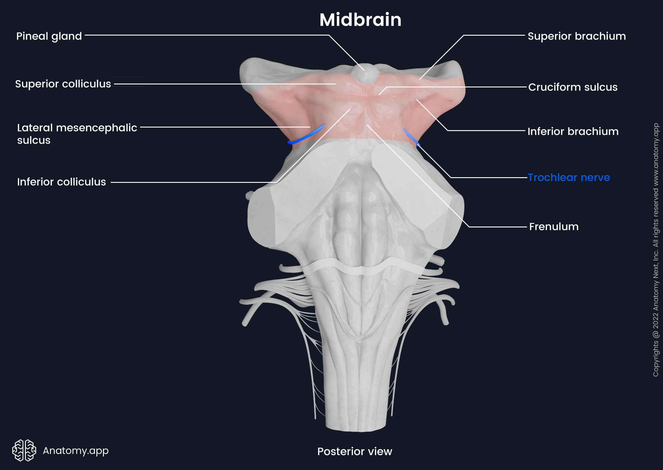
The colliculi are separated from each other by the cruciform sulcus. The cruciform sulcus is a cross-shaped groove, and its vertical part stretches from the rostrally located depression of the pineal gland to the caudally located frenulum. The frenulum is a part of the medullary velum, and it separates both inferior colliculi from each other. The horizontal part of the cruciform sulcus separates the superior colliculi from the inferior ones.
Due to the arrangement of four colliculi, the tectum is also known as the quadrigeminal plate. It comes from Latin, where quadri is a combining form used as a prefix meaning "four." The four colliculi together are named corpora quadrigemina. Lateral from each colliculus, extends a white matter containing eminence called a brachium (pl. brachia). Therefore, there are two superior and two inferior brachia and they continue forward and upward.
The superior brachia connect the superior colliculi of the midbrain with the retinae of the eyes via the lateral geniculate body and the optic tract. The inferior brachia connect the lateral lemniscus and inferior colliculus of the midbrain to the medial geniculate bodies of the thalami, which are connected to the auditory cortex.
On the lateral surfaces of the midbrain, between the cerebral peduncles and the dorsal surface, is another sulcus – the lateral mesencephalic sulcus. On the dorsal surface of the midbrain emerge the right and left trochlear nerves (CN IV). After appearing, both nerves continue forward and laterally along the border between the midbrain and pons.
Midbrain internal anatomy
The internal anatomy of the midbrain can be better understood when viewed from the sagittal and transverse planes. In the sagittal cross-section, the midbrain is divided into the crura cerebri, tegmentum and tectum. The crura cerebri form the ventral part, the tegmentum is the middle portion, and the tectum is the dorsal aspect of the midbrain. Various inner features can be seen by dissecting the midbrain and looking at it in the transverse plane cross-section at different levels.
To get a better understanding, let's rewind. The midbrain is separated into the anterior and posterior parts. The border between the anterior and posterior regions is a 1.5 - 2 cm long narrow canal called the cerebral aqueduct of Sylvius, and it connects the third ventricle with the fourth. The anterior part consists of cerebral peduncles. Within the cerebral peduncles is the substantia nigra. It separates the cerebral peduncles into the anteriorly located crus cerebri and the posteriorly situated tegmentum. Moreover, the tegmentum is the middle aspect of the midbrain. The posterior portion, located behind the aqueduct, is the tectum.
As mentioned before, inner features can be inspected by dissecting the midbrain and looking at it in the transverse plane cross-section at different levels. This allows us to better understand the structures, features, and characteristics of the nuclei and tracts located in the midbrain. The most common dissection points are at the level of the superior colliculi and the level of the inferior colliculi. Specific structures are only seen in one of those cross-sections.
The midbrain is made up of white and gray matter. Gray matter forms the nuclei. The structures of the gray matter located in the midbrain include the nuclei of the cranial nerves (oculomotor nerve nuclei, accessory oculomotor nuclei, trochlear nerve nuclei, mesencephalic trigeminal nerve nuclei), substantia nigra, red nucleus, periaqueductal gray matter, and parts of the reticular formation.
The white matter of the midbrain is concentrated in the ascending and descending tracts. The ascending tracts of the midbrain are the superior cerebellar peduncles, medial longitudinal fasciculus, spinal lemniscus, medial lemniscus, trigeminothalamic tract with trigeminal lemniscus, and lateral lemniscus. The descending tracts include the frontopontine and temporopontine tracts, as well as the corticospinal and corticobulbar tracts.
Crus cerebri
The crus cerebri are two symmetrical stalks found at the most ventral part of the cerebral peduncles and separated by the interpeduncular fossa. They appear semilunar-shape when viewed in a cross-section. The crus cerebri contains white matter fibers that form four separate descending tracts. The four pathways are located in each crus cerebri.
Tracts of crus cerebri
The crus cerebri of the midbrain contains the following four tracts:
- Frontopontine tract
- Temporopontine tract
- Corticospinal tract
- Corticobulbar tract
The frontopontine and temporopontine tracts are part of the corticopontine pathway, which connects the cerebral cortex and pons. The frontopontine tract fibers are situated most medially, while the temporopontine tract fibers are located most posterolaterally. Between the frontopontine and temporopontine fibers, the corticospinal and corticobulbar tracts are situated. They contain motor fibers that come from the primary motor cortex.
The frontopontine tract fibers extend from the premotor and motor cortices of the frontal lobe and then pass through the anterior limb of the internal capsule to reach the nuclei of the pons. The temporopontine tract is formed of fibers that predominantly originate from the superior and middle temporal gyri of the temporal lobe and terminate in the pontine nuclei of the pons.
The corticospinal tract is one of two pyramidal tracts that connect the cerebral cortex to the spinal cord. It is a major neuronal pathway that provides voluntary motor function. The other pyramidal tract is the corticobulbar tract. It connects the motor cortex to the pyramids of the medulla oblongata. It provides motor innervation to the muscles of the head and neck.
Substantia nigra
Posterior to the crus cerebri is the substantia nigra. The substantia nigra is a darkly pigmented stripe of dopaminergic neurons. The dark color comes from a pigment called neuromelanin that is found in the dopaminergic neurons. The substantia nigra is located on both sides of the interpeduncular fossa, and it separates the crus cerebri from the tegmentum. It is a part of the extrapyramidal motor system, communicating with the basal ganglia.
The substantia nigra is composed of two separate components: the ventral pars reticulata and dorsal pars compacta. The ventral pars reticulata receives information predominantly from the striatum and further communicates with the thalamus. It contains neurons that produce inhibitory GABA (gamma-aminobutyric acid) neurotransmitters. The pars compacta supplies the striatum (part of the basal ganglia) with dopamine.
Tegmentum
The tegmentum is the central part of the midbrain, situated between the crus cerebri and tectum. The substantia nigra separates the tegmentum from the crus cerebri, and posteriorly, the aqueduct of Sylvius separates it from the tectum. At various levels of the tegmentum, different structures can be seen. Namely, these include the cranial nerve nuclei, red nucleus, periaqueductal gray matter, ascending pathways and the reticular formation.
Cranial nerve nuclei
Cranial nerve nuclei are paired bilaterally located sensory and motor nuclei found at all brainstem levels. They are made up of neuronal cell bodies and give rise to axons - nerve fibers that form the corresponding cranial nerves. The nuclei vary in size and length. The following four cranial nerve nuclei are located in the tegmentum of the midbrain:
- Oculomotor nerve nucleus (2)
- Accessory oculomotor nucleus (2)
- Trochlear nerve nucleus (2)
- Mesencephalic trigeminal nerve nucleus (2)
The oculomotor nerve nucleus is located centrally at the level of the superior colliculi. It is a paired structure situated anterior to the periaqueductal gray matter. Laterally, the oculomotor nerve nuclei are bordered by the medial longitudinal fascicles. It is a motor nucleus from which general somatic efferent fibers that form the oculomotor nerve (CN III) originate and further innervate almost all extraocular muscles, except for the lateral rectus and superior oblique muscles.
The accessory oculomotor nucleus often called the Edinger-Westphal nucleus, is a paired motor nucleus located posterior to the oculomotor nerve nucleus. It is a parasympathetic preganglionic nucleus. The preganglionic nerve fibers enter the orbit together within the oculomotor nerve (CN III) and innervate the ciliary muscle and sphincter pupillae muscles.
The trochlear nerve nucleus is a general somatic efferent nucleus situated in the ventral part of the periaqueductal gray matter at the level of the inferior colliculi. The nerve fibers (axons) that form the trochlear nerve (CN IV) originate from it. The nerve fibers leave the nucleus, pass laterally and posteriorly and span posteriorly around the periaqueductal gray area. Further, they decussate (cross) at the midline of the superior medullary velum and exit the midbrain.
The trochlear nerves emerge on the posterior surface of the midbrain, near its border with the underlying pons. The right and left nerves leave the midbrain at the base of the corresponding inferior colliculi. The trochlear nerve is the only cranial nerve that appears from the dorsal surface of the brainstem. Fibers from this motor nucleus are responsible for the motor innervation of the superior oblique muscle.
The mesencephalic trigeminal nerve nucleus is a general somatic afferent nucleus located along both lateral sides of the periaqueductal gray matter. The elongated sensory nuclei ascend from the pons into the midbrain up to the level of the superior colliculi. They are one of the nuclei from which the trigeminal nerve (CN V) originates. Overall, the trigeminal nerve originates from four different nuclei - one motor and three sensory nuclei.
The mesencephalic trigeminal nerve nucleus is a sensory nucleus responsible for the proprioceptive reflex of the jaw. It works together with the muscles of mastication (except the lateral pterygoid) and periodontium to regulate the strength of biting to prevent the use of excessive force and damage to the teeth.
Red nucleus
The red nucleus, also called the nucleus ruber, is a prominent oval-shaped nucleus located in the upper part of the midbrain. It is a paired structure seen in cross-section at the level of the superior colliculi. The red nucleus is made up of gray matter. It got its name from Latin, where the word ruber means "red". This is because, in fresh dissections, the red nuclei look very pinkish-red. The red color comes from the extensive capillary network and ferric iron pigment deposited in the cells of the red nuclei.
Both nuclei connect with the cerebral cortex, cerebellum and spinal cord via several pathways. The red nuclei are involved in motor control and maintenance of muscle tone. The rostral part of each nucleus is the parvocellular portion, and the caudal region is the magnocellular portion. The red nucleus communicates with the primary somatomotor and somatosensory areas of the cerebral cortex via the corticorubral tract.
Moreover, the rostrally lying parvocellular portions of the red nuclei contribute to the dentato(cerebello)-rubro-olivary circuit (also called the Guillain-Mollaret triangle). Fibers from the dentate nucleus in the cerebellum travel to the red nucleus. From there, they pass through the central tegmental tract to reach the inferior olivary nucleus. Subsequently, fibers from the inferior olivary nucleus return back to the dentate nucleus. This circuit is involved in regulating motor control, steadiness and coordination.
The rubrospinal tract originates in the caudal magnocellular portion of the red nucleus. It further decussates and descends as part of the spinal cord. The rubrospinal tract terminates in the lateral column of the spinal cord's cervical portion. It ensures motor regulation of the upper limbs and balance. In the parvocellular portion, the oculomotor (CN III) nerve fibers pass, while fibers from the superior cerebellar peduncle pass in the magnocellular portion.
Periaqueductal gray
The periaqueductal gray (PAG), also termed central gray, is situated around the aqueduct of Sylvius. It is made up mainly of neuron cell bodies - gray matter. The aqueduct of Sylvius connects the third and fourth ventricles and separates the tegmentum from the tectum. Axons coming from the periaqueductal gray communicate with the cerebral cortex and spinal cord. The primary function of the periaqueductal gray is pain modulation.
The periaqueductal gray is involved in forming the integrated behavioral response to internal (pain) and external (threat) stressors. It releases endogenous opioids and further communicates with various other parts of the brain, including the somatosensory areas of the cerebral cortex. The primary functions of the PAG include pain perception and regulation, protection from harm and general survival.
Ascending pathways
Many pathways ascend through the tegmentum of the midbrain. They connect the spinal cord and cerebellum to the cerebrum and transmit sensory information from the peripheral nerves to the cerebral cortex and other brain parts. The ascending pathways of the tegmentum include the following paired structures:
- Tracts of the superior cerebellar peduncle:
- Cerebellothalamic tract
- Cerebellorubral tract
- Spinocerebellar tract
- Medial longitudinal fasciculus
- Medial lemniscus
- Trigeminal lemniscus
- Spinal lemniscus
- Lateral lemniscus
The superior cerebellar peduncle is a paired structure composed of white matter. It connects the cerebellum to the midbrain at the level of the inferior colliculi. The superior cerebellar peduncle contains afferent and efferent fibers. Upon reaching the midbrain, the fibers of both peduncles decussate and form two bundles - descending and ascending bundles. The superior cerebellar peduncles assist in proprioception, motor regulation, and learning of new movements.
The ascending bundle is made up of two efferent tracts - cerebellothalamic and cerebellorubral tracts. The cerebellothalamic tract advances via the midbrain to the ventrolateral nucleus of the thalamus. And the cerebellorubral tract connects to the contralateral red nucleus. Additionally, the ascending bundle contains separate ascending fibers that directly link up with the periaqueductal gray and reticular formations of the midbrain.
The descending bundle communicates with the below lying reticular formation and olivary nuclei of the medulla oblongata. Moreover, the anterior spinocerebellar tract is also a part of the superior cerebellar peduncles. It is a paired tract that conveys proprioceptive information. The afferent anterior spinocerebellar tract ascends from the spinal cord and terminates in the corresponding side of the cerebellum.
The medial longitudinal fasciculus is a paired tract found in the central part of the midbrain. The medial longitudinal fasciculus primarily interconnects the oculomotor, accessory oculomotor, trochlear, abducens, and vestibular nuclei, as well as the reticular formation. It is involved in the regulation of eye movements and therewith associated head and neck movements.
On both lateral sides of the tegmentum are positioned the four lemnisci. They are part of the sensory pathways. From anterior to posterior, the four lemnisci are situated in the following order: the medial lemniscus, trigeminal lemniscus, spinal lemniscus, and lateral lemniscus. They originate from lower levels of the nervous system, and each carries nerve fibers responsible for their specific functions. The fibers further advance to other brain parts.
The fibers of the medial lemniscus originate from the dorsal column of the spinal cord. Then the fibers travel upward, and at the level of the medulla oblongata, they reach the cuneatus and gracilis nuclei. The axons of the nuclei decussate at the sensory decussation and form the internal arcuate fibers that continue upwards as the medial lemniscus. The medial lemniscus is part of the posterior (dorsal) column - medial lemniscus pathway. It transmits sensory information about vibration, stretch, texture, pressure, and touch felt by receptors of the skin to the thalamus.
The trigeminal lemniscus, also called the trigeminothalamic tract, is located posterior to the medial lemniscus. It consists of the ventral and dorsal trigeminal tracts that convey sensory information about touch, pain and temperature from the regions of the head. It connects the trigeminal nerve (CN V) nuclei to the thalamus.
The spinal lemniscus is formed in the medulla oblongata by merging of the following tracts - the anterior and lateral spinothalamic and spinotectal pathways. It conveys sensory information about pain, touch, pressure, and temperature to the thalamus. The lateral lemniscus is located most posteriorly. It is a sensory tract located in the brainstem. It originates from the cochlear nucleus and travels further to reach the superior olivary nucleus. Fibers arising from the superior olivary nucleus transmit auditory information to the contralateral inferior colliculus.
Tectum
The tectum, also known as the quadrigeminal plate, is the most posterior part of the midbrain, and it is separated from the tegmentum by the aqueduct of Sylvius. The dorsal surface of the midbrain is the superficial part of the tectum. Internally, the tectum is formed by the corpora quadrigemina. The corpora quadrigemina is made of the same formations that can be clearly seen on the dorsal surface of the midbrain, namely, the four colliculi (two superior and two inferior colliculi).
Superior colliculi
The superior colliculi are multisensory midbrain structures located just beneath the diencephalon. Externally, they appear as the two upper tubercles - the right and left superior colliculi. Internally, the paired superior colliculus is a structure composed of neuronal cell bodies and synapses located posterior to the externally visible tubercules.
The superior colliculi receive input from the retina and further transmit information to the lateral geniculate bodies. The superior colliculi integrate visual, auditory, and somatosensory spatial information to initiate protective ocular reflexes and movements of the eyes and head in relation to objects and other focus points.
Inferior colliculi
Beneath the superior colliculi are the inferior colliculi - the two lower tubercles. Internally, they are also composed of neuronal cell bodies and synapses. The inferior colliculi serve as the main channel for all auditory information of the body. They further transmit the auditory information to the medial geniculate bodies of the thalamus.
The inferior colliculi receive the auditory information from the cochlear nucleus, which is situated in the vestibular area of the rhomboid fossa at the junction site of the medulla oblongata and pons. They are further involved in the procession of auditory information through signal integration, frequency recognition, and pitch discrimination. The inferior colliculi are also involved in generating reflexive motor movements in response to auditory stimuli.
Cross-section of midbrain
The internal anatomy of the midbrain can be better understood when viewed in a transverse cross-section. At different levels, several inner features can be seen. This allows us to better understand the structures, features, and characteristics of the nuclei and tracts located in the midbrain. The most common dissection levels are at the superior colliculi level and the inferior colliculi level. Certain structures are only seen in one of those cross-section levels.
At both the level of the superior and the level of the inferior colliculi, the crus cerebri remains unchanged and contains fibers of the frontopontine, temporopontine, corticospinal and corticobulbar tracts. Moreover, the cerebral aqueduct, periaqueductal gray, medial longitudinal fascicles, mesencephalic nuclei of the trigeminal nerve, the four lemnisci and substantia nigra are expected to be distinguished.
Level of superior colliculi
At the level of the superior colliculi in the anterior part of the tegmentum, two very prominent red nuclei can be seen. In front and medially to the red nuclei, the decussation of the rubrospinal tracts is positioned. Posteriorly to the red nuclei, in the anterior part of the periaqueductal gray, the oculomotor nuclei can be visualized. In some instances, when the cross-section is done at the appropriate level, it is also possible to see the exiting oculomotor nerve fibers.
Level of inferior colliculi
At the level of the inferior colliculi, some structures are no longer present, yet others have appeared. In the anteromedial part of the tegmentum, instead of the red nuclei and rubrospinal decussation, a prominent decussation of the superior cerebellar peduncles is present. Posterior to it, in the anterior part of the periaqueductal gray, the trochlear nuclei are situated. Typically, at this level, the trochlear nerve fibers leave the nuclei anterolaterally and then, after laterally curbing around the periaqueductal gray, exit the midbrain posteriorly.
Vasculature of midbrain
Arterial blood supply to the midbrain is provided by the posterior choroidal artery and the basilar artery and its branches, namely, the posterior cerebral artery and its peduncular branch, the superior cerebellar artery, and the interpeduncular perforating and paramedian branches of the basilar artery. The interpeduncular vein and two basal veins collect venous blood from the midbrain and drain into the great cerebral vein.
The anterior medial part of the crus cerebri receives oxygenated blood from the posterior choroidal artery. The middle portion of the crus cerebri and anterior aspect of the tegmentum are supplied by the paramedian branches of the basilar artery and peduncular branch of the posterior cerebral artery. The lateral part of the crus cerebri is directly provided by the latter artery.
The anteromedial part of the tegmentum is supplied by the interpeduncular perforating branches of the basilar artery. The superior cerebellar artery brings oxygenated blood to the lateral aspects of the tegmentum. The superior cerebellar artery primarily supplies the posterior surface, tectum, and part of the posterolateral tegmentum. Besides the superior cerebellar artery, the tectum also receives arterial blood by the branches of the posterior cerebral artery.
References:
- Haines, D. E. (2018). The midbrain. In P. J. May & G. A. Mihailoff (Eds.), Fundamental Neuroscience for Basic and Clinical Applications (Fifth Edition, pp. 183–194). Elsevier.
- Joyce, C., Le, P. H., & Peterson, D. C. (2021). Neuroanatomy, Cranial Nerve 3 (Oculomotor). NCBI. https://www.ncbi.nlm.nih.gov/books/NBK537126/
- Kurkcuoglu, A. (2017). Mesencephalon; Midbrain. Intechopen. https://www.intechopen.com/chapters/55330
- Miller, L. E., & Gibson, A. R. (2009). Red Nucleus. Encyclopedia of Neuroscience, 55–62. https://www.sciencedirect.com/science/article/pii/B9780080450469013346
- Ruchalski, K., & Hathout, G. M. (2012). A Medley of Midbrain Maladies: A Brief Review of Midbrain Anatomy and Syndromology for Radiologists. Hindawi: Radiology Research and Practice. https://www.hindawi.com/journals/rrp/2012/258524/
- Waitzman, D. M., & Oliver, D. L. (2002). Midbrain. Encyclopedia of the Human Brain, 43–68. https://www.sciencedirect.com/science/article/pii/B0122272102002089
Anatomy.app
Contact information
- For questions regarding business inquiries. Please contact:
- info@anatomy.app