- Anatomical terminology
- Skeletal system
- Skeleton of trunk
-
Skull
- Neurocranium
- Viscerocranium
- Auditory ossicles
- Sutures of skull
- Topography of skull
- Skeleton of upper limb
- Skeleton of lower limb
- Joints
- Muscles
- Heart
- Blood vessels
- Lymphatic system
- Nervous system
- Respiratory system
- Digestive system
- Urinary system
- Female reproductive system
- Male reproductive system
- Endocrine glands
- Eye
- Ear
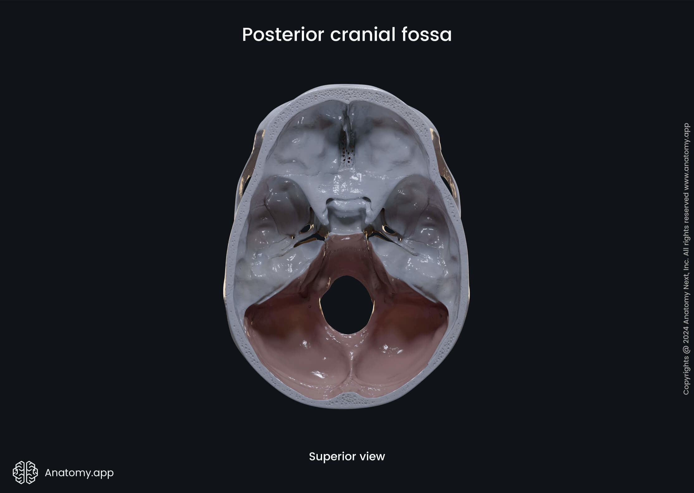
Posterior cranial fossa
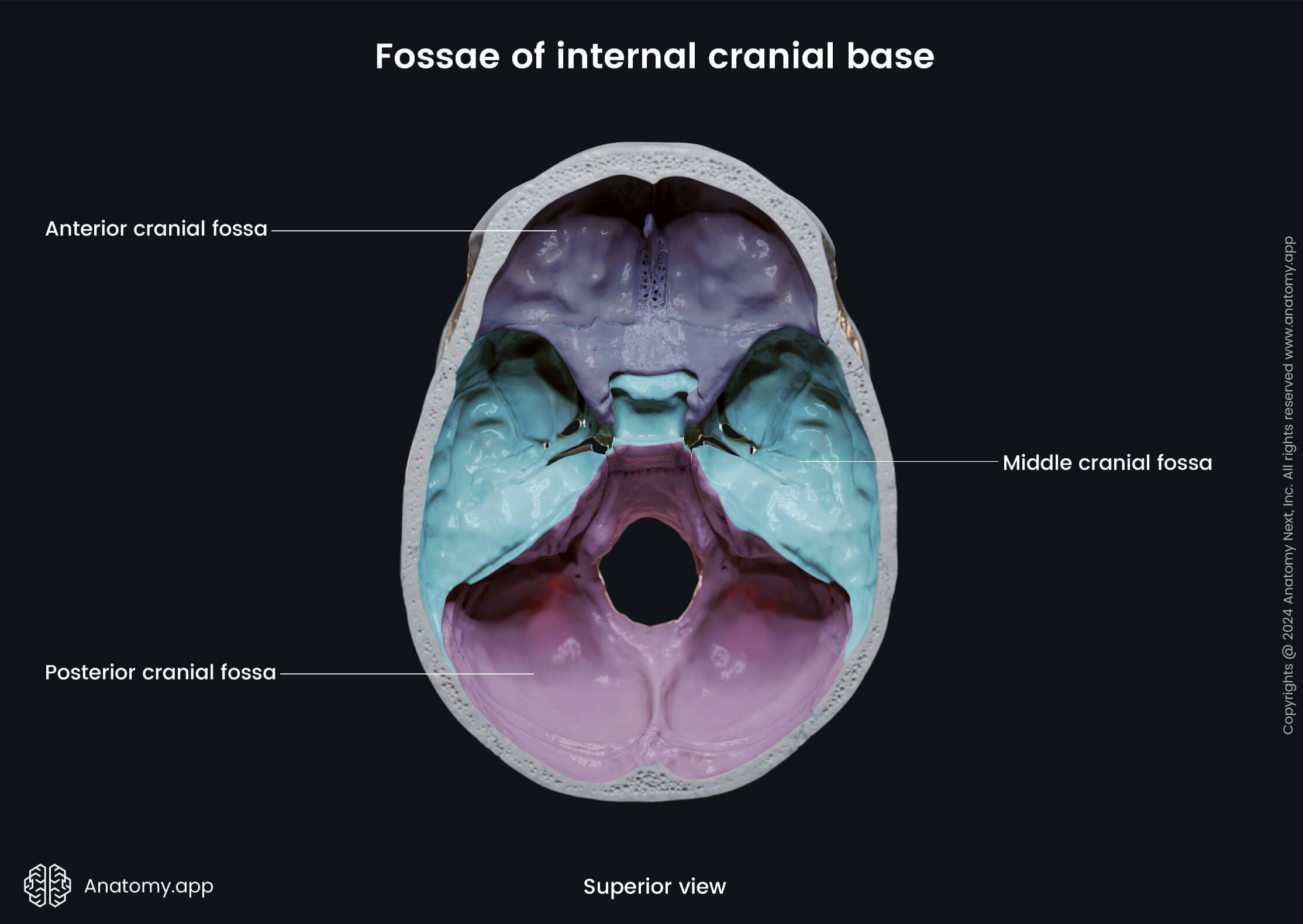
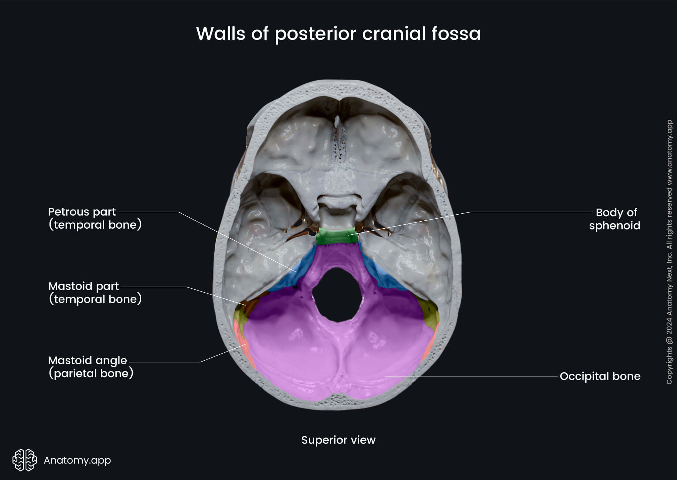
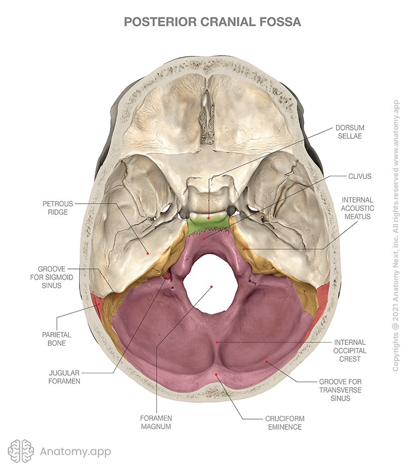
The posterior cranial fossa (Latin: fossa cranii posterior) lies at the lowest level of the internal cranial base and is the largest of the three cranial fossae. Its is formed by the sphenoid, temporal and occipital bones.
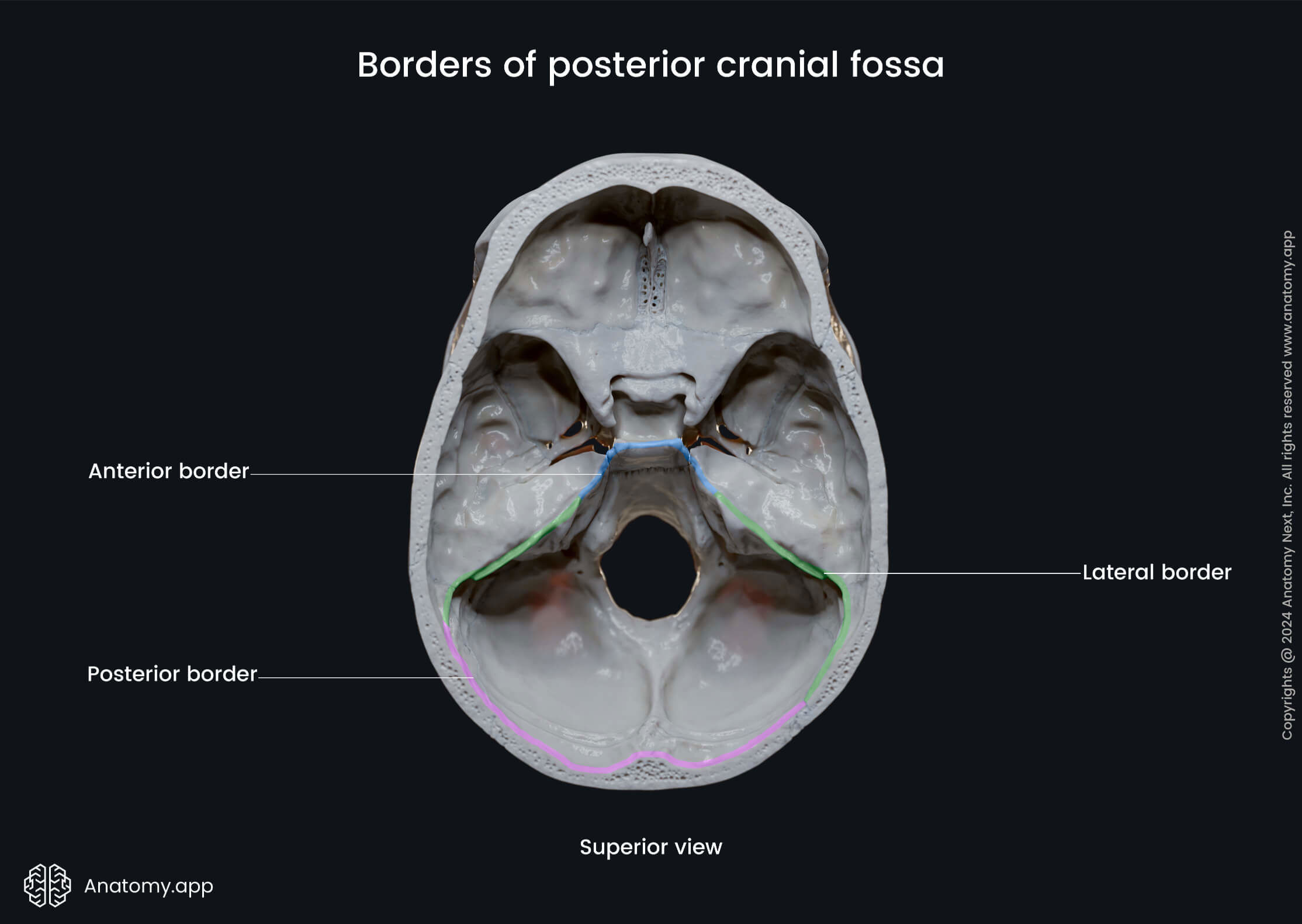
Borders of posterior cranial fossa
The borders of the posterior cranial fossa are the following:
- Anteromedially - dorsum sellae of the sphenoid bone;
- Anterolaterally - superior border of the petrous part of the temporal bone;
- Posteriorly - squamous part of the occipital bone, groove for transverse sinus, internal occipital protuberance;
- Inferiorly - mastoid part of the temporal bone, the squamous, condylar and basilar parts of the occipital bone.
Contents of posterior cranial fossa
The posterior cranial fossa accommodates two parts of the brain:
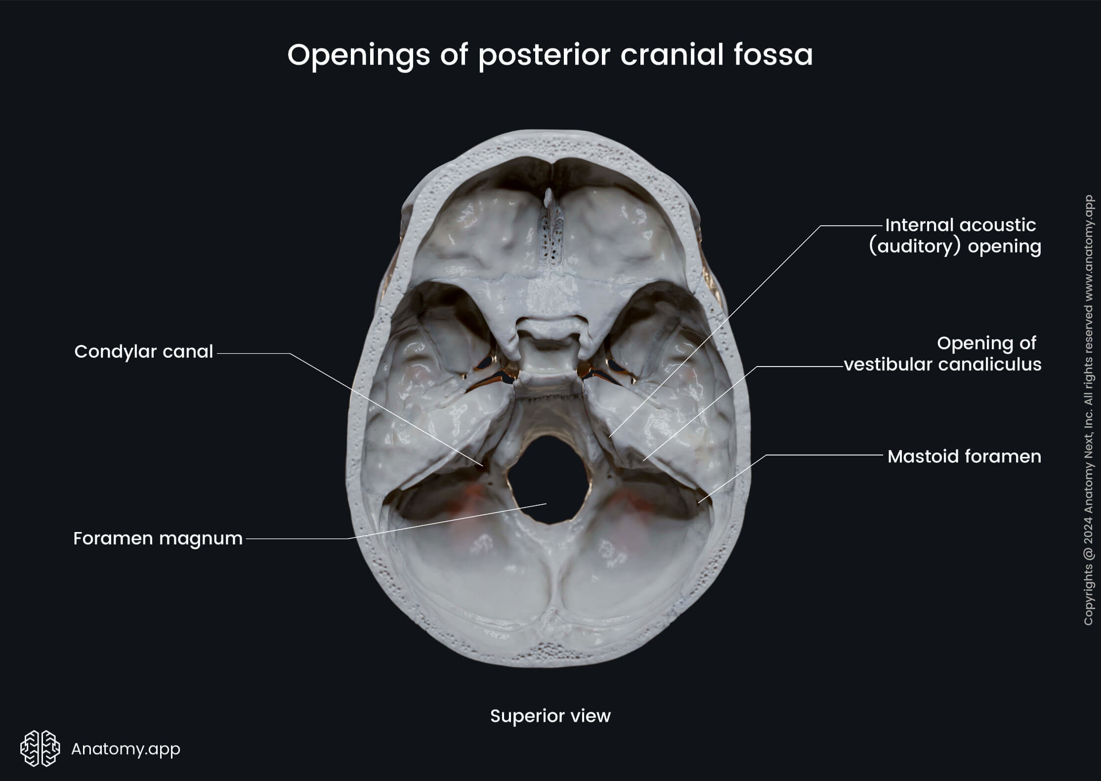
Openings of posterior cranial fossa
There are several opening connecting the posterior cranial fossa with other parts of the skull, and these are as following:
- Inner acoustic meatus
- Opening of vestibular canaliculus
- Mastoid foramen
- Foramen magnum
- Hypoglossal canal
- Condylar canal
- Jugular foramen
The first three openings on the list - the inner acoustic meatus, opening of the vestibular canaliculus and mastoid foramen - are all located in the temporal bone.
The inner acoustic meatus is a paired canal connecting the posterior cranial fossa and the facial canal. It transmits the facial nerve (CN VII) and the vestibulocochlear nerve (CN VIII), and also the labyrinthine artery.
The opening of the vestibular canaliculus is an opening that leads into the vestibular canaliculus which connects the posterior cranial fossa with the vestibule of the internal ear. It carries the vestibular aqueduct.
The mastoid foramen leads from the posterior cranial fossa to the external cranial base and transmits the mastoid emissary vein, a small branch of the occipital artery, and the posterior meningeal artery.
Three openings of the posterior cranial fossa are located in the occipital bone, these are the foramen magnum, hypoglossal canal, and condylar canal.
The foramen magnum is the largest opening in the skull connecting the posterior cranial fossa with the external cranial base. It transmits the lower end of the medulla oblongata, the vertebral artery, the accessory nerve (CN XI), and the anterior and posterior spinal arteries.
The paired hypoglossal canal is found on the lateral part of the occipital bone, superiorly and medially to each occipital condyle. It leads to the external cranial base and carries the twelfth cranial nerve - the hypoglossal nerve (CN XII).
The paired condylar canal also leads to the external surface of the cranial base. It is located behind the occipital condyle and conducts the occipital emissary vein.
The jugular foramen forms bilaterally between the occipital and temporal bones in the posterior cranial fossa and leads to the external cranial base. This opening transmits the inferior petrosal and sigmoid sinuses, and three cranial nerves - the glossopharyngeal (CN IX), vagus (CN X) and accessory (CN XI) nerves.
Anatomy.app
Contact information
- For questions regarding business inquiries. Please contact:
- info@anatomy.app