- Anatomical terminology
- Skeletal system
- Joints
- Muscles
- Heart
- Blood vessels
- Lymphatic system
- Nervous system
- Respiratory system
- Digestive system
- Urinary system
- Female reproductive system
- Male reproductive system
- Endocrine glands
- Eye
- Ear
Larynx
The larynx (Latin: larynx) is a flexible passageway for air between the oropharynx and the trachea. It is a part of the upper respiratory tract and plays an essential role in sound production. It also protects the lower airways against food inspiration. The larynx is primarly built of a cartilaginous skeleton and muscles. The inner surface of the larynx is lined with a mucosal membrane.
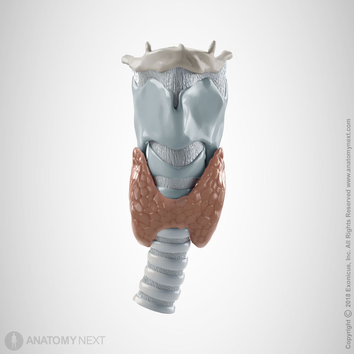
Cartilages of larynx
The cartilaginous skeleton of the larynx is formed by nine irregularly shaped plates of hyaline and elastic cartilage - three large unpaired (cricoid, thyroid, epiglottis) and three pairs of smaller cartilages (arytenoids, corniculate, cuneiform). The most important two are the epiglottis and the arytenoid cartilage.
During swallowing, the pharynx and larynx both elevate due the contraction of the extrinsic laryngeal muscles. The epiglottis moves down and forms a lid over the glottis closing it off, thereby protecting airways against blockade from foods and drinks. The arytenoid cartilages are able to influence the position and tension of the vocal folds.
Laryngeal muscles
The muscles of the larynx (laryngeal muscles) are skeletal muscles that participate in forming the walls of the larynx and in providing movements that are involved in breathing, phonation, and swallowing processes. The laryngeal muscles can be categorized into two groups: extrinsic (external) and intrinsic (internal) muscles.
The extrinsic muscles of the larynx act to elevate or depress the larynx, most importantly, during swallowing. The extrinsic muscles of the larynx include the suprahyoid and infrahyoid muscles of the neck, as well as the stylopharyngeus, which is a muscle of the pharynx.
The intrinsic muscles activate individual components of the larynx, playing a vital role in breathing and phonation. In general, they control the shape of the rima glottis, as well as the length and tension of the vocal cords. This muscle group includes the cricothyroid, thyroarytenoid, posterior cricoarytenoid, lateral cricoarytenoid, and transverse and oblique arytenoids.
Mucosa of larynx
The mucosa of the larynx forms the inner layer of the laryngeal wall, and is continuous with mucosa of the pharynx above and the trachea below. The laryngeal mucosa is the chief component of the vestibular folds, where it is the thickest. It is thinner over the vocal folds, where it is firmly fixed to the underlying vocal ligaments. It is loosely attached to the anterior surface of the epiglottis, but firmly attached to the anterior surface and the floor of the valleculae. On the aryepiglottic folds the mucosa is strengthened by large amount of fibrous connective tissue, and it attaches closely to the laryngeal surfaces of the cuneiform and arytenoid cartilages.
The laryngeal mucosa is lined mostly by the respiratory epithelium - ciliated, pseudostratified epithelium. It provides the mucociliary clearance mechanism similarly to other parts of the respiratory tract. However, there are specific areas of the larynx requiring a different functional type of epithelium. The vocal folds are covered by non-keratinized, stratified squamous epithelium, which is much more durable and can protect the tissue from the effects of mechanical stresses that affect the surfaces of the vocal folds. This type of epithelium also lines the upper parts of larynx, which merge with the laryngopharynx and continues as the oropharynx. This is because these surfaces are affected by the abrasive effects of swallowed food.
The lamina propria of the laryngeal mucosa (connective tissue layer) contains numerous mucous glands, especially over the epiglottis, and along the margins of the aryepiglottic folds anterior to the arytenoid cartilages (arytenoid glands). There are many large glands in the saccules of the larynx that secrete periodically over the vocal folds during phonation. The free edges of the folds do not contain glands and the stratified squamous epithelium here requires the secretions of the neighbouring glands to keep the vocal folds lubricated.
Note that taste buds, like those in the tongue, are also present in the laryngeal mucosa. More specifically, they can most commonly be found in the mucosal membrane covering the posterior epiglottic surface, aryepiglottic folds, and less often in other parts of the larynx.
Blood supply of larynx
The arterial blood supply to the larynx comes mainly from the paired superior and inferior laryngeal arteries, with rich anastomoses between the corresponding contralateral, and between the ipsilateral arteries. The superior laryngeal artery is a branch of the superior thyroid artery that arises from the external carotid artery. The inferior laryngeal artery, on the other hand, arises from the inferior thyroid artery, which usually branches off the thyrocervical trunk.
Innervation of larynx
The innervation of the larynx is provided by the vagus nerve (CN X) through its branches - the superior laryngeal nerve and recurrent laryngeal nerve. The branches of the vagus nerve provides innervation to the mucosa and intrinsic muscles of the larynx. However, the extrinsic laryngeal muscles receive separate innervation, mostly, from the facial nerve (CN VII) and cervical spinal nerves.
The superior laryngeal nerve divides into two branches: internal (sensory) and external (motor). The internal branch (also known as the internal laryngeal nerve) innervates the mucosa of the larynx down to the vocal folds. It also provides branches that communicate with the recurrent laryngeal nerve. The external branch of the superior laryngeal nerve provides innervation to the cricothyroid muscle.
The recurrent laryngeal nerve is a mixed nerve that provides motor innervation to all intrinsic laryngeal muscles (except the cricothyroid muscle), and sensory innervation to the laryngeal mucosa below the vocal cords. It gives branches that anastomose with the internal laryngeal nerve. Therefore, there is an overlap at the vocal cords between the areas innervated by the two mentioned nerves.
Anatomy.app
Contact information
- For questions regarding business inquiries. Please contact:
- info@anatomy.app