- Anatomical terminology
- Skeletal system
- Joints
- Muscles
- Heart
- Blood vessels
- Lymphatic system
-
Nervous system
- Central nervous system
-
Peripheral nervous system
- Cranial nerves
- Spinal nerves
- Respiratory system
- Digestive system
- Urinary system
- Female reproductive system
- Male reproductive system
- Endocrine glands
- Eye
- Ear
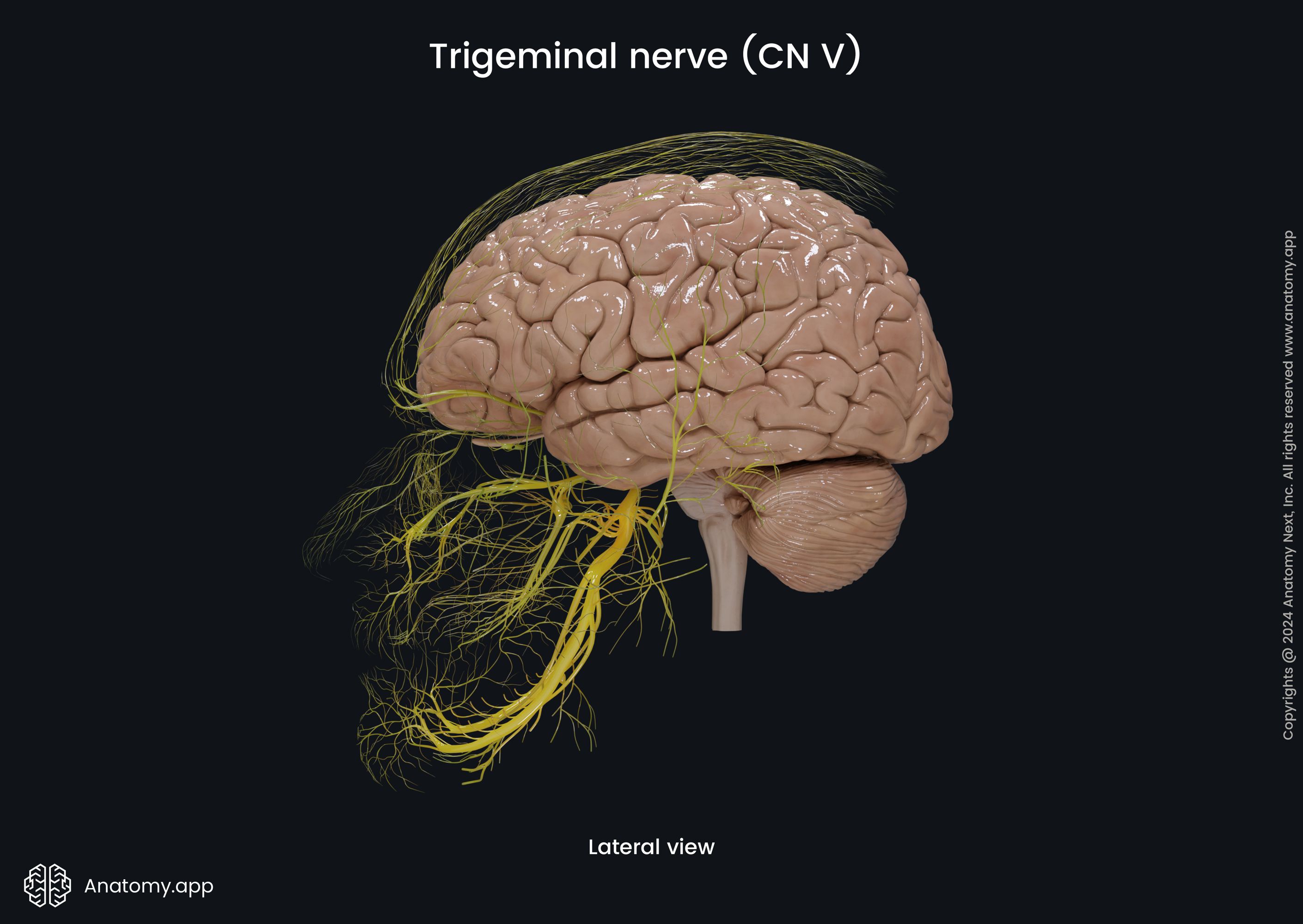
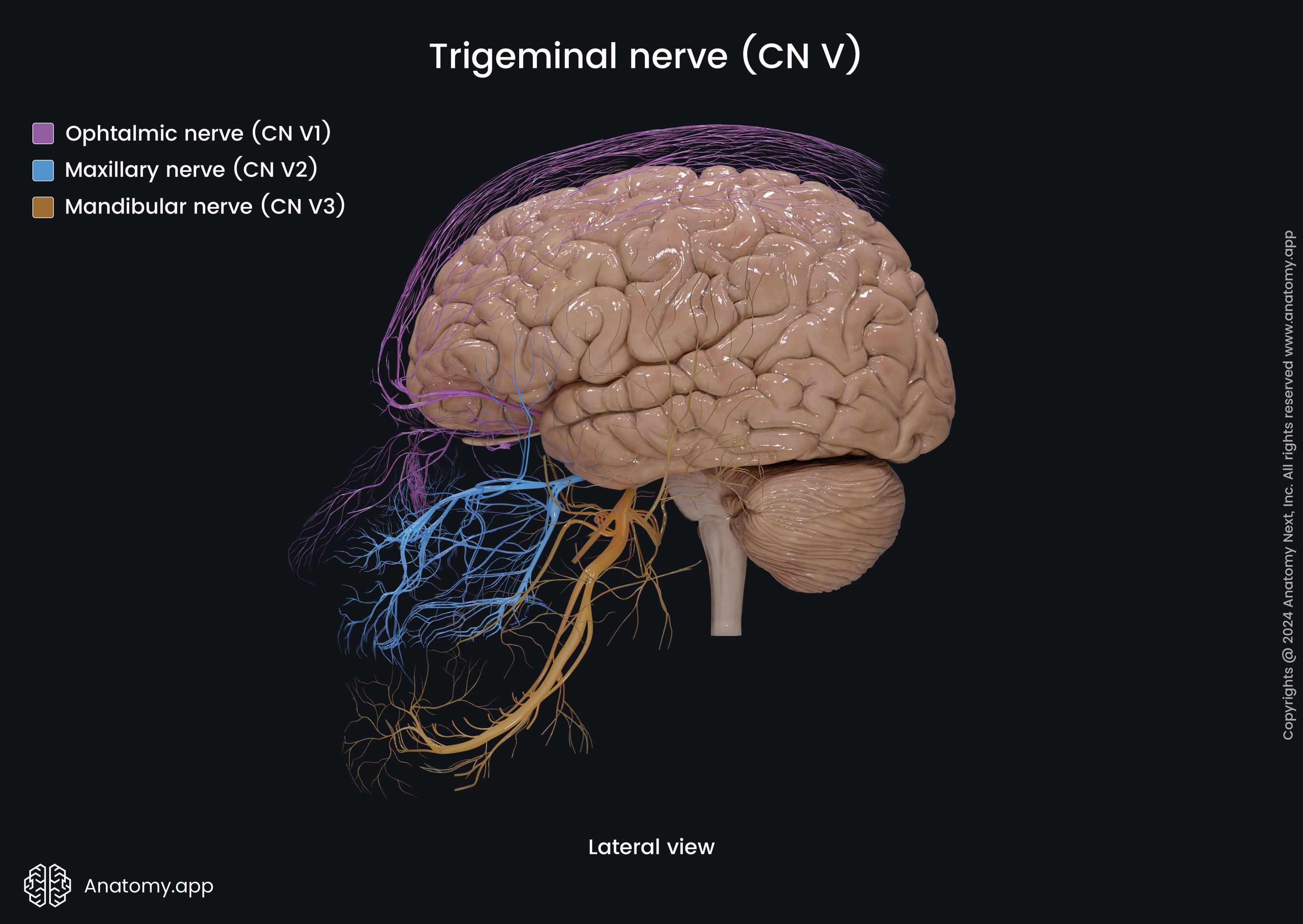
Ophthalmic nerve (CN V1)
The ophthalmic nerve (CN V1, Latin: nervus ophthalmicus) is the first branch or division of the trigeminal nerve (CN V). It is a sensory nerve transmitting sensory information to the central nervous system mostly from structures of the upper face region, including structures of the orbit, nasal cavity and paranasal sinuses, as well as from part of the dura mater. Some of the ophthalmic nerve branches also convey parasympathetic fibers.
Ophthalmic nerve course
The ophthalmic nerve arises from the trigeminal ganglion in the middle cranial fossa and passes forward along the lateral wall of the cavernous sinus, traveling below the trochlear nerve (CN IV). The maxillary nerve (CN V2) is located inferior and lateral to it. Before entering the sinus, the ophthalmic nerve gives off a recurrent tentorial branch that goes backward to innervate the cerebellar tentorium. The ophthalmic nerve reaches the orbit by passing through the superior orbital fissure and divides into smaller branches.
Ophthalmic nerve branches
In the superior orbital fissure or just before reaching the fissure, the ophthalmic nerve gives off three major branches:
Frontal nerve
The frontal nerve is a sensory nerve and the thickest branch of the ophthalmic nerve. It runs just a little bit below the roof of the orbit and, while in the orbit, divides into two terminal branches - the supratrochlear and supraorbital nerves. Both branches arise on the face and innervate the skin of the forehead.
The supraorbital nerve is the lateral branch, and it leaves the orbit through the supraorbital notch of the frontal bone. It distributes in the skin of the forehead supplying the conjunctiva and the skin of the upper eyelid. The supraorbital nerve has two terminal branches named the lateral and medial branches.
The supratrochlear nerve is located more medially and travels through the frontal notch. This branch of the frontal nerve supplies the skin of the forehead and radix of the nose, as well as the skin of the upper eyelid.
Lacrimal nerve
The lacrimal nerve is the lateral branch of the ophthalmic nerve going forward along the lateral wall of the orbit. It passes in the direction of the lacrimal gland, located in the upper lateral aspect of the orbital rim. The lacrimal nerve provides sensory innervation for the lacrimal gland, conjunctiva and skin covering the lateral part of the upper eyelid.
The lacrimal nerve also connects with the postganglionic parasympathetic fibers coming from the zygomatic nerve (a branch of the maxillary division of the trigeminal nerve (CN V2)) with the help of the communicant branch. Therefore, parasympathetic fibers reach the lacrimal gland via the lacrimal nerve.
Nasociliary nerve
The nasociliary nerve is the medial branch of the ophthalmic nerve. The main areas supplied by this sensory nerve and its branches are the mucosa of the nasal cavity, skin of the nasal root, as well as the skin and conjunctiva of the medial corner of the eye. This nerve ends with two terminal branches at the anterior ethmoid foramen - the infratrochlear nerve and the anterior ethmoid nerve.
The infratrochlear nerve passes forward in the direction of the upper medial aspect of the orbital rim. It innervates the skin of the forehead, radix of the nose, and the skin and conjunctiva of the medial aspect of the upper eyelid. The anterior ethmoid nerve leaves the bony orbit through the anterior ethmoid foramen and reaches the anterior cranial fossa, giving the meningeal branch to the dura mater of the fossa.
From the anterior cranial fossa, the anterior ethmoid nerve further continues to the nasal cavity via the foramina of the cribriform plate. It innervates the mucous membrane of the nasal cavity and mucosa of the frontal sinus and ethmoidal air cells. The terminal part of the anterior ethmoid nerve goes through the nasal foramen of the nasal bone and ends on the outer nasal surface as the external nasal branch, innervating the skin of the apex and nasal dorsum.
Besides the terminal branches, the nasociliary nerve also gives rise to several other branches on its course. The side branches of the nasociliary nerve include a sensory communicating branch for the ciliary ganglion, posterior ethmoid nerve, as well as short and long ciliary nerves. The sensory communicating branch for the ciliary ganglion is a branch that travels to the ciliary ganglion located in orbit lateral to the optic nerve (CN II).
The posterior ethmoid nerve reaches the nasal cavity via the posterior ethmoid foramen reaches and innervates the mucosa of the nasal cavity and the mucosal lining of the ethmoidal air cells and sphenoid sinus. Finally, the short and long ciliary nerves penetrate the posterior aspect of the sclera, pass between the outer and middle layers of the eyeball. These nerves provide innervation for the mentioned eyeball layers, mainly for the sclera and choroid.
Anatomy.app
Contact information
- For questions regarding business inquiries. Please contact:
- info@anatomy.app