- Anatomical terminology
- Skeletal system
- Skeleton of trunk
-
Skull
- Neurocranium
- Viscerocranium
- Auditory ossicles
- Sutures of skull
- Topography of skull
- Skeleton of upper limb
- Skeleton of lower limb
- Joints
- Muscles
- Heart
- Blood vessels
- Lymphatic system
- Nervous system
- Respiratory system
- Digestive system
- Urinary system
- Female reproductive system
- Male reproductive system
- Endocrine glands
- Eye
- Ear
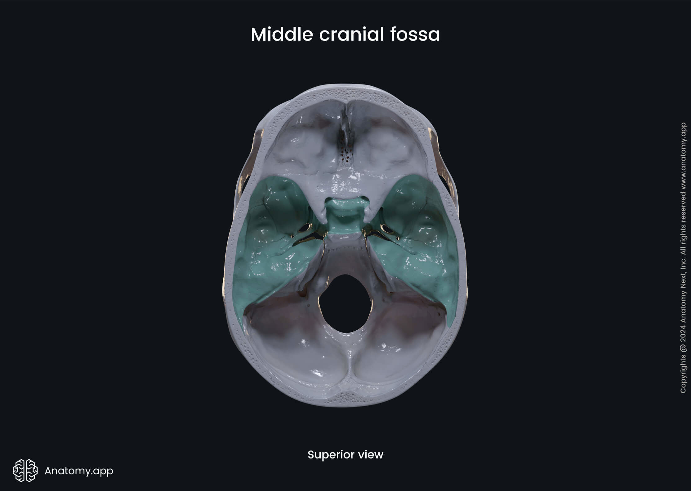
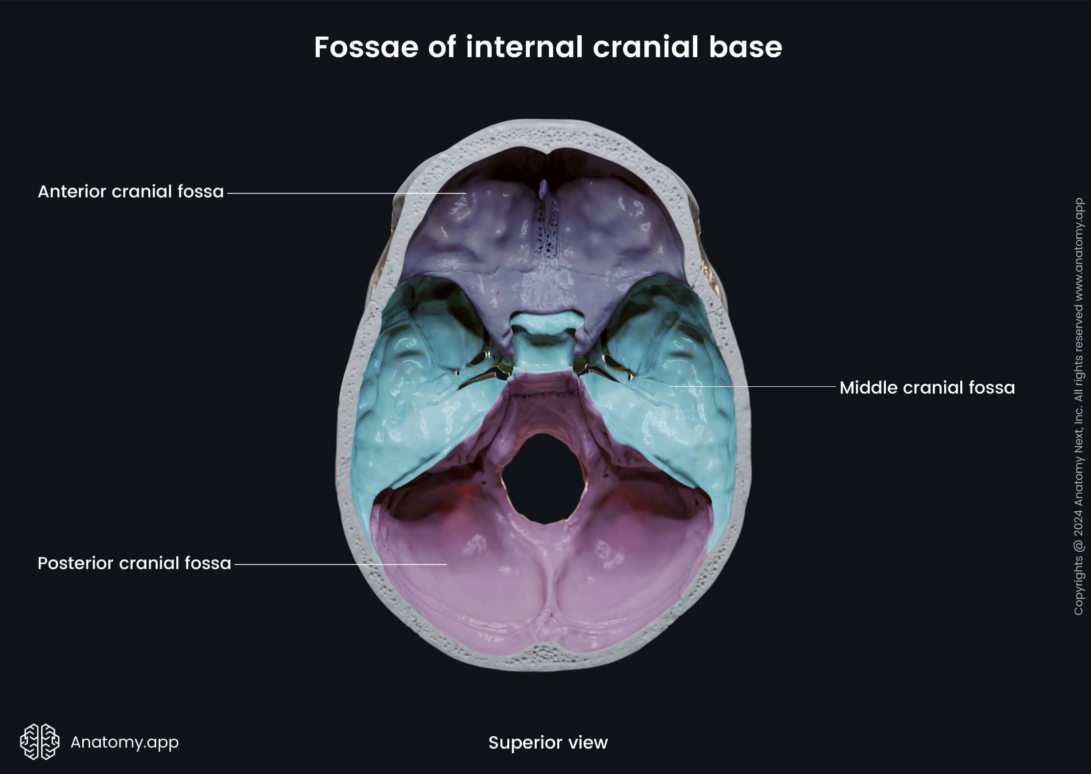
Middle cranial fossa
The middle cranial fossa (Latin: fossa cranii media) is a region of the internal cranial base between its other two parts - the anterior and posterior cranial fossae. It lies deeper and is wider than the anterior cranial fossa. The middle cranial fossa is created by the sphenoid, temporal and parietal bones.
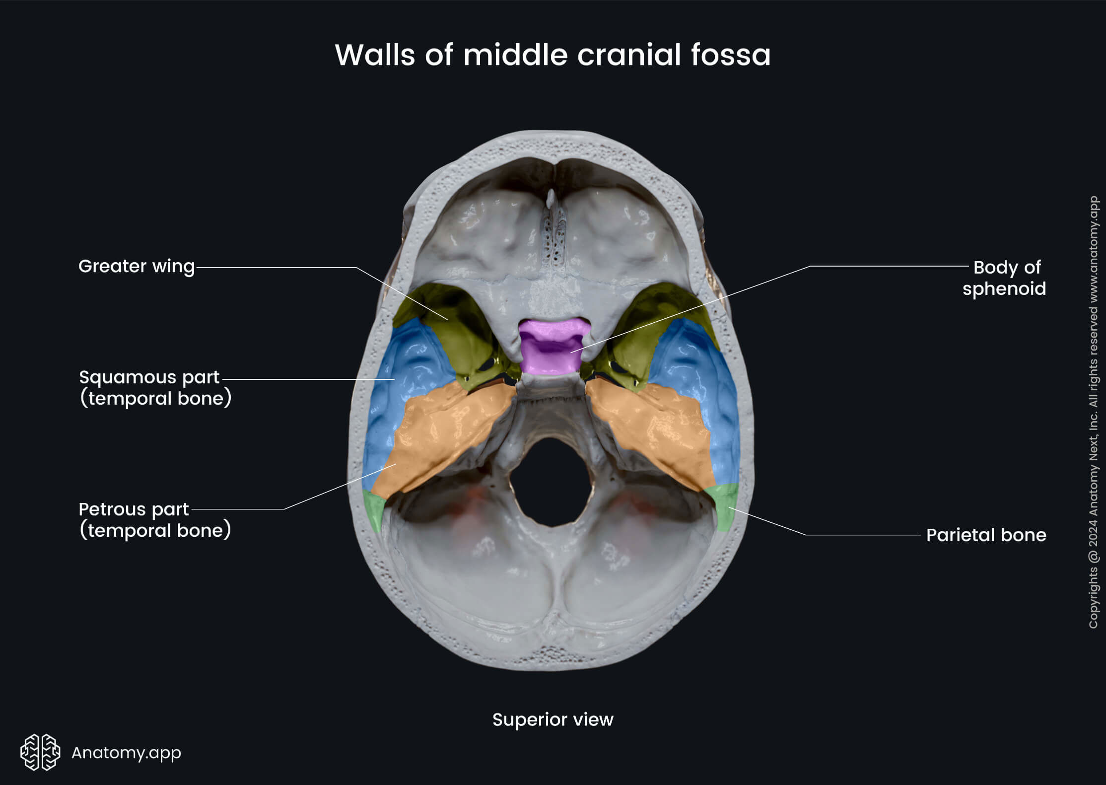
Borders of middle cranial fossa
These are the structures forming borders of the middle cranial fossa:
- Anteromedially - limbus of the sphenoid bone;
- Anterolaterally - lesser wings and part of the body of the sphenoid;
- Posteromedially - dorsum sellae of the sphenoid;
- Posterolaterally - superior margin of the petrous part of the temporal bone;
- Laterally - squamous parts of the temporal and parietal bones;
- Inferiorly - body and greater wings of the sphenoid, squamous and petrous parts of the temporal bone.


Contents of middle cranial fossa
The middle cranial fossa accommodates the following anatomical structures:
- Pituitary gland
- Temporal lobes of the cerebral hemispheres
Openings of middle cranial fossa
There are many openings in the middle cranial fossa connecting it to other parts of the skull:
- Optic canal
- Superior orbital fissure
- Foramen rotundum
- Foramen ovale
- Foramen spinosum
- Foramen lacerum
- Carotid canal
- Hiatus for lesser petrosal nerve
- Hiatus for greater petrosal nerve
The pair of optic canals connect the middle cranial fossa with the orbits. The optic canal transmits the second cranial nerve - the optic nerve (CN II), and the ophthalmic artery.
The paired superior orbital fissure also connects this fossa with the orbit. This opening transmits four cranial nerves - the oculomotor (CN III), trochlear (CN IV), ophthalmic (CN V1) and abducens (CN VI) nerves, the ophthalmic veins, and sympathetic nerve fibers.
The foramen rotundum is a paired opening serving as a connection between the middle cranial fossa and the pterygopalatine fossa. It transmits the maxillary nerve (CN V2) - the second branch of the trigeminal nerve (CN V).
The foramen ovale is another paired opening that connects the middle cranial fossa with the external surface of the cranial base and the infratemporal fossa. It transmits the mandibular nerve (CN V3), the lesser petrosal nerve, the accessory meningeal branch of the maxillary artery, and an emissary vein.
The paired foramen spinosum connects the middle cranial fossa with the infratemporal fossa. It transmits the middle meningeal artery and vein, and the meningeal branch of the mandibular nerve (CN V3).

The foramen lacerum is a paired opening between the middle cranial fossa and the external cranial base. The foramen lacerum is filled with cartilage after birth and it transmits the artery and nerve of the pterygoid canal.
The carotid canal and the hiatuses for the lesser and greater petrosal nerves all are paired openings in the temporal bone. The carotid canal connects the middle cranial fossa to the external cranial base and carries the internal carotid artery.
The hiatus for the lesser petrosal nerve transmits the named nerve to the tympanic cavity. And the hiatus for the greater petrosal nerve transmits the greater petrosal nerve and the petrosal branch of the middle meningeal artery from the facial canal to the middle cranial fossa.
Anatomy.app
Contact information
- For questions regarding business inquiries. Please contact:
- info@anatomy.app