- Anatomical terminology
- Skeletal system
- Joints
- Muscles
- Heart
- Blood vessels
- Lymphatic system
-
Nervous system
- Central nervous system
-
Peripheral nervous system
- Cranial nerves
- Spinal nerves
- Respiratory system
- Digestive system
- Urinary system
- Female reproductive system
- Male reproductive system
- Endocrine glands
- Eye
- Ear
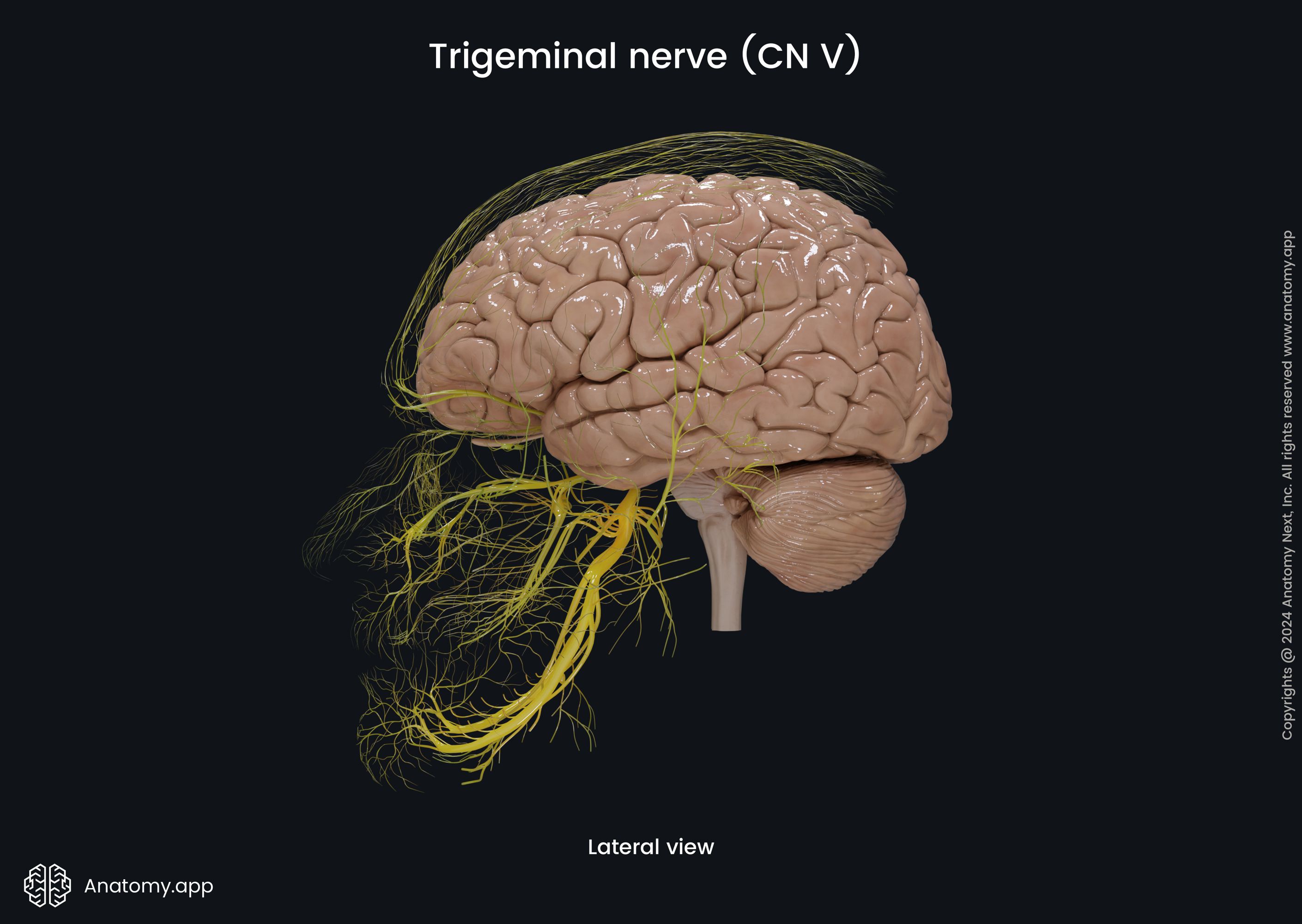
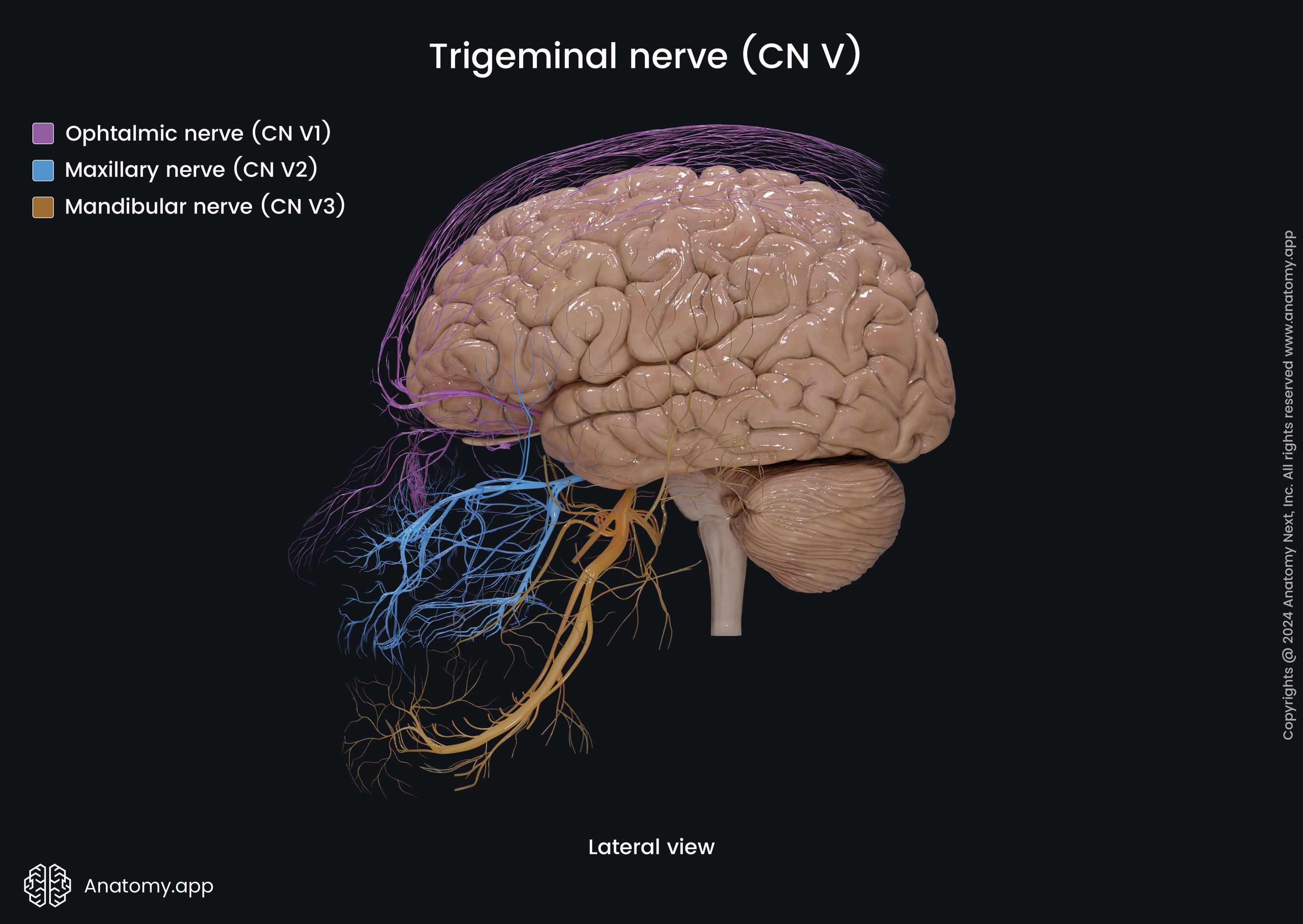
Mandibular nerve (CN V3)
The mandibular nerve (CN V3, Latin: nervus mandibularis) is the third and the largest branch of the trigeminal nerve (CN V), also known as the third or mandibular division of the trigeminal nerve. It is a large mixed nerve consisting of motor and sensory fibers. The sensory fibers of the mandibular nerve innervate several skin regions of the face, mostly its lower part, as well as the mucosa of the oral cavity, lower teeth and gingiva. Its motor branches supply the first branchial arch muscles, including all of the muscles of mastication.
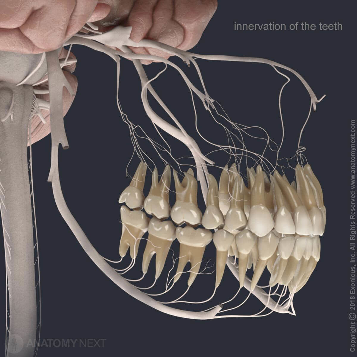
The mandibular nerve carries general somatic afferent fibers that provide sensory innervation for the skin of the temporal region of the scalp, skin of the external acoustic meatus, tympanic membrane, and lower part of the face. The sensory fibers of the mandibular nerve also supply the mucosa lining the floor of the oral cavity, anterior two-thirds of the tongue, and the teeth and gingiva of the lower jaw (mandible).
The motor branches of the mandibular nerve carrying general somatic efferent fibers that supply all of the muscles that developed from the first branchial arch, which are:
- Masseter muscle
- Medial pterygoid muscle
- Lateral pterygoid muscle
- Temporal muscle
- Tensor tympani muscle
- Tensor veli palatini muscle
- Mylohyoid muscle
- Anterior belly of the digastric muscle
Mandibular nerve course
The mandibular nerve emerges from the lateral part of the trigeminal ganglion with two roots - sensory and motor. The sensory root of the mandibular nerve arises from the lateral part of the trigeminal ganglion, while the smaller motor root passes below the trigeminal ganglion. Both roots exit the cranial cavity through the foramen ovale and reach the infratemporal fossa, and both unite just outside of the skull creating one main trunk.
Mandibular nerve branches
In the infratemporal fossa, the main trunk gives several branches. The first one is a sensory meningeal branch that through the foramen spinosum enters the middle cranial fossa and provides nerve supply to the dura mater of the fossa and calvaria. The other branch is a muscular motor branch that supplies the medial pterygoid muscle (medial pterygoid nerve) and, via the otic ganglion, provides the innervation to the tensor veli palatini and tensor tympani muscles.
After the main trunk has given the mentioned branches, it divides into even smaller divisions, named the anterior and posterior divisions or trunks. The smaller anterior portion of the mandibular nerve gives off motor efferent branches to the three muscles of mastication (these branches are called the lateral pterygoid, masseteric and deep temporal nerves) and an afferent sensory branch to the cheek (buccal nerve).
The posterior trunk of the mandibular nerve gives off motor branches to supply the mylohyoid muscle (mylohyoid nerve) and the anterior belly of the digastric muscle, and three main sensory branches: auriculotemporal, lingual and inferior alveolar nerve. The last two are the terminal branches of the posterior division. To sum up, the major branches of the mandibular nerve include the following:
- Medial pterygoid nerve (motor)
- Lateral pterygoid nerve (motor)
- Masseteric nerve (motor)
- Deep temporal nerves (motor)
- Nerve to the tensor veli palatini muscle (motor)
- Nerve to the tensor tympani muscle (motor)
- Meningeal branch of the mandibular nerve (sensory)
- Buccal nerve (sensory)
- Auriculotemporal nerve (sensory)
- Lingual nerve (sensory)
- Inferior alveolar nerve (motor and sensory), ending with the mental nerve (sensory)
Meningeal branch
The meningeal branch of the mandibular nerve arises in the infratemporal fossa and returns to the cranial cavity via the foramen spinosum. This nerve carries sensory fibers to innervate the dura mater of the middle cranial fossa and a small part of the anterior cranial fossa.
Medial pterygoid nerve
The medial pterygoid nerve is a motor branch of the mandibular nerve that arises in the infratemporal fossa just before the main trunk of the mandibular nerve divides into two portions. The medial pterygoid nerve travels through the otic ganglion (without synapsing there) to innervate the medial pterygoid muscle, supplying the tensor veli palatini (nerve to the tensor veli palatini muscle) and tensor tympani muscles (nerve to the tensor tympani muscle) with branches along the way.
Lateral pterygoid nerve
The lateral pterygoid nerve also arises in the infratemporal fossa from the anterior division of the main trunk - lower than the medial pterygoid nerve, and it carries motor fibers. It is a short nerve that perforates the lateral pterygoid muscle and innervates it.
Masseteric nerve
The masseteric nerve is another motor branch of the mandibular nerve that arises in the infratemporal fossa from the anterior division of the main trunk. It runs above the mandibular notch, passing posterior to the tendon of temporalis muscle, and penetrates the masseter muscle from inside, innervating it.
Deep temporal nerves
The deep temporal nerves are two branches named the anterior and posterior deep temporal nerves arising in the infratemporal fossa from the anterior division of the main mandibular nerve trunk. They run laterally above the infratemporal crest, then upward to the temporal fossa, and reach the temporalis muscle to innervate it.
Buccal nerve
The buccal nerve is a sensory branch arising from the anterior division of the main trunk of the mandibular nerve. It passes through the buccinator muscle (does not innervate it!) and transmits sensory information from the skin covering the buccinator muscle, as well from the mucosa of the cheek, and from the second and third molar teeth. The nerve passes between the heads of the lateral pterygoid muscle, courses through the anterior surface of the masseter muscle, and runs with the buccal branches of the facial nerve (CN VII).
Auriculotemporal nerve
The auriculotemporal nerve is a sensory nerve that originates from the posterior division of the mandibular nerve. It emerges onto the face at the back of the temporomandibular joint (TMJ). The auriculotemporal nerve begins with two roots surrounding the middle meningeal artery, and then both join together in one trunk.
The nerve wraps around the neck of the mandible and goes postero-superiorly upward together with the superficial temporal artery behind the TMJ. The auriculotemporal nerve ascends and runs behind the superficial temporal vessels, crosses over the posterior root of the zygomatic process of the temporal bone, and reaches the temporal region, where it ends with its terminal branches - superficial temporal branches. These branches innervate the skin of the posterior aspect of the temporal area.
During its course, the auriculotemporal nerve gives small branches that innervate the auricle, external acoustic meatus, outer aspect of the tympanic membrane, TMJ and parotid gland. Before the auriculotemporal nerve gives off its parotid branches, it unites with the lesser petrosal nerve of the glossopharyngeal nerve (CN IX), which carries postganglionic fibers from the otic ganglion. Therefore, the parotid branches of the auriculotemporal nerve provide secretomotor innervation to the parotid gland.
Lingual nerve
The lingual nerve is a sensory nerve arising from the posterior division of the main mandibular nerve trunk. It passes between the lateral and medial pterygoid muscles and arches forward and downward to reach the tongue, where it distributes into small lingual branches. These branches innervate the dorsum of the tongue from the terminal sulcus up to the apex providing transmission of general sensation (touch, pain, temperature, but not taste). Also, they supply the filiform papillae.
At the beginning of the lingual nerve course, the chorda tympani of the intermediate nerve containing special sensory (taste) and parasympathetic preganglionic fibers joins the lingual nerve. On its way, the lingual nerve gives rise to the sublingual nerve, which innervates the mucosa lining the floor of the oral cavity and gingiva, as well as fibers to the submandibular ganglion providing the innervation for the submandibular and sublingual glands.
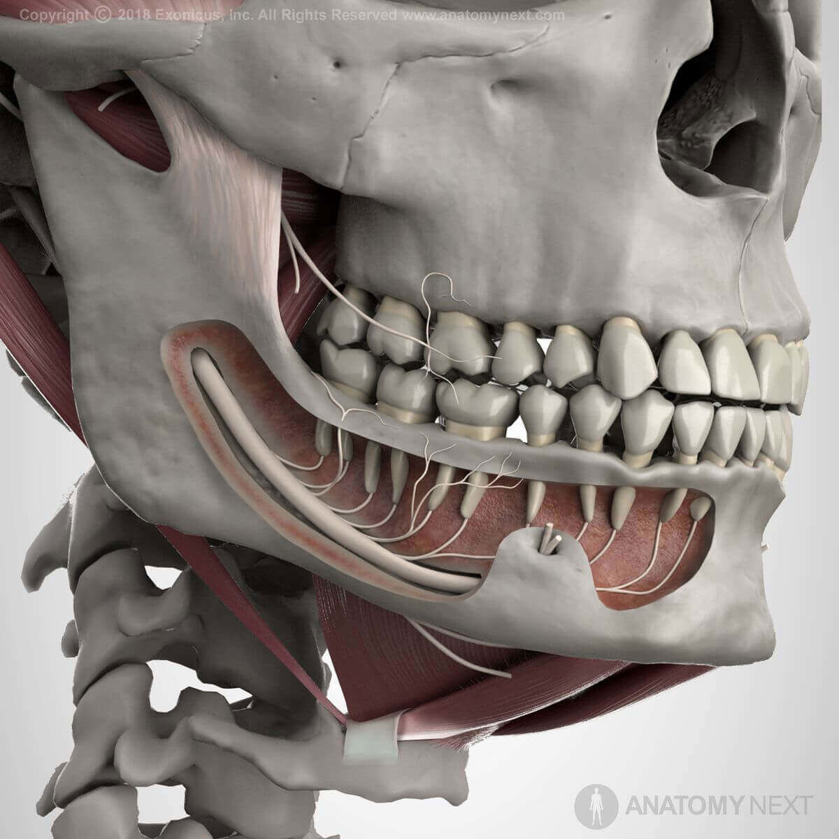
Inferior alveolar nerve
The inferior alveolar nerve is the only nerve of the mandibular nerve containing sensory and motor fibers. It arises from the posterior division of the main trunk and passes on the medial aspect of the lateral pterygoid muscle going downward. The inferior alveolar nerve via the mandibular foramen of the mandible reaches the mandibular canal and passes through the canal along its entire length.
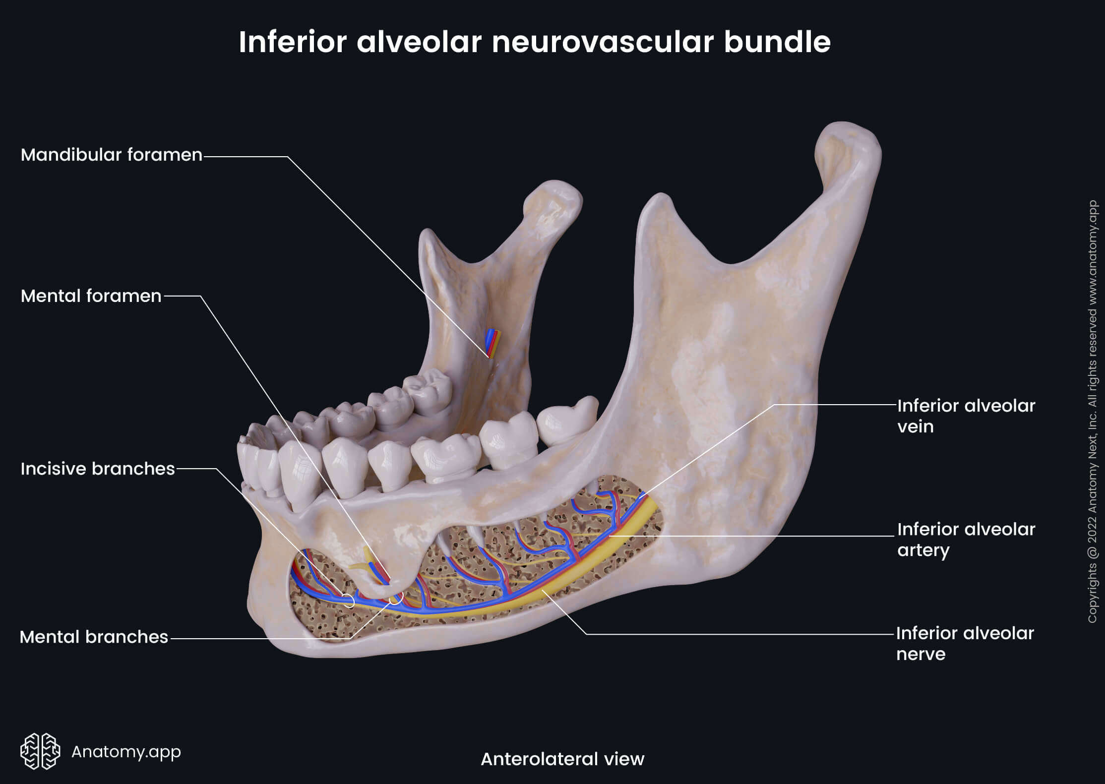
The nerve emerges on the face through the mental foramen and ends on the chin as the mental nerve (sensory) that innervates the skin of the chin and skin and mucosa of the lower lip. Above the mandibular foramen, just before entering it, the inferior alveolar nerve gives off some motor fibers that all together form the mylohyoid nerve (motor) supplying the mylohyoid muscle and the anterior belly of the digastric muscle.
In the mandibular canal, the inferior alveolar nerve gives many small branches that form the inferior alveolar (dental) plexus. From the plexus arise inferior dental branches to the lower teeth (primarily supplying mandibular molars and premolars) and inferior gingival branches to the buccal gingiva.
Anatomy.app
Contact information
- For questions regarding business inquiries. Please contact:
- info@anatomy.app