- Anatomical terminology
- Skeletal system
- Joints
- Muscles
- Heart
- Blood vessels
- Lymphatic system
-
Nervous system
- Central nervous system
-
Peripheral nervous system
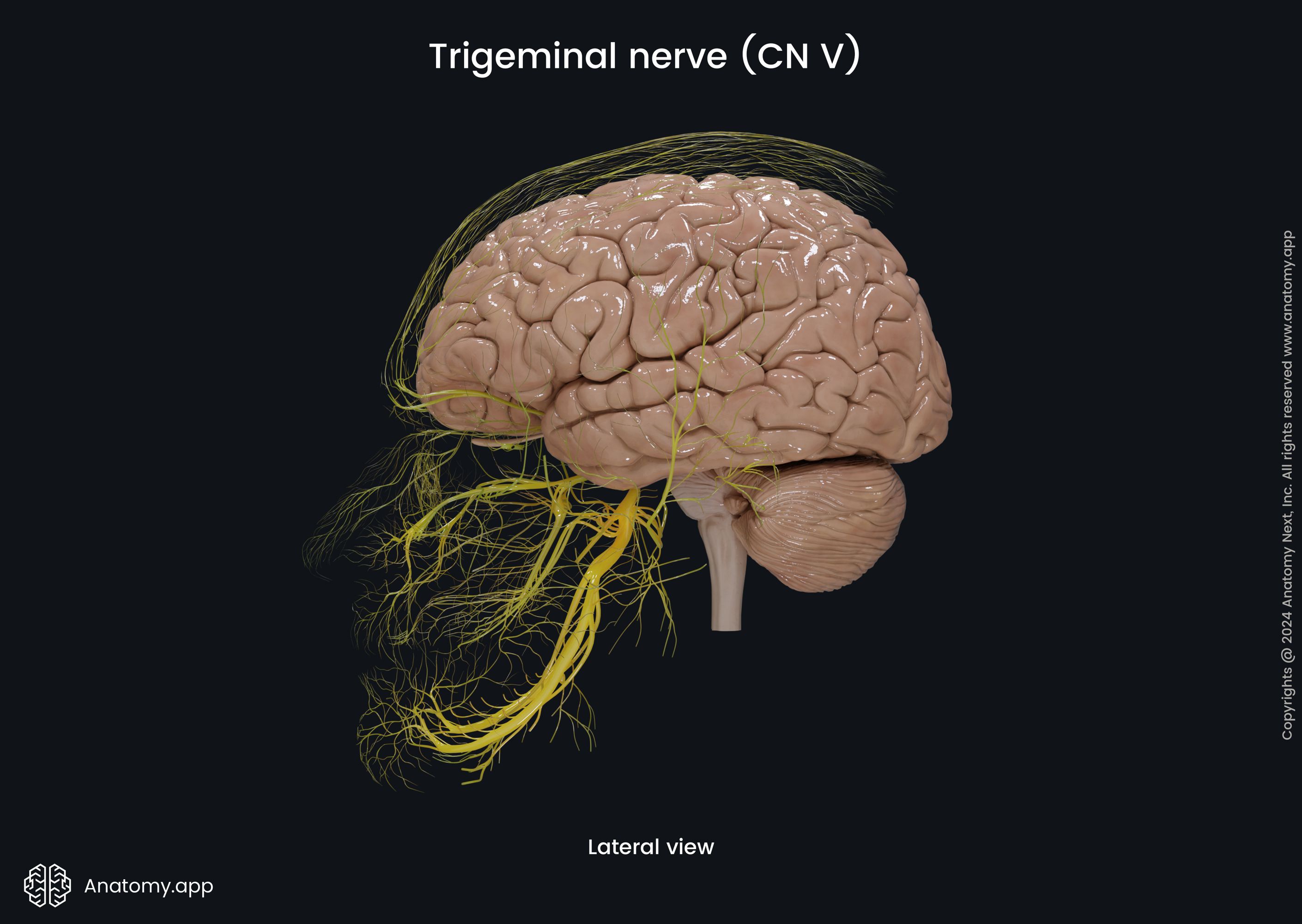
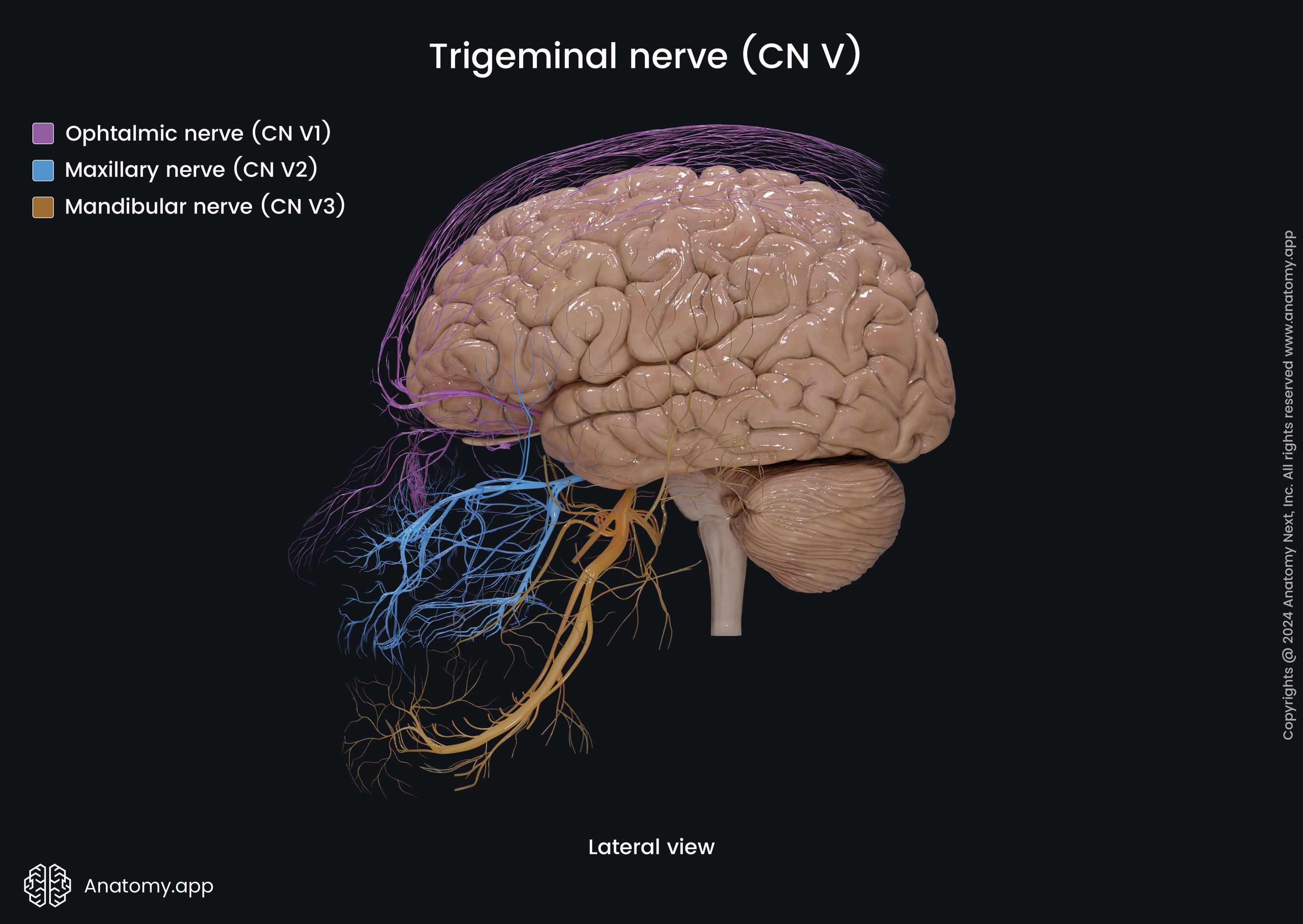
- Cranial nerves
- Spinal nerves
- Respiratory system
- Digestive system
- Urinary system
- Female reproductive system
- Male reproductive system
- Endocrine glands
- Eye
- Ear
Maxillary nerve (CN V2)
The maxillary nerve (CN V2, Latin: nervus maxillaris) is the second branch or division of the trigeminal nerve (CN V), also known as the maxillary division of the trigeminal nerve. It conveys sensory information to the central nervous system from the middle parts of the face, including skin in this area, upper gingiva and upper teeth, mucosa of the palate, nasal cavity, nasopharynx, upper lip and cheek.
Maxillary nerve course
The maxillary nerve arises from the trigeminal ganglion in the middle cranial fossa, travels within the lateral wall of the cavernous sinus, and leaves the cranial cavity through the foramen rotundum, emerging in the pterygopalatine fossa. Before leaving the cranial cavity, the maxillary nerve gives off a meningeal branch to the meninges of the middle cranial fossa.
After emerging in the pterygopalatine fossa, the maxillary nerve gives rise to most of its branches, including two branches to the pterygopalatine ganglion (pterygopalatine nerves). The maxillary nerve further passes forward through the pterygopalatine fossa, giving off several more sensory branches, and turns laterally, reaching the posterior surface of the maxilla.
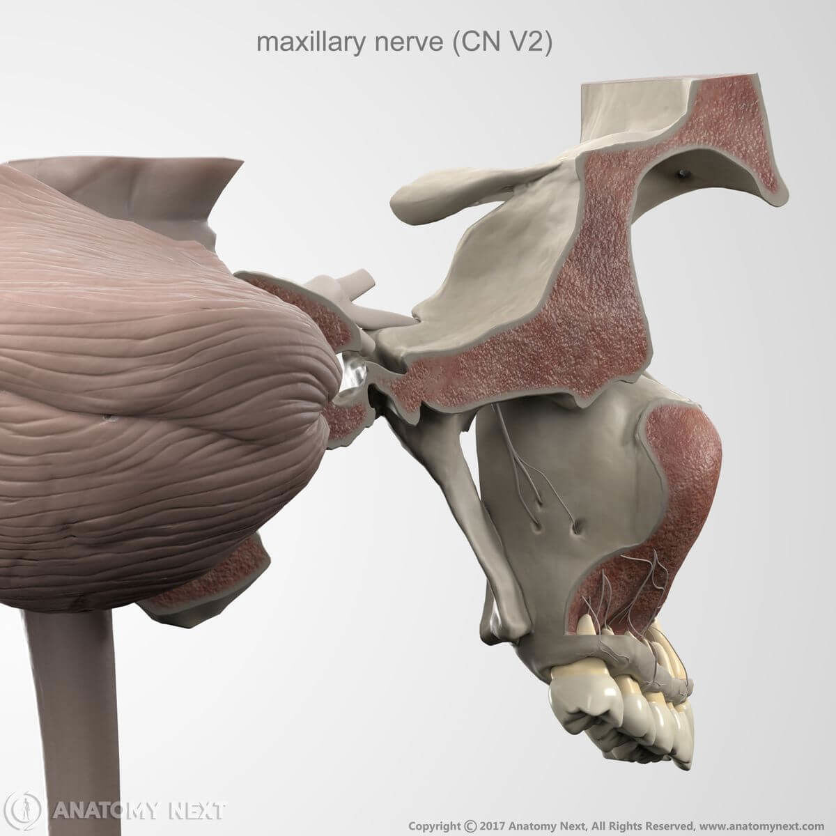
The nerve then travels through the pterygomaxillary fissure and reaches the infratemporal fossa, where it goes medially and through the inferior orbital fissure to reach the orbit. Further, the maxillary nerve passes anteriorly in the inferior wall or floor of the orbit - first in the infra-orbital groove and, further, in the infra-orbital canal. Within this canal, the maxillary nerve continues as the infraorbital nerve.
The infraorbital nerve gives off several branches within the canal and then passes through the infra-orbital foramen, emerging on the face. Right after leaving the infra-orbital foramen, the infraorbital nerve divides into several sets of sensory branches to innervate the skin from the level of the eyes to the mouth.
Maxillary nerve branches
Branches of the maxillary nerve can be categorized in groups by the location of their origins:
- Originating in the cranial cavity:
- Originating in the pterygopalatine fossa:
- Originating in the infra-orbital canal or groove - branches of the infraorbital nerve:
- Originating on the face - branches of the infraorbital nerve:
Meningeal branch of maxillary nerve
The meningeal branch of the maxillary nerve, also referred to as the middle meningeal nerve, arises in the cranial cavity just before the maxillary nerve reaches the cavernous sinus and leaves the cranial cavity. This nerve supplies the dura mater in the anterior part of the middle cranial fossa with sensory fibers.
Pterygopalatine nerves
The pterygopalatine nerves (also known as the sphenopalatine branches) are two to three nerves that arise from the maxillary nerve within the pterygopalatine fossa. They descend to the pterygopalatine ganglion. These nerves give sensory afferent branches to the ganglion and receive back efferent branches. Branches of pterygopalatine nerves innervate the mucous membrane of the palate and nasal cavity.
Zygomatic nerve
The zygomatic nerve is mainly sensory as it carries sensory fibers to the CNS from the skin of the face. However, it also carries parasympathetic fibers that innervate the lacrimal gland as in orbit this nerve has an anastomosis with the lacrimal nerve of the ophthalmic nerve (CN V1).
The zygomatic nerve arises in the pterygopalatine fossa. From the fossa, via the pterygomaxillary fissure, it reaches the infratemporal fossa. From there, this nerve enters the orbit through the inferior orbital fissure, goes along the lateral wall of the orbit, and divides into two branches - the zygomaticotemporal and zygomaticofacial nerves.
The zygomaticotemporal nerve arises in a canal in the zygomatic bone and emerges on the face through the zygomaticotemporal foramen. It innervates the skin on the face in the anterior area of the temporal region. The other branch of the zygomatic nerve, the zygomaticofacial nerve, emerges on the face and supplies the skin of the lateral cheek region with sensory nerve fibers.
The zygomaticofacial nerve passes along the inferolateral angle of the orbit, enters the zygomatic bone through the zygomatico-orbital foramen, and emerges on the face through the zygomaticofacial foramen. The nerve perforates the orbicularis oculi muscle and supplies the skin of the cheek with sensory fibers.
The zygomaticofacial nerve forms a nerve plexus together with the zygomatic branches of the facial nerve (CN VII) and the palpebral branches of the maxillary nerve (CN V2), specifically, with the inferior palpebral branches of the infraorbital nerve.
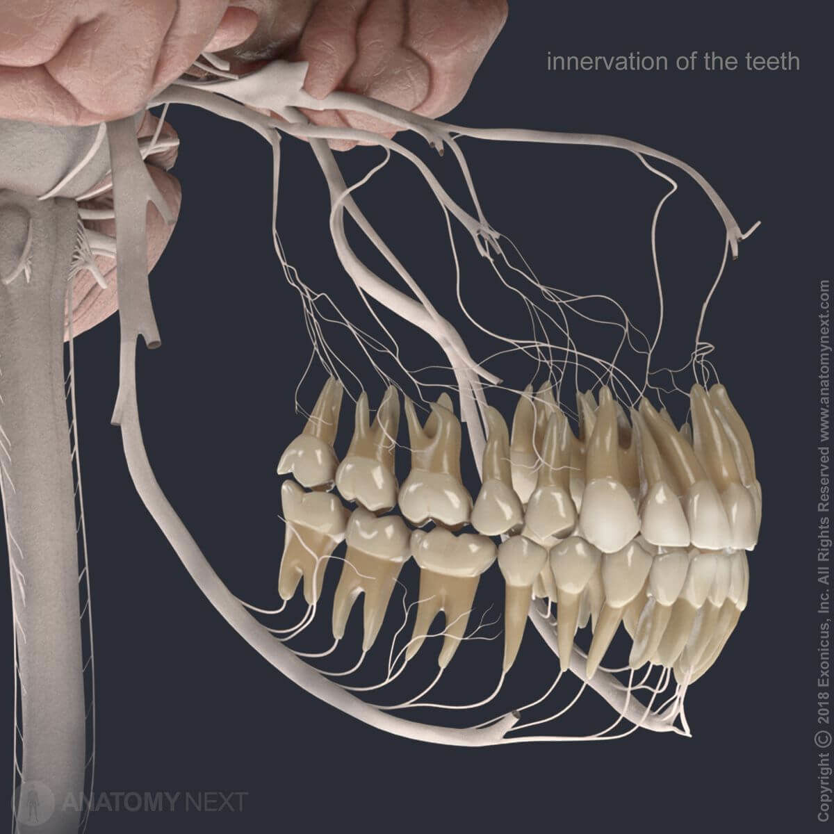
Posterior superior alveolar nerve
The posterior superior alveolar nerve is a branch of the maxillary nerve, which invests its sensory branches in the superior alveolar (dental) plexus. The posterior superior alveolar nerve arises in the pterygopalatine fossa and travels antero-inferiorly. Through the pterygomaxillary fissure, the nerve reaches the infratemporal fossa, and there it pierces the infratemporal surface of the maxilla.
Through the alveolar foramina of the maxilla, the nerve enters the alveolar canals and descends beneath the mucosa of the maxillary sinus, which it also supplies. Then the posterior superior alveolar nerve divides into smaller nerves, which link up and form a part of the superior alveolar (dental) plexus. The posterior superior alveolar nerve provides sensory innervation for the maxillary molar teeth, as well as the upper gingiva and the adjoining part of the cheek.
Pharyngeal nerve
The pharyngeal nerve is a small branch that arises from the maxillary nerve right after it emerges through the foramen rotundum in the pterygopalatine fossa and posterior to the pterygopalatine ganglion. This nerve is connected to the pterygopalatine ganglion, as it receives parasympathetic fibers from this ganglion (these fibers reach the ganglion via the greater petrosal nerve).
The pharyngeal nerve leaves the pterygopalatine fossa through the palatovaginal canal (also known as the pharyngeal canal; located between the palatine bone and the sphenoid bone), where it is accompanied by the pharyngeal branch of the maxillary artery (or pharyngeal artery).
The nerve reaches the superior-posterior aspect of the nasal cavity and supplies the mucosa of the nasopharynx with sensory fibers (that are carried by this nerve, but do not synapse in the pterygopalatine ganglion), and pharyngeal mucosal glands with its parasympathetic fibers (that come from neurons located within the ganglion).
Greater palatine nerve
The greater palatine nerve (also called the anterior palatine nerve) branches off the maxillary nerve right after it emerges in the pterygopalatine fossa, and it is connected to the pterygopalatine ganglion. It receives parasympathetic and special sensory (taste) fibers from this ganglion (the fibers reach the ganglion via the greater petrosal nerve).
The nerve travels through the greater palatine canal (a canal that starts inferiorly in the fossa and goes through the maxilla and palatine bones, reaching the posterior hard palate). In this canal, it gives rise to the lateral posterior inferior nasal nerve, which supplies the postero-inferior lateral wall of the nasal cavity.
Afterward, the greater palatine nerve emerges in the oral cavity via the greater palatine foramen, then curves anteriorly and travels along the hard palate (it may create a groove here). With its sensory fibers, this nerve supplies the mucosa of the posterior hard palate, with its parasympathetic fibers - the palatine mucosal glands, and with the special sensory (taste) fibers - this nerve supplies taste buds of the palate.
Lesser palatine nerves
The lesser palatine nerves (also known as the posterior palatine nerves) are usually 2 to 3 nerves that arise from the maxillary nerve right after it emerges in the pterygopalatine fossa. These nerves are also connected to the pterygopalatine ganglion. Like the greater palatine nerve, these nerves receive parasympathetic and special sensory fibers (that convey taste sensation) from the pterygopalatine ganglion to innervate palatine mucosal glands and taste buds.
The lesser palatine nerves travel inferiorly and leave the pterygopalatine fossa through the lesser palatine canals, which are found posterior to the greater palatine canal (these canals are found in the palatine bones and sometimes are described as arising from the greater palatine canal). The nerves emerge on the roof of the oral cavity through the lesser palatine foramina, innervating mucosa of the uvula, soft palate and the palatine tonsils.
Nasopalatine nerve
The nasopalatine nerve (also called the long sphenopalatine nerve) is another branch that emerges in the pterygopalatine fossa right after the maxillary nerve appears through the foramen rotundum and this nerve is also connected to the pterygopalatine ganglion. It also receives parasympathetic and special sensory (taste) fibers from this ganglion via the greater petrosal nerve. With these fibers it innervates nasal and palatine mucosal glands and palatine taste buds, respectively.
The nerve travels inferomedially through the sphenopalatine foramen along with the posterior superior nasal nerves and the sphenopalatine artery, emerging in the nasal cavity posterior to the superior nasal meatus. Then it passes along the superior wall of the nasal cavity and reaches the nasal septum (note: the nerve may create a groove in the vomer).
On its way, the nasopalatine nerve innervates the mucosa of the postero-inferior part of the nasal septum, then travels further antero-inferiorly along the nasal septum, reaching the incisive canal. The nasopalatine nerve passes through this canal, entering the roof of the oral cavity via the incisive foramen. The nerve then supplies the mucosa lining the anterior hard palate and gingiva posterior to the maxillary incisor teeth.
Infraorbital nerve
The infraorbital nerve is the terminal branch of the maxillary nerve. It arises from the maxillary nerve, and through the inferior orbital fissure, it reaches the orbit, where it goes forward and medially on the inferior wall of the orbit. It goes into the infra-orbital groove and from there into the infra-orbital canal. The nerve emerges on the face through the infra-orbital foramen, and then it branches into groups of nerves forming the so-called pes anserinus minor on the face.
The branches of the pes anserinus minor (inferior palpebral branches, external nasal branches, superior labial branches) innervate the skin of the lower eyelid, the nose (specifically, the skin of ala nasi), and the skin of the upper lip and lateral aspect of the cheek. The infraorbital nerve also gives the anterior and middle superior alveolar nerves within the infra-orbital groove and canal.
Middle superior alveolar nerve
The middle superior alveolar nerve is a branch of the infraorbital nerve, which provides sensory fibers to the superior alveolar (dental) plexus. This nerve arises in the infra-orbital groove (or in the infra-orbital canal), and goes antero-inferiorly within the lateral wall of the maxillary sinus.
The nerve divides into smaller branches, which link up with other branches of the superior alveolar (dental) plexus supplying sensory fibers to the upper premolar teeth. The middle superior alveolar nerve is a variable nerve, it can be absent, or there can be one or two of them.
Anterior superior alveolar nerve
The anterior superior alveolar nerve is also a branch of the infraorbital nerve, which provides sensory fibers to the superior dental plexus. It arises near the middle of the infra-orbital canal, crosses the anterior wall of the maxillary sinus, curves under the infra-orbital foramen, then goes medially towards the nose and finally turns downwards.
The nerve then divides into smaller branches which link with the superior alveolar (dental) plexus to supply upper teeth - the upper incisors and canines - with sensory fibers. The anterior superior alveolar nerve usually innervates all anterior teeth of the upper jaw, then loops backwards to join the middle superior alveolar nerve to form the superior dental plexus.
The anterior superior alveolar nerve also gives off a nasal branch, which goes through a small canal in the lateral wall of the inferior nasal meatus innervating mucous membranes on the anterior aspect of the inferior meatus and the floor of the nasal cavity. The anterior superior nerve also communicates with the nasal branches arising from the pterygopalatine ganglion, and supplies the adjoining part of the nasal septum with its terminal nerve endings.
Inferior palpebral branches
The inferior palpebral branches are a set of sensory nerves arising from the infraorbital nerve when it emerges on the face. These branches supply the skin of the lower eyelid and conjunctiva with sensory fibers.
External nasal branches
The external nasal branches are another set of sensory nerves arising from the infraorbital nerve when it emerges on the face. These nerves supply the skin of the alae (wings) of the nose, nasal vestibule, and cheeks.
Superior labial branches
The superior labial branches form the third set of sensory nerves arising from the infraorbital nerve after it emerges on the face. These branches provide sensory nerve fibers to the skin of the upper lip, cheeks, and alae of the nose.
Anatomy.app
Contact information
- For questions regarding business inquiries. Please contact:
- info@anatomy.app