- Anatomical terminology
- Skeletal system
- Joints
- Muscles
- Heart
- Blood vessels
- Lymphatic system
- Nervous system
- Respiratory system
- Digestive system
- Urinary system
- Female reproductive system
- Male reproductive system
- Endocrine glands
- Eye
- Ear
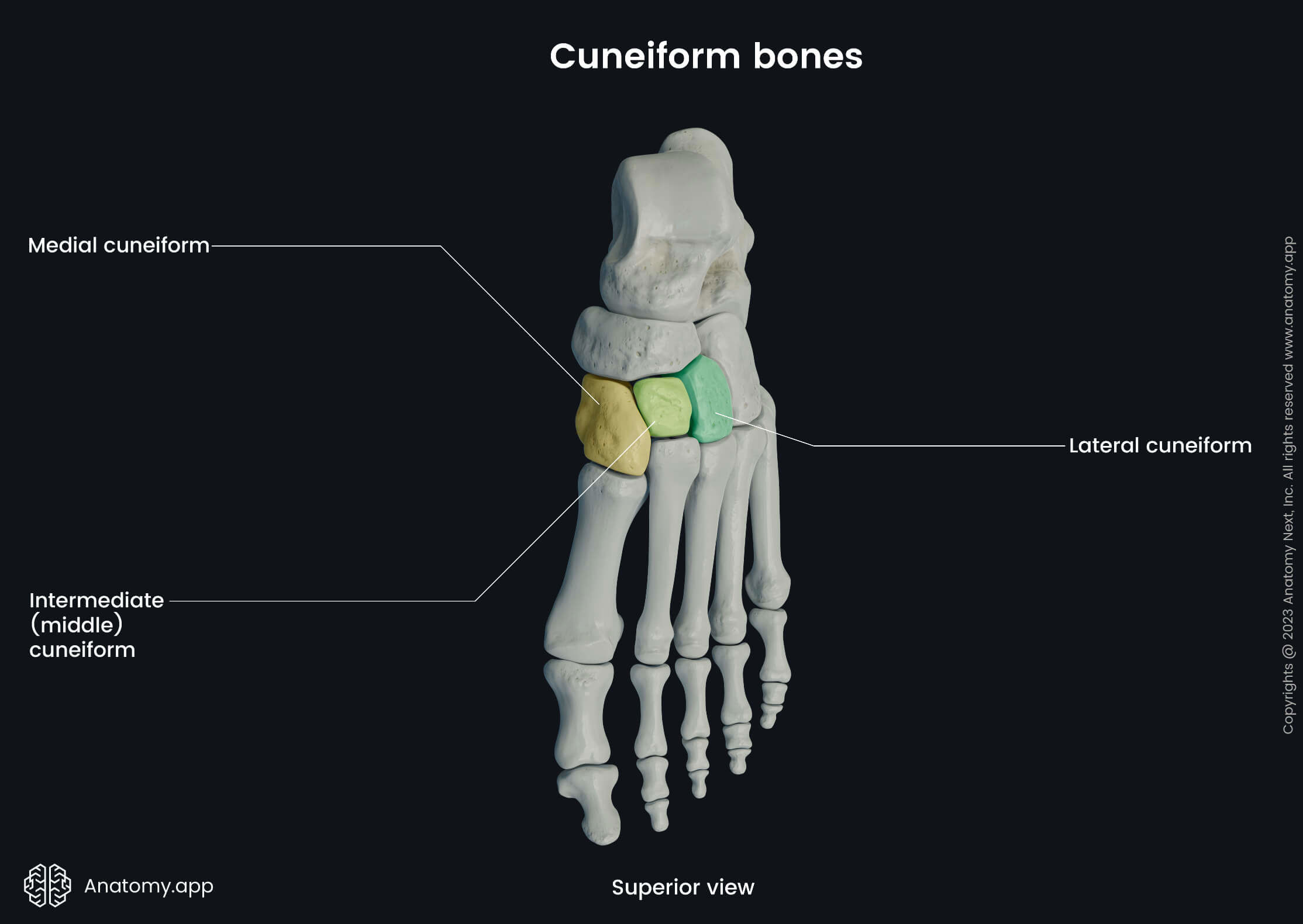
Cuneiform bones
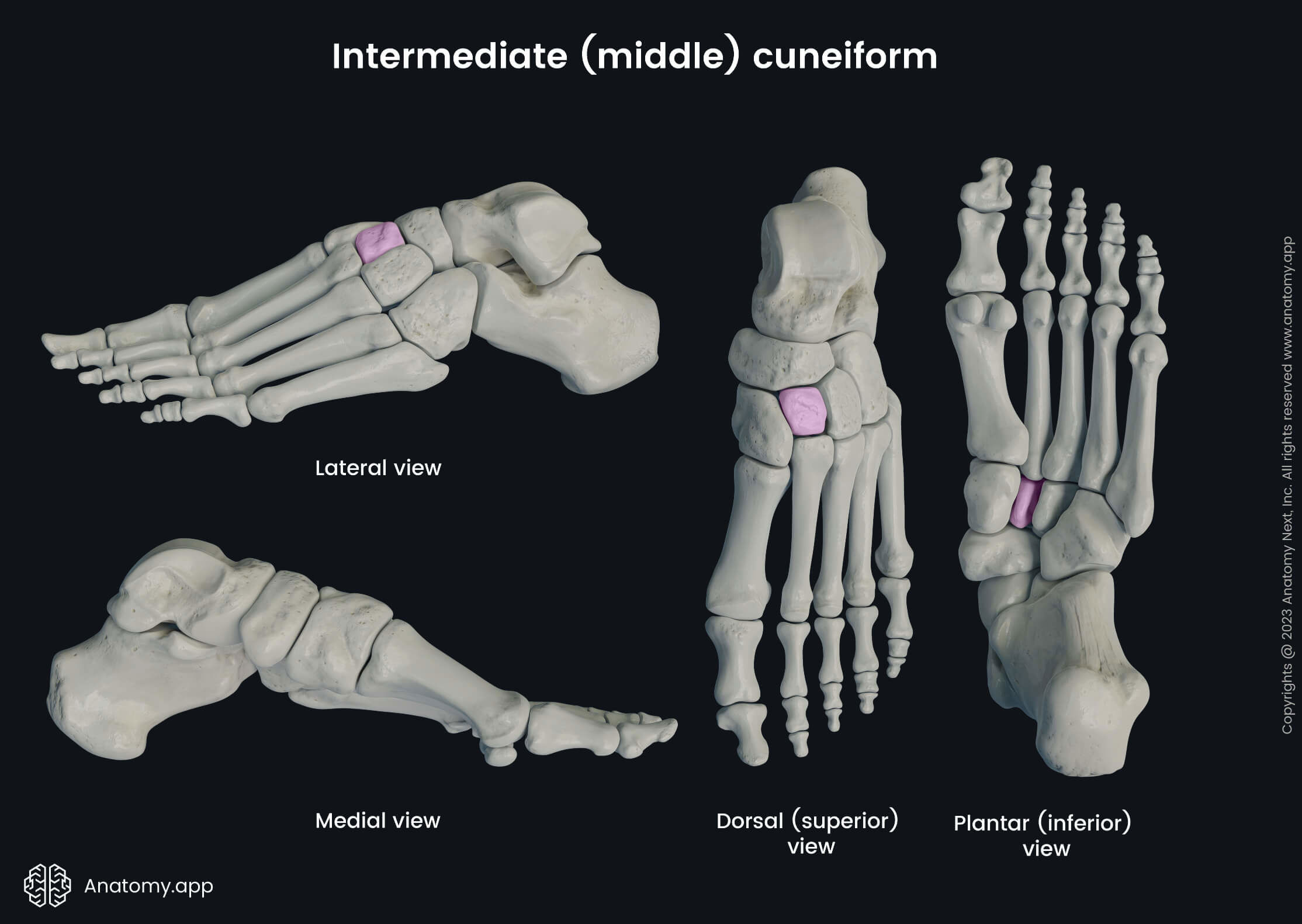
The cuneiform bones (Latin: ossa cuneiformia) are a set of three tarsal bones located between the proximally situated navicular bone and distally positioned first three metatarsal bones. Lateral to the lateral cuneiform bone is the cuboid bone.
Each foot is composed of three cuneiform bones located on its medial side. All cuneiform bones participate in forming the shape of the transverse arch of the foot. The cuneiform bones are named as follows:
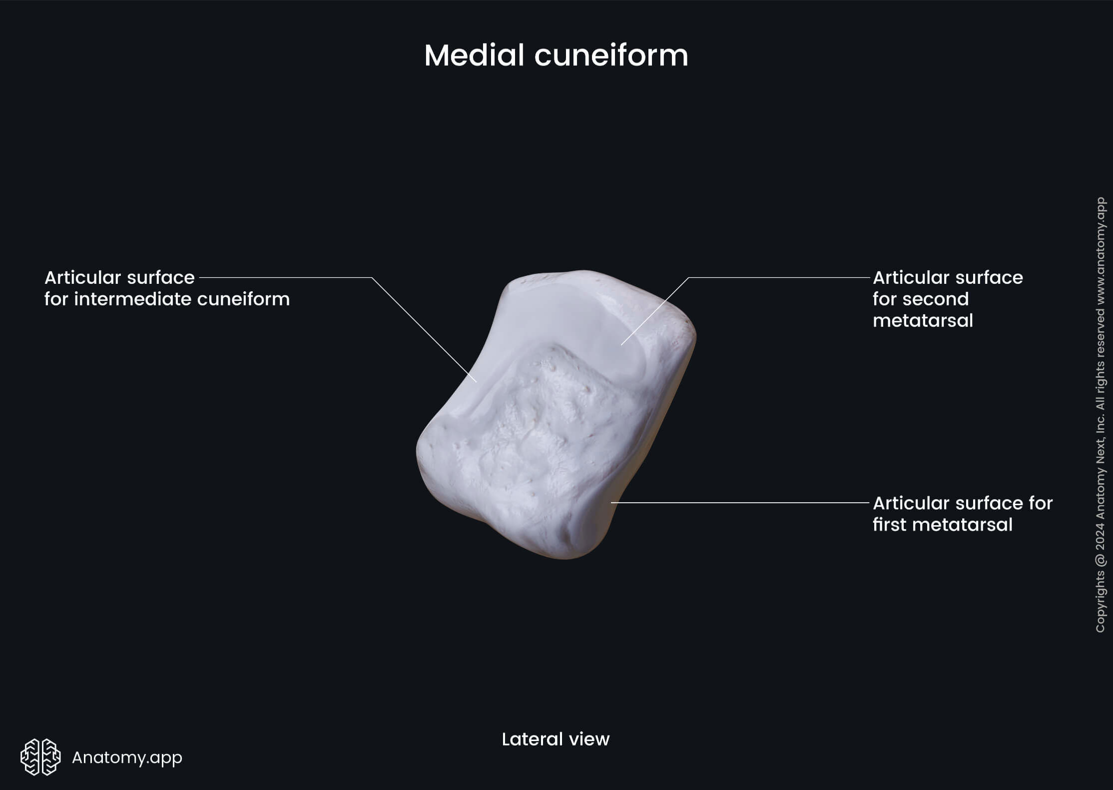
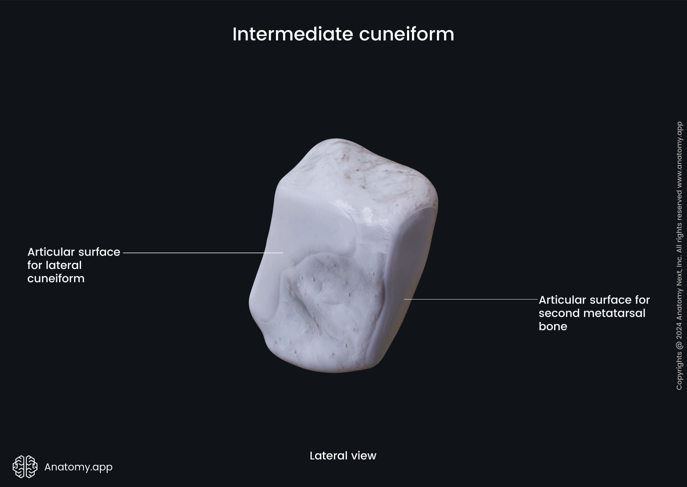
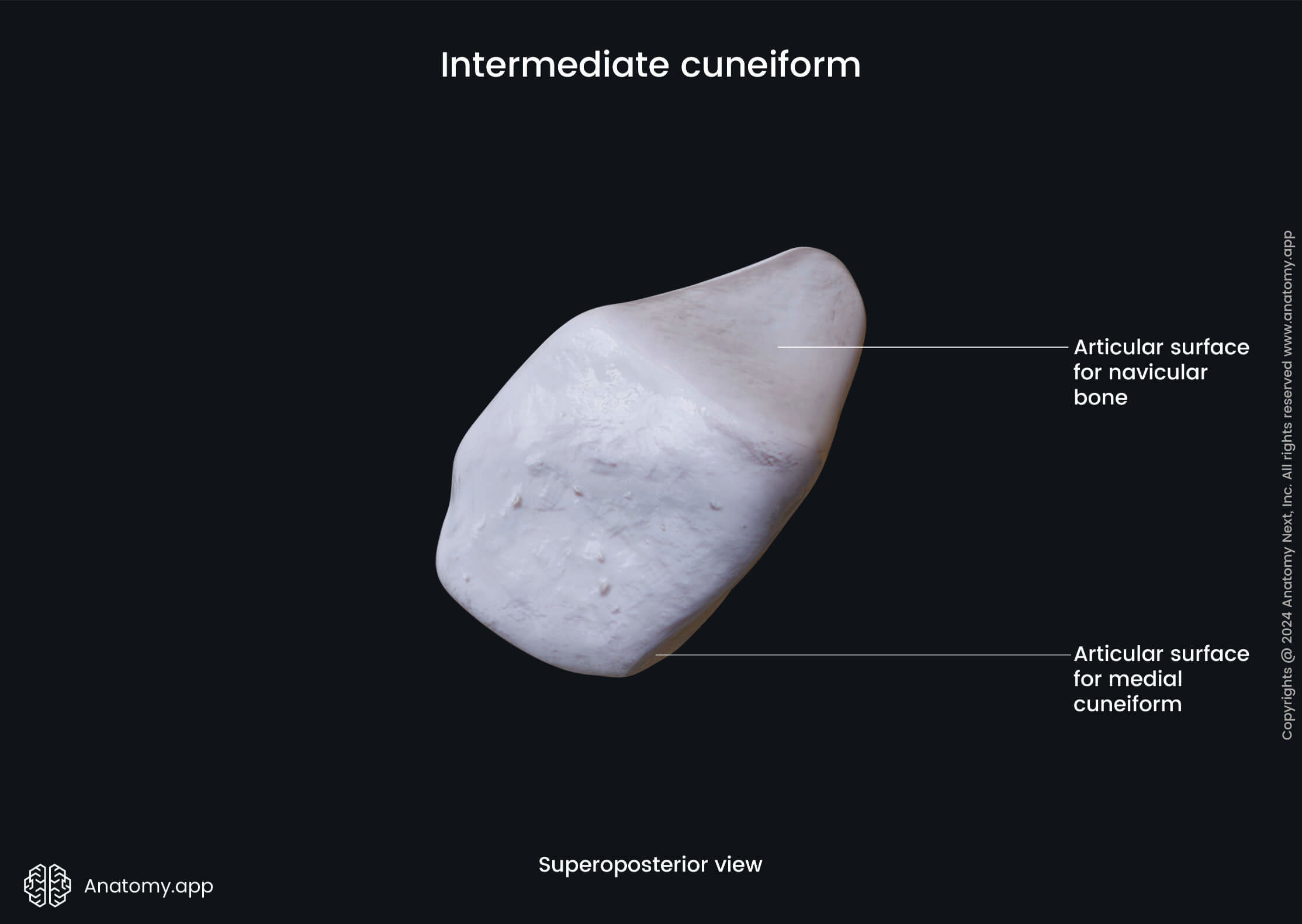
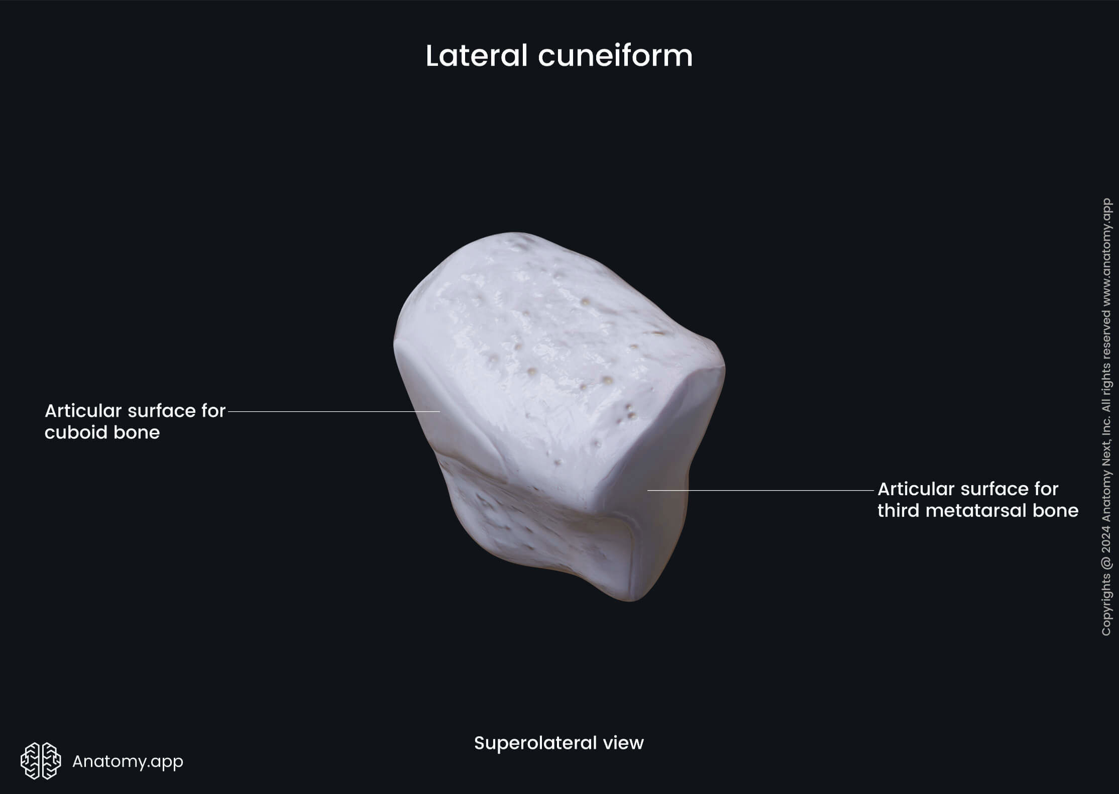
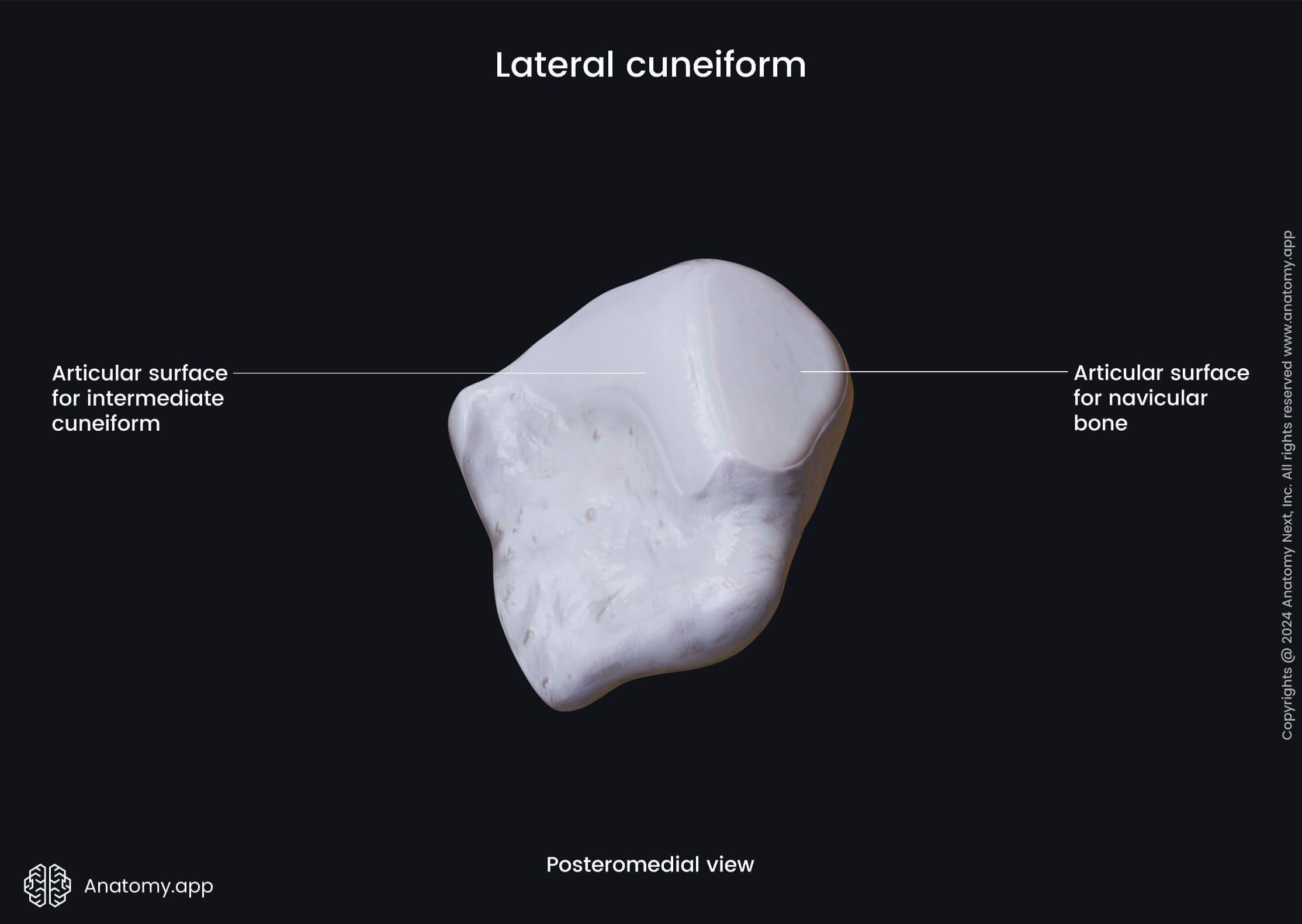
Each cuneiform bone articulates with each other and the adjacent bones, and all of them present four articular surfaces:
- Posterior articular surface
- Anterior articular surface
- Lateral articular surface
- Medial articular surface
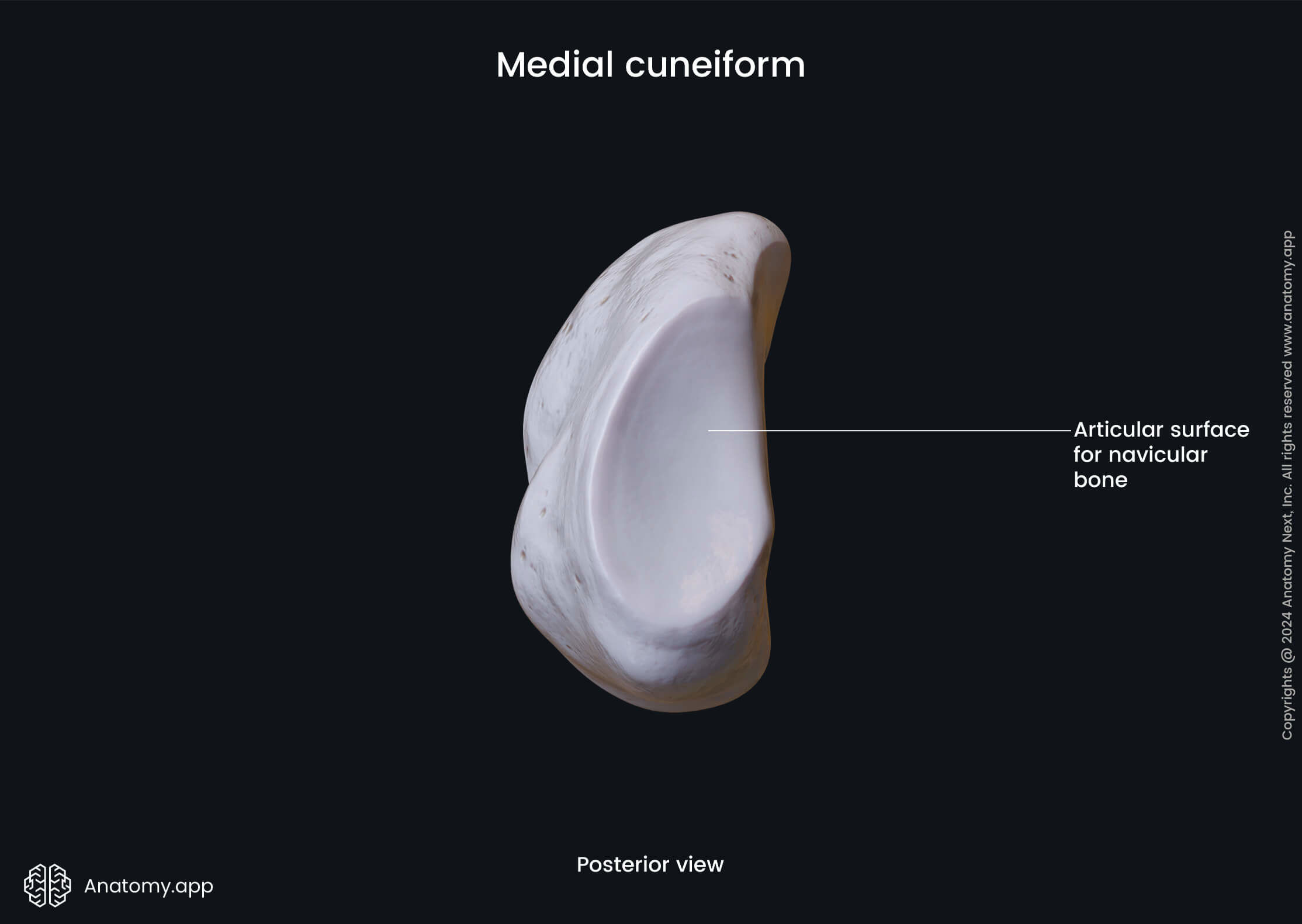
The posterior articular surfaces of all cuneiform bones articulate with the navicular bone, while the anterior articular surfaces with the corresponding metatarsal bones. The lateral and medial articular surfaces articulate with the surfaces of the adjacent cuneiform bones. But the lateral surface of the lateral cuneiform bone articulates with the cuboid bone.
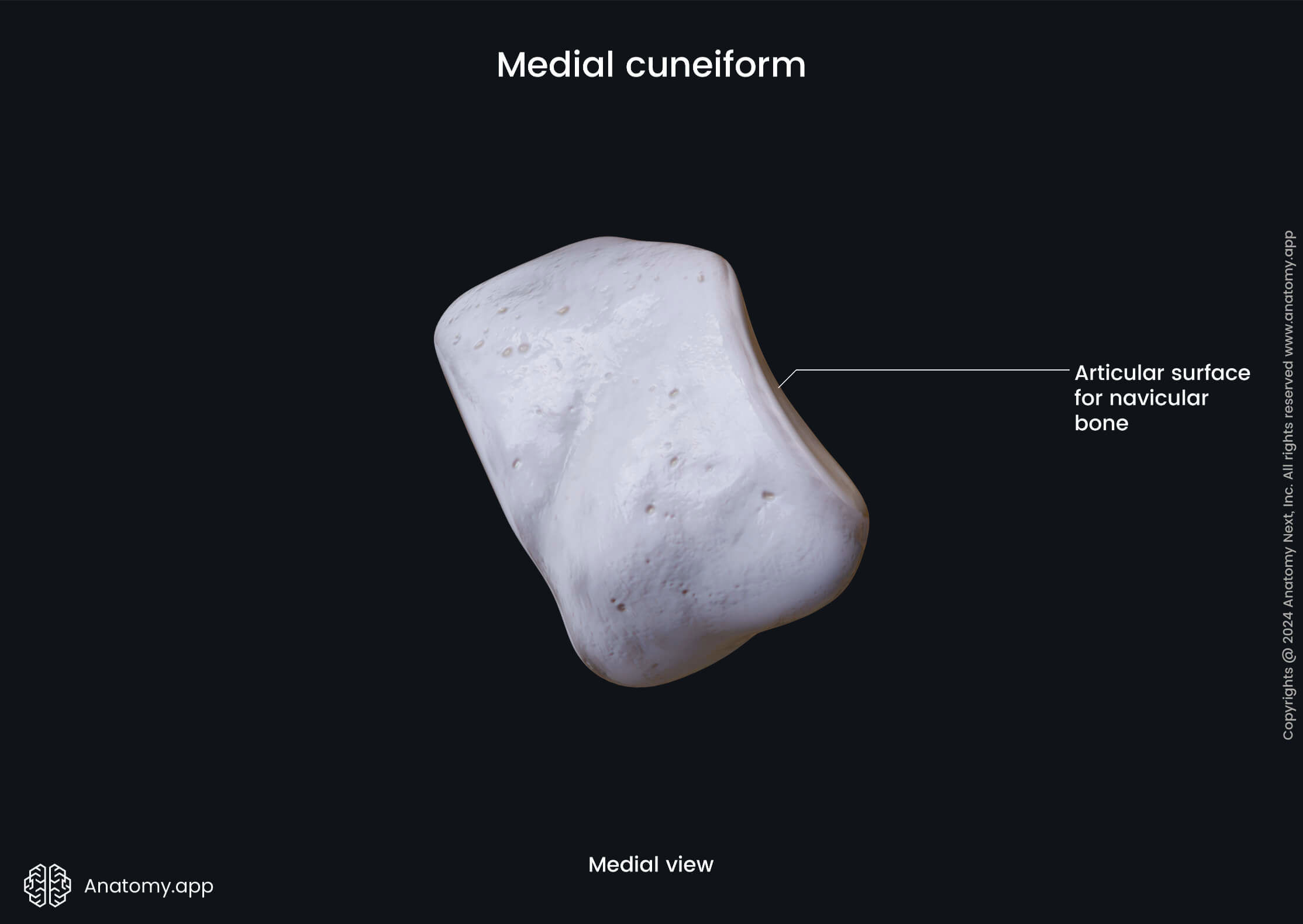
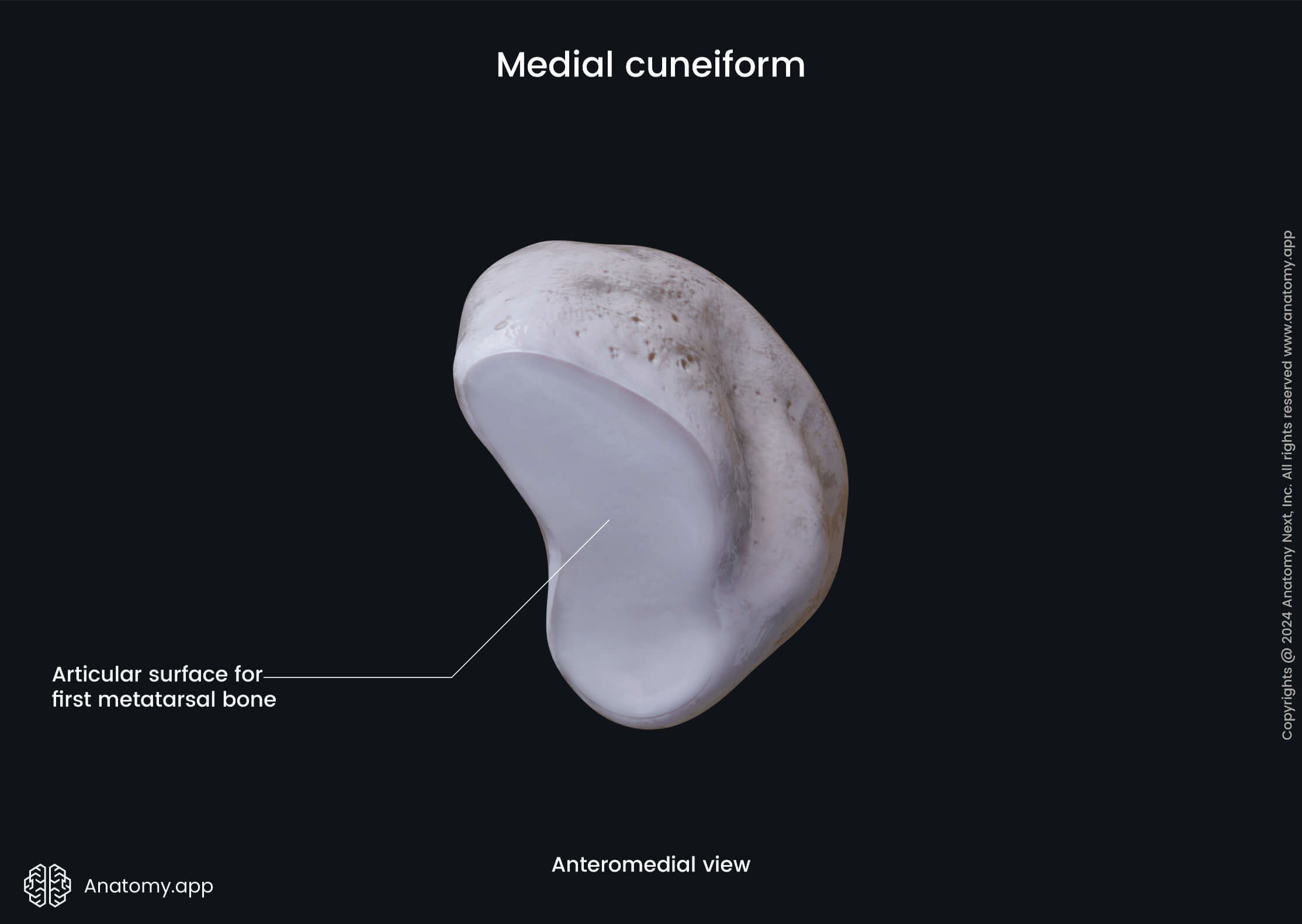
Medial cuneiform bone
The medial cuneiform bone (Latin: os cuneiforme mediale), or simply the first cuneiform, is the largest of all three cuneiform bones. It articulates with four bones - navicular bone, intermediate cuneiform bone, and the first and second metatarsal bones. The medial cuneiform bone serves as an insertion site for two muscles - tibialis anterior and peroneus longus muscles.

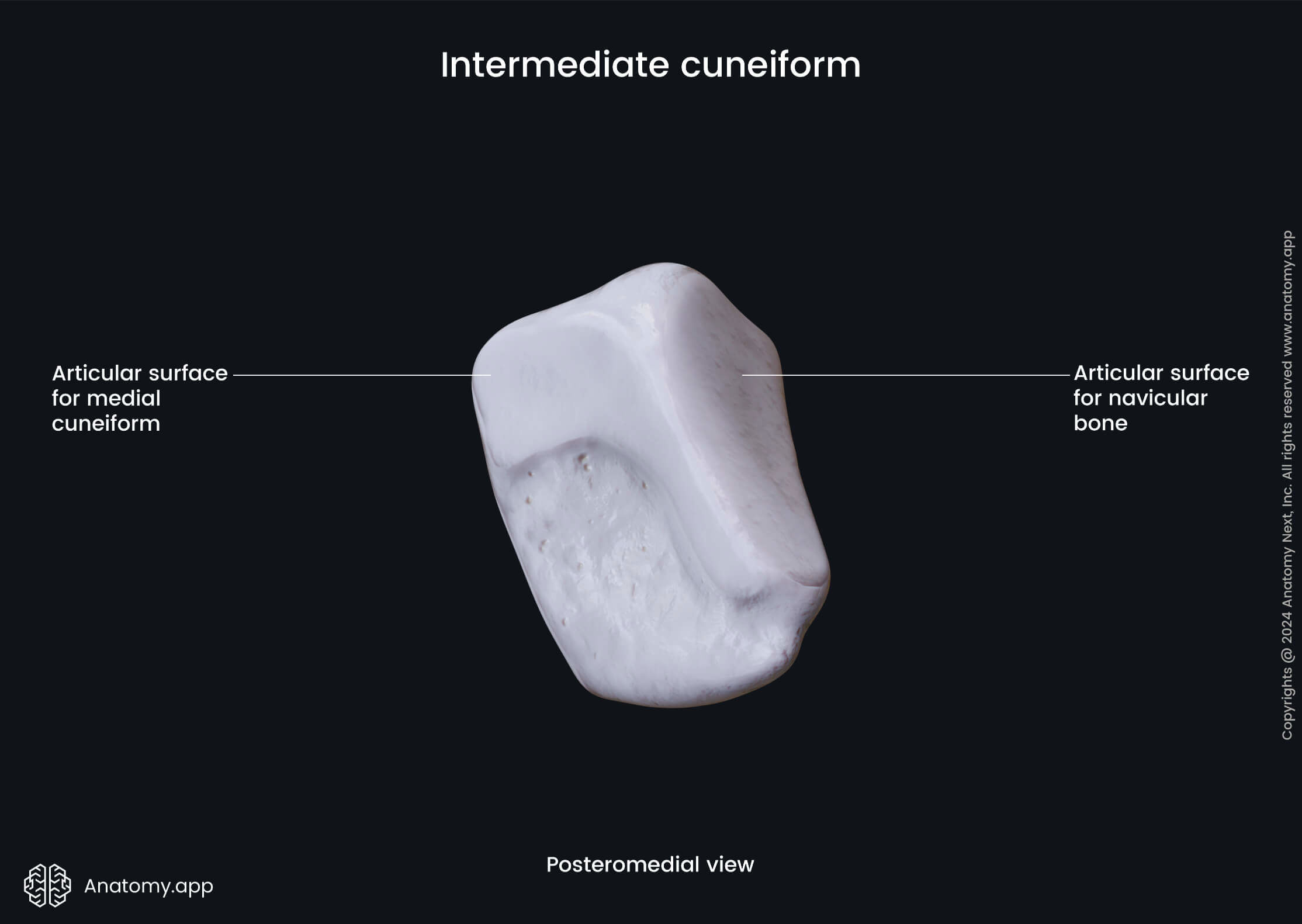
Intermediate cuneiform bone
The intermediate cuneiform bone (Latin: os cuneiforme intermedium), also known as the second cuneiform or the middle cuneiform, is the smallest cuneiform bone. It is situated between the lateral and medial cuneiform bones. The intermediate cuneiform articulates with four bones - navicular bone, medial and lateral cuneiform bones, and the second metatarsal bone.

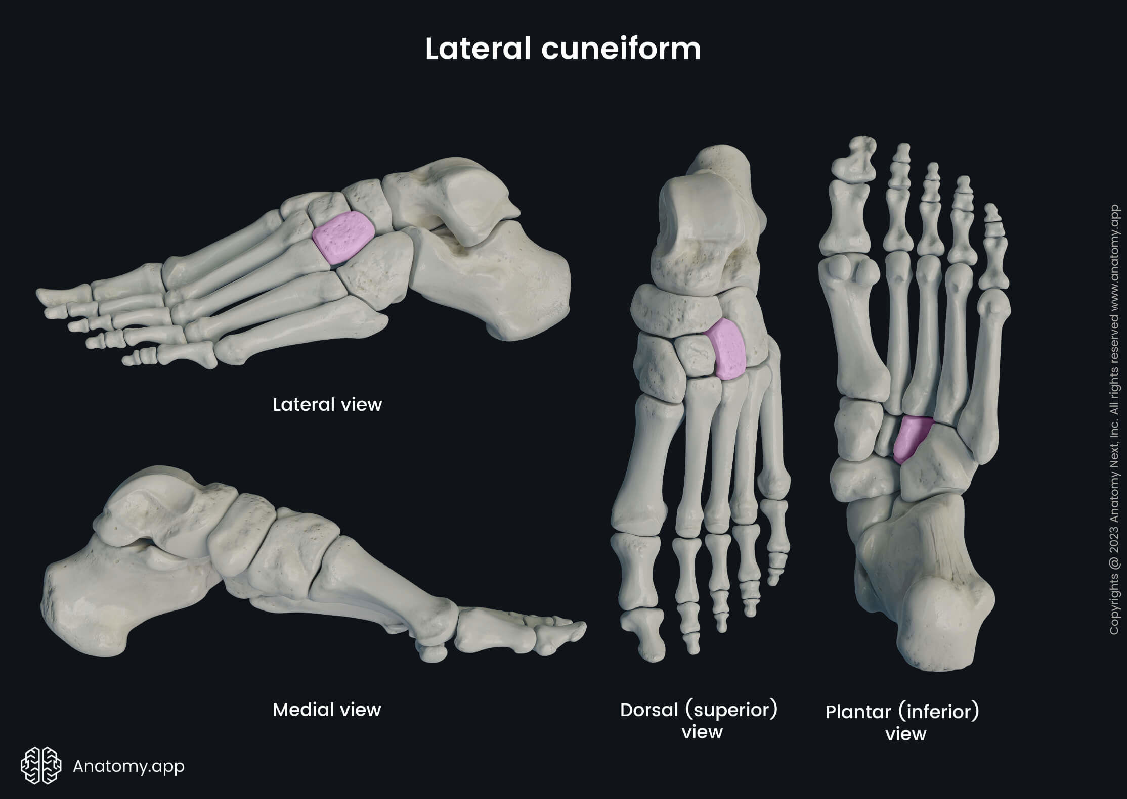
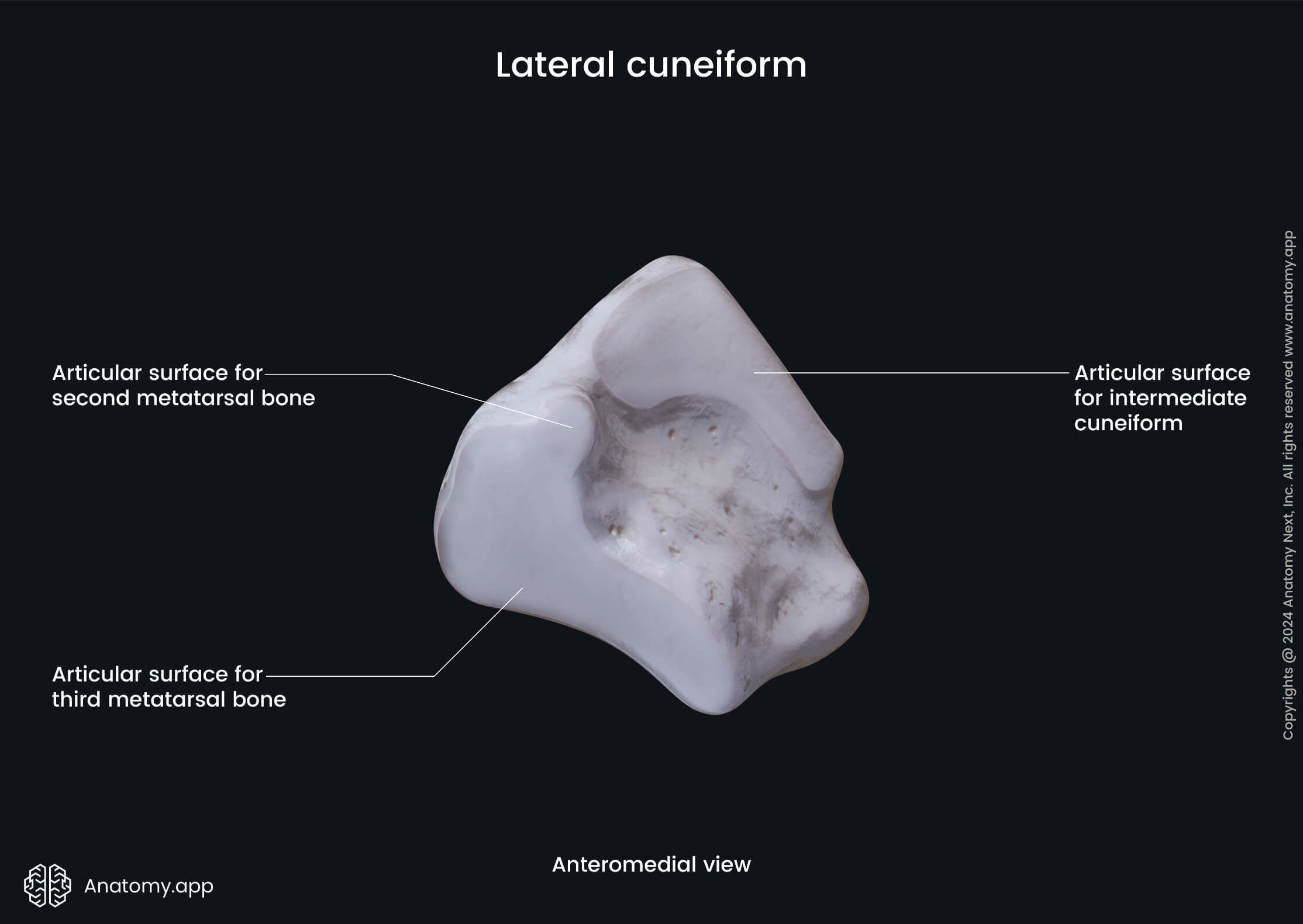
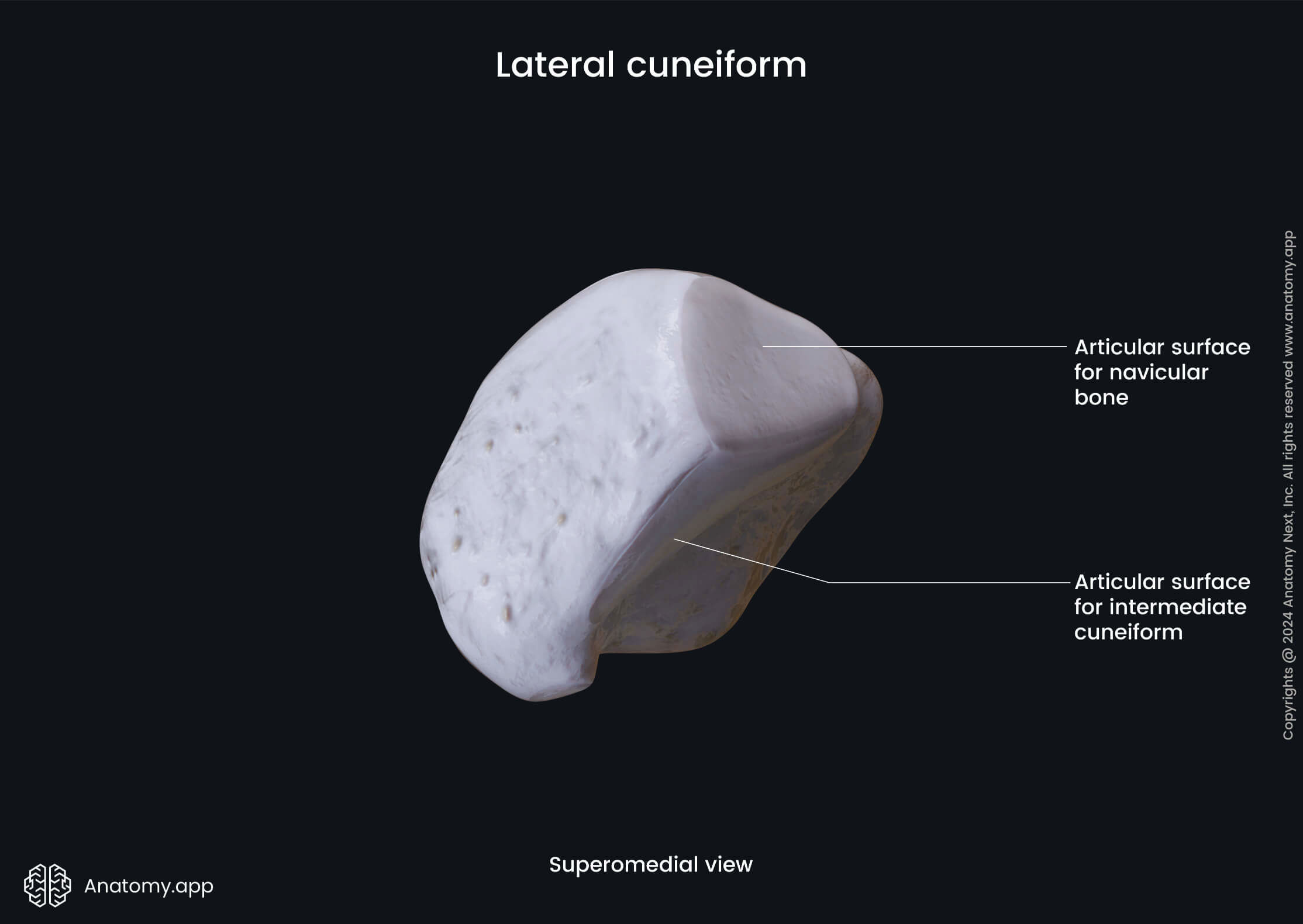
Lateral cuneiform bone
The lateral cuneiform bone (Latin: os cuneiforme laterale) is also known as the third or external cuneiform. It articulates with four bones - intermediate cuneiform, cuboid bone, navicular bone, and the third metatarsal bone. The lateral cuneiform bone serves as an attachment site for two muscles. The tibialis posterior muscle inserts onto the bone, but the flexor hallucis brevis muscle originates from it.
Anatomy.app
Contact information
- For questions regarding business inquiries. Please contact:
- info@anatomy.app