- Anatomical terminology
- Skeletal system
- Joints
- Muscles
- Heart
- Blood vessels
- Lymphatic system
- Nervous system
- Respiratory system
- Digestive system
- Urinary system
- Female reproductive system
- Male reproductive system
- Endocrine glands
- Eye
- Ear
Calcaneus
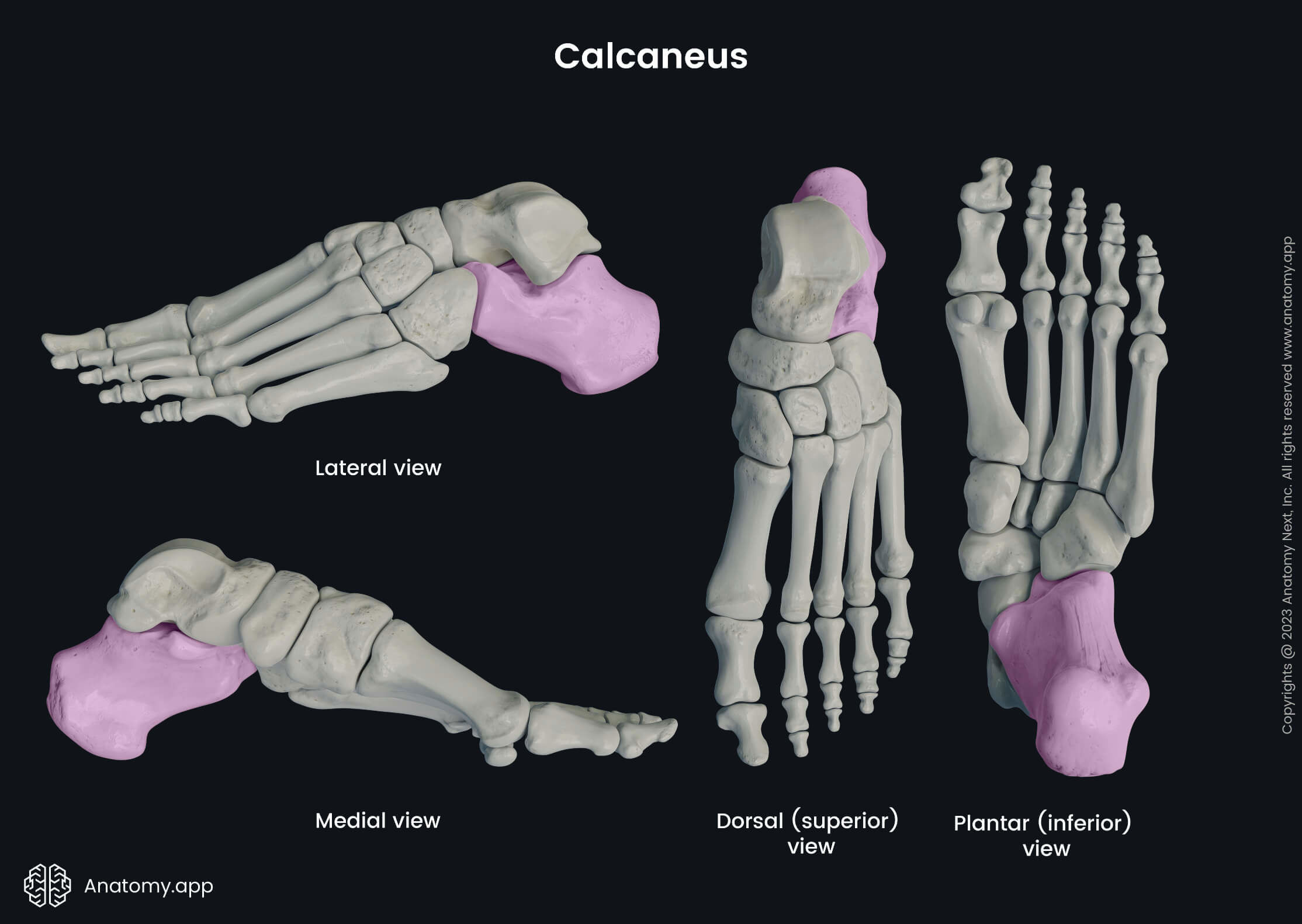
The calcaneus (Latin: calcaneus) is the largest bone of the tarsal bones, and it forms the heel. It is also the largest bone of the foot. The calcaneus articulates with the adjacent located cuboid bone and talus.

The calcaneus acts as a short lever for the triceps surae muscles that insert onto its posterior surface via the Achilles tendon. The calcaneus also provides attachment for other muscles and ligaments, participates in weight-bearing, and provides the stability of the food.
Landmarks of calcaneus
The surfaces of the calcaneus contain several essential anatomical landmarks, including:

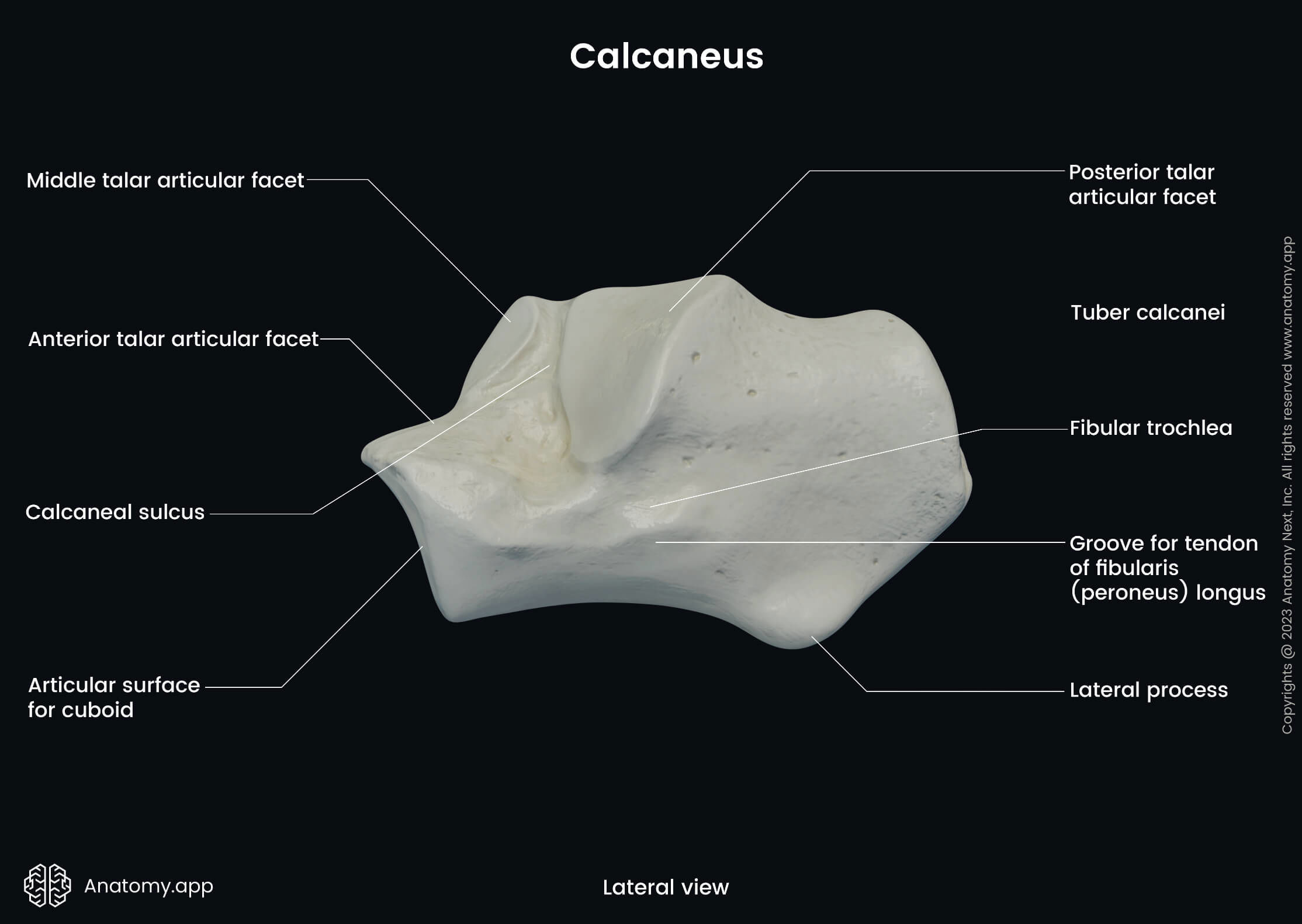
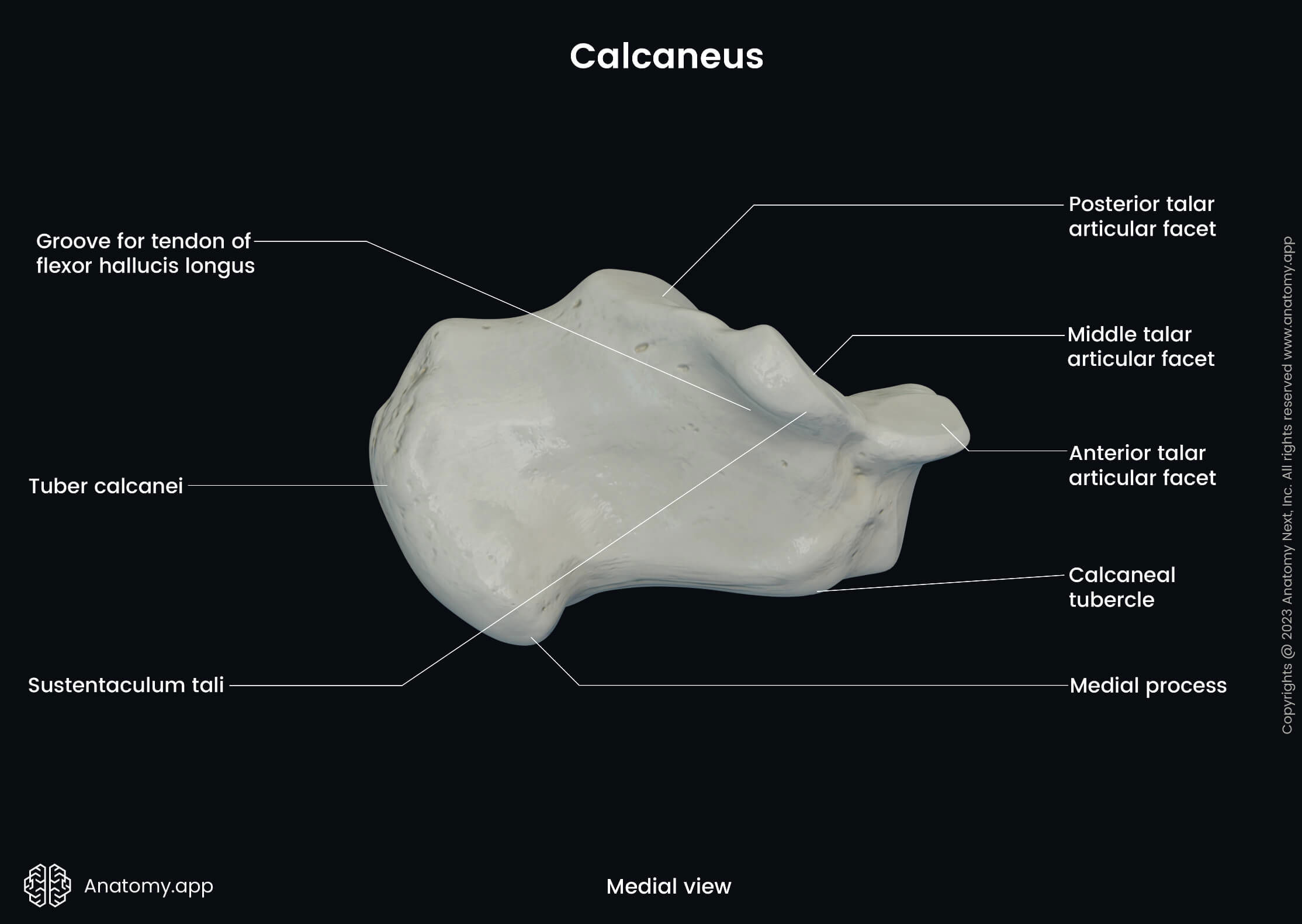
The tuber calcanei (calcaneal tuberosity) is the posterior thickened part of the calcaneus. Also, it is the half of the bone that participates in forming the heel. This tuberosity serves as an origin site for the abductor hallucis and abductor digiti minimi muscles.
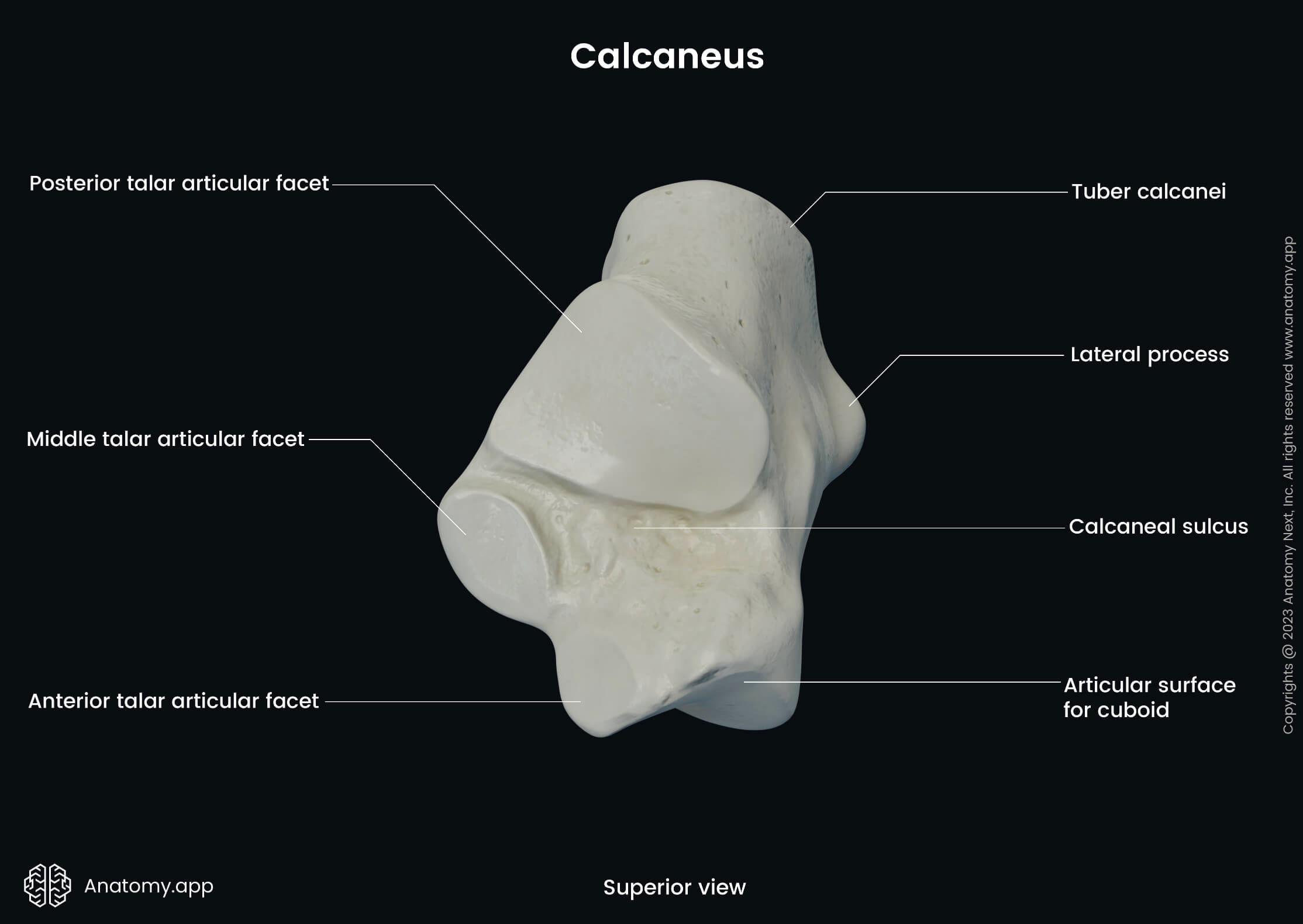
The calcaneal sulcus is a deep groove located on the superior aspect of the calcaneus between the middle and posterior superior articular surfaces. It provides attachments for the interosseous, talocalcaneal and cervical ligaments and a part of the inferior extensor retinaculum.

The calcaneal sulcus forms the tarsal sinus with a corresponding groove on the inferior aspect of the talus called the sulcus tali. It is a cylindrical-shaped cavity that opens to the sustentaculum tali as a funnel-shaped tarsal canal. The tarsal sinus contains blood vessels, nerves and ligaments.
The sustentaculum tali or the talar shelf is a horizontal eminence arising from the anteromedial part of the calcaneus. It serves as an attachment site for the plantar calcaneonavicular, tibiocalcaneal and medial talocalcaneal ligaments.
Articular surfaces of calcaneus
The calcaneus has four articular surfaces, and they are known as follows:

The superior aspect of the calcaneus contains three superior articular surfaces for articulation with the talus. These surfaces include the anterior, middle and posterior articular surfaces.
The anterior aspect of the bone presents one anterior articular surface. This surface articulates with the posterior articular surface of the cuboid bone, forming the calcaneocuboid joint.