- Anatomical terminology
- Skeletal system
- Joints
- Muscles
- Heart
- Blood vessels
- Lymphatic system
- Nervous system
- Respiratory system
- Digestive system
- Urinary system
- Female reproductive system
- Male reproductive system
- Endocrine glands
- Eye
- Ear
Talus
The talus (Latin: talus), also known as the ankle bone, is an irregularly shaped tarsal bone that links the foot and the lower leg through the ankle joint. The talus articulates not only with the lower leg bones (tibia and fibula) at the ankle joint but also with the calcaneus located inferior to it and the anteriorly situated navicular bone, forming the talocalcaneonavicular joint.
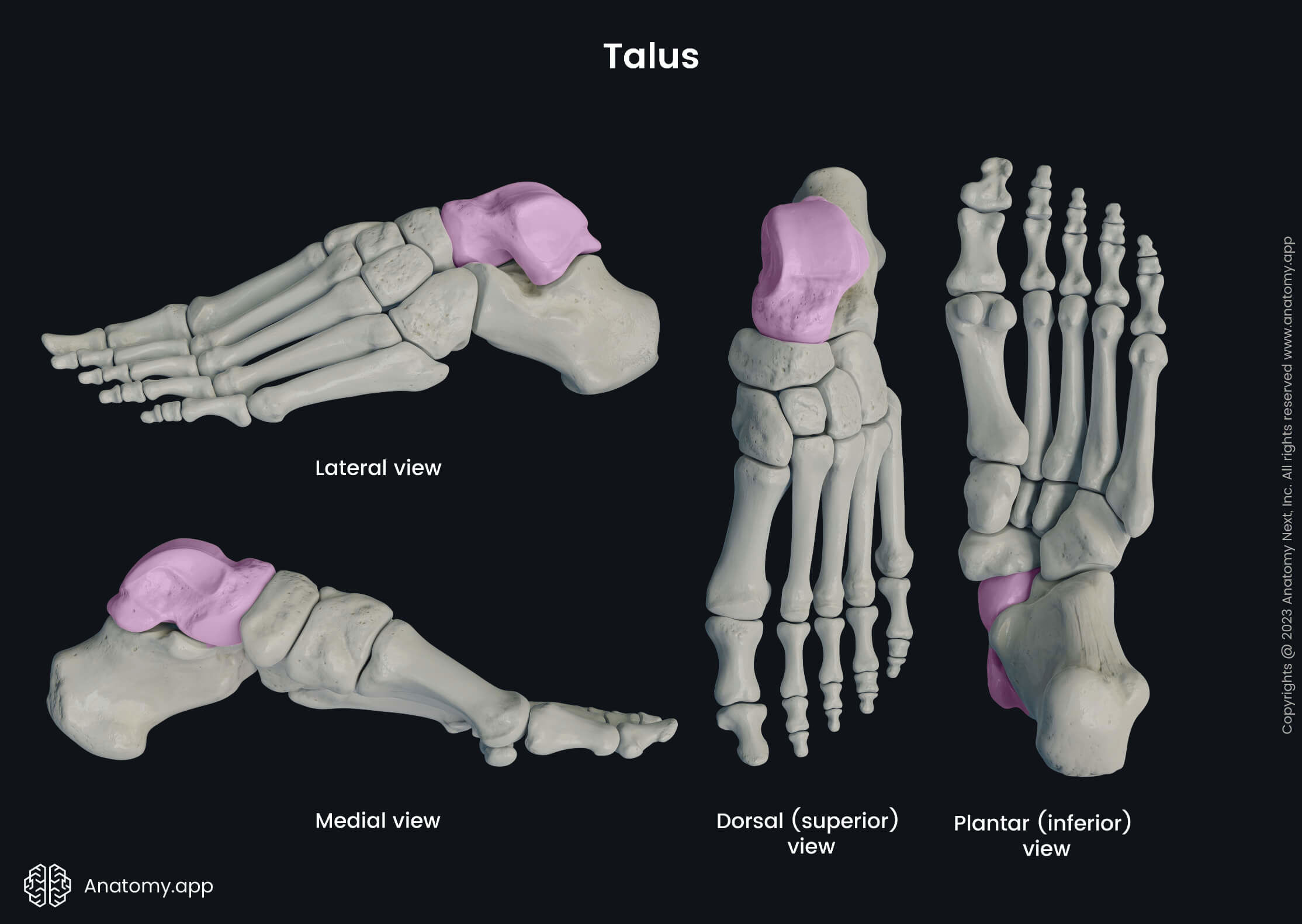
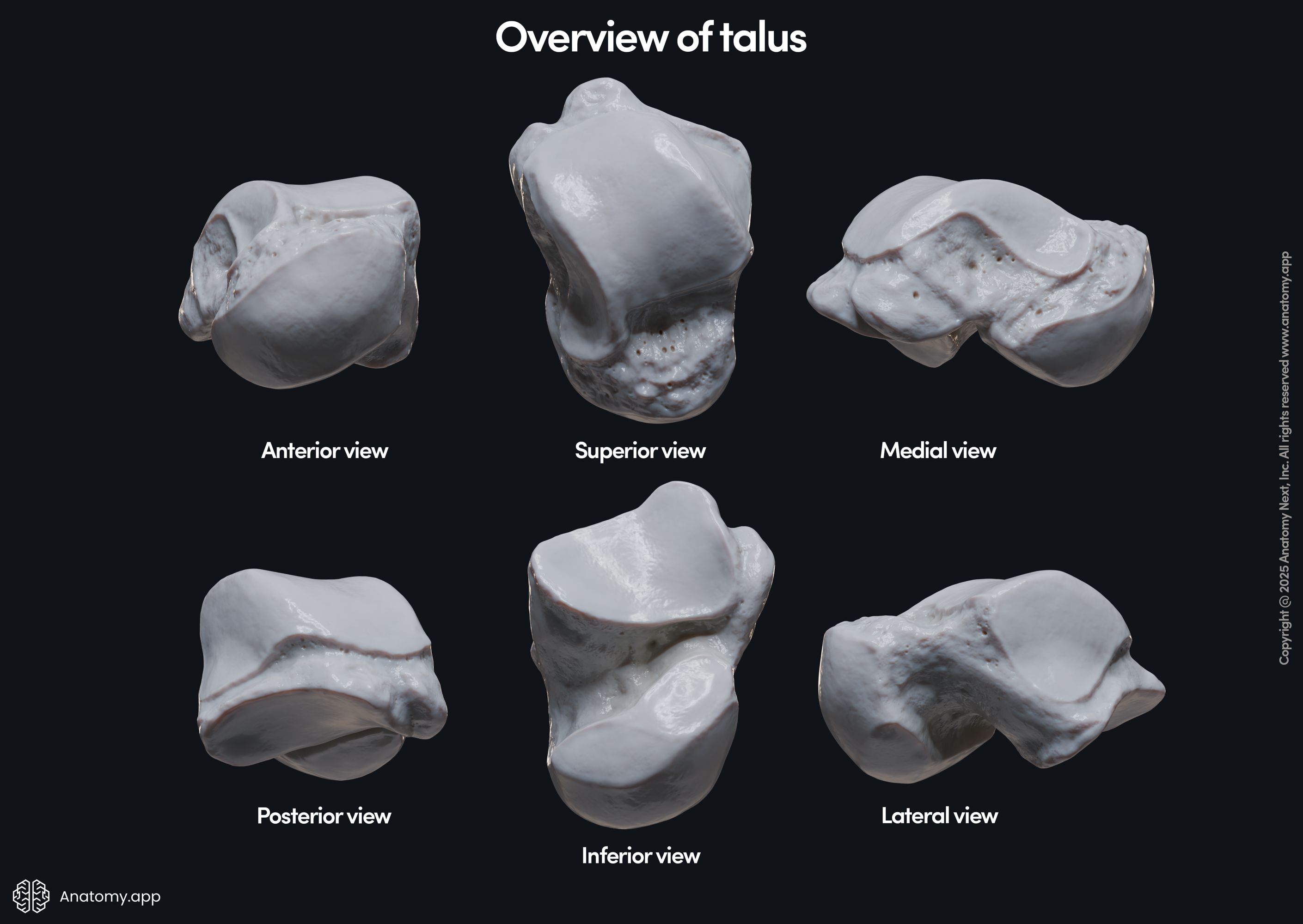
The talus is the second largest tarsal bone, and at the same time, it is also the most proximally located tarsal bone. The talus has several articular surfaces. It serves as an attachment site for numerous ligaments as it is a crucial bone involved in stabilizing the ankle joint and foot. The talus does not have any muscular attachments, and it has three parts:
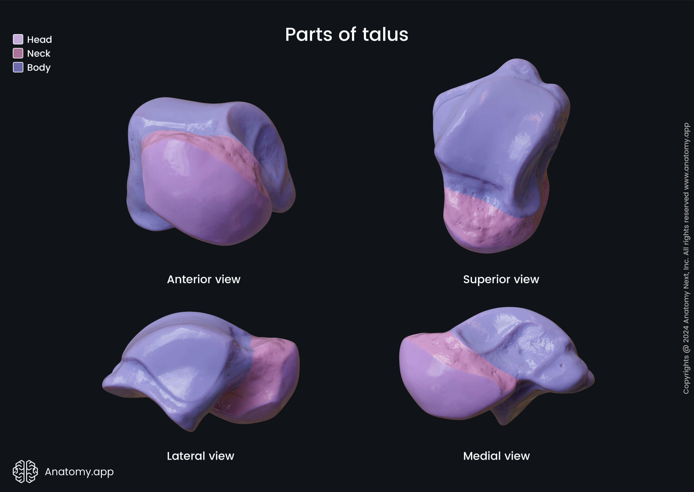
Head and neck of talus
The head of the talus is a spherical-shaped anteriorly and medially directed part of the bone. It contains an articular surface that articulates with the navicular bone. Proximally the head of the talus narrows, and this part is known as the neck of the talus.
Body of talus
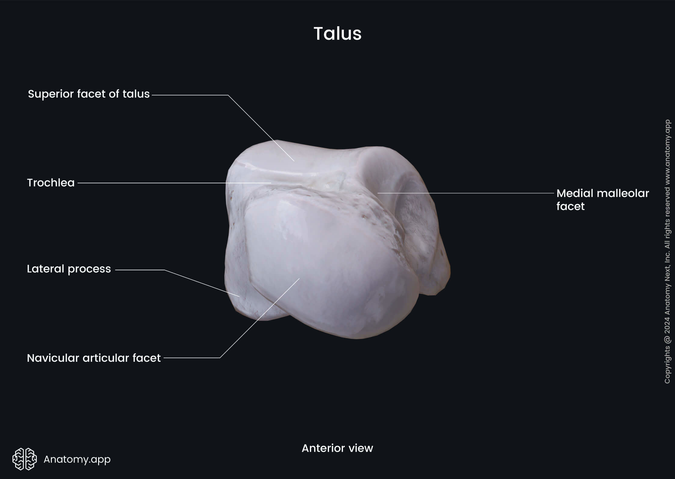
The body of the talus is the most proximal and most significant part of the bone situated posterior to the neck of the talus. It is crucial because it contains several surfaces and serves as an attachment site for ligaments. The body of the talus features the following landmarks and articular surfaces:
- Trochlea of the talus containing three surfaces
- Inferior articular surfaces (anterior, middle and posterior)
- Sulcus tali
- Tarsal sinus
The trochlea of the talus is the largest and posterior part of the body. It has a semi-cylindrical form, and its superior surface articulates with the tibia. The trochlea of the talus has three surfaces - the medial and lateral malleolar surfaces for articulation with both malleoli and the superior articular surface.
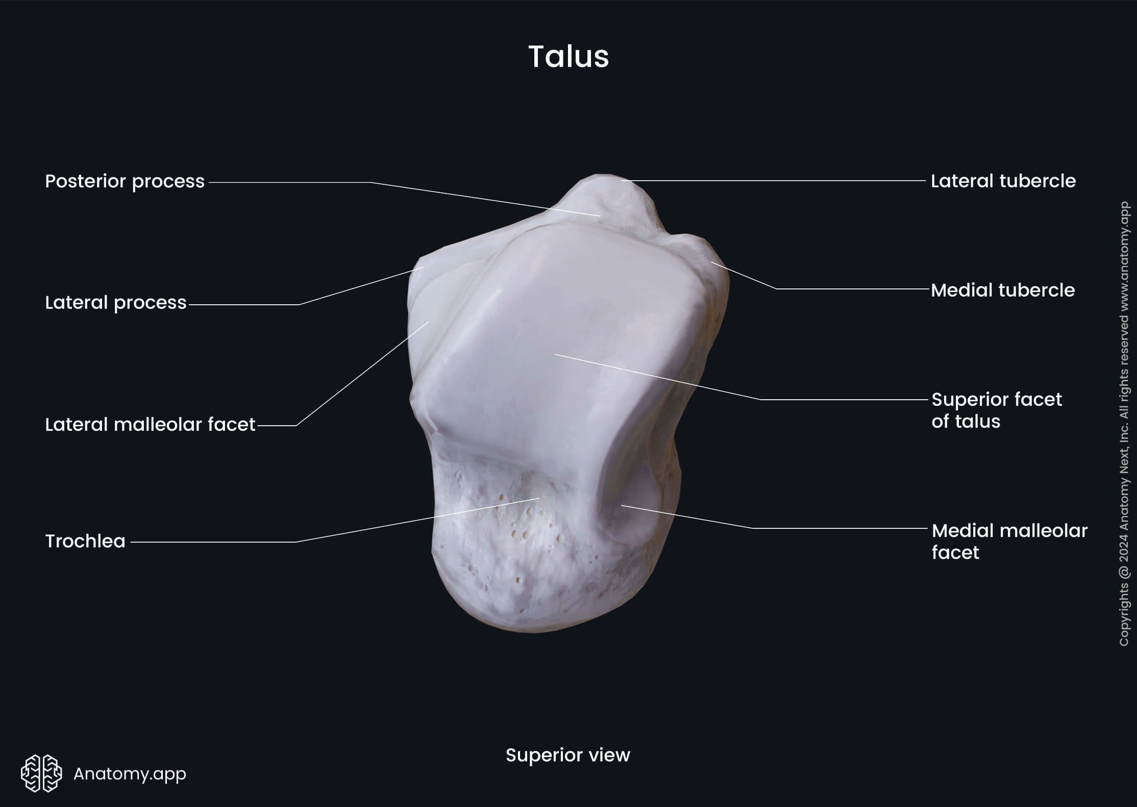

The medial malleolar surface is an almost sagittally oriented surface that articulates with the medial malleolus of the tibia. The lateral malleolar surface is also a sagittally oriented surface on the lateral side of the talus articulating with the lateral malleolus of the fibula. The superior articular surface articulates with the inferior articular surface of the tibia.

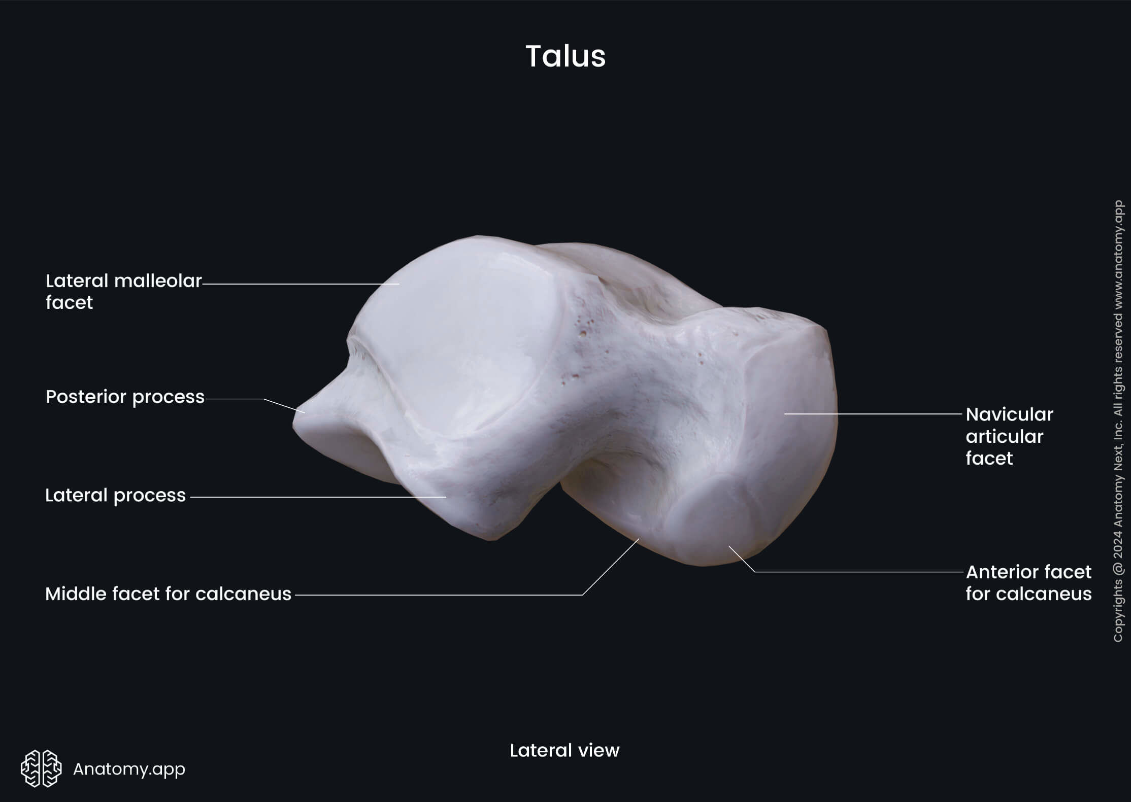
The inferior aspect of the body of talus contains three inferior articular surfaces known as the anterior, middle and posterior articular surfaces. All these surfaces face calcaneus and articulate with the bone, forming joints and connecting the talus with the calcaneus.
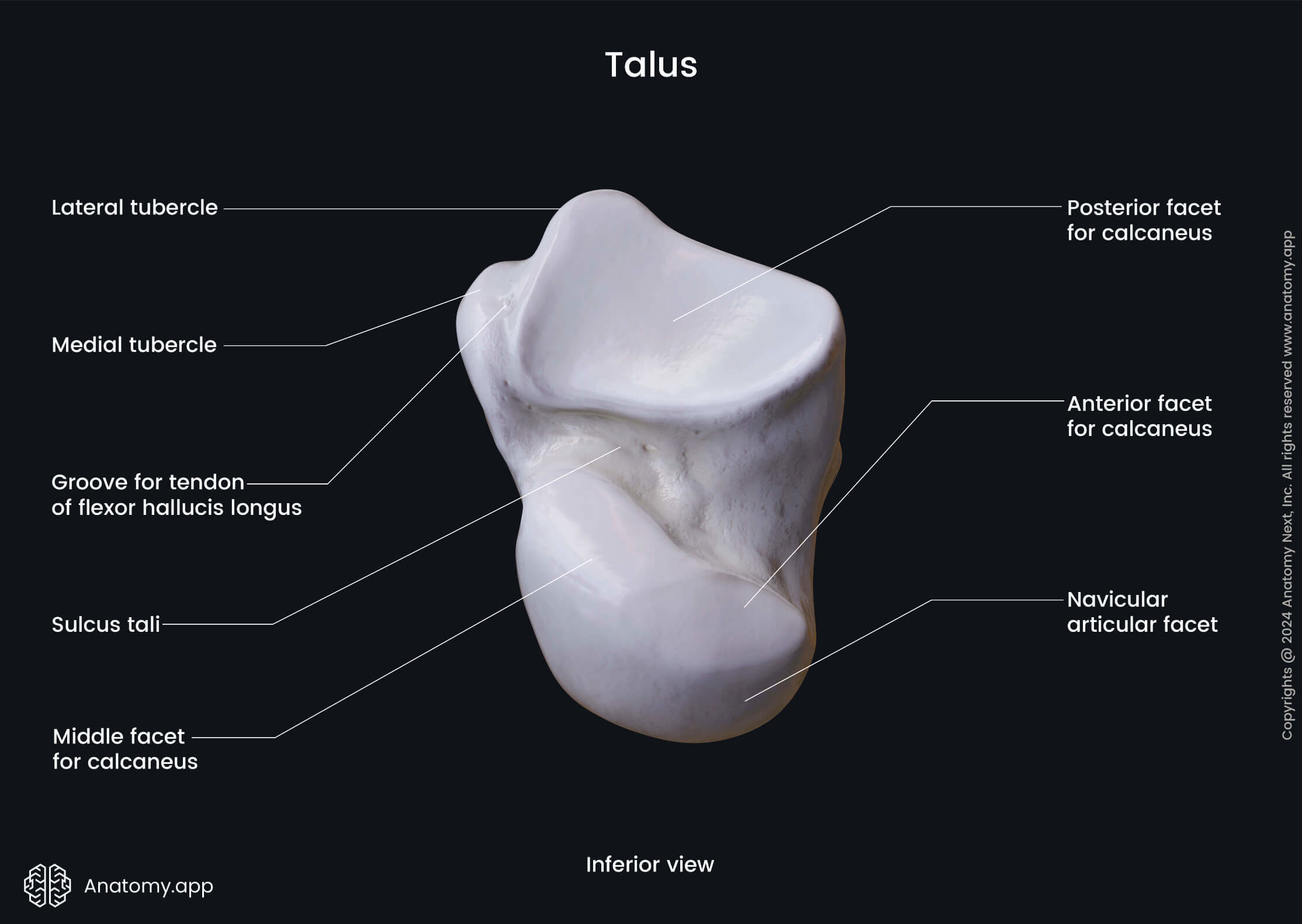
The sulcus tali is a deep groove located on the inferior portion of the talus between the posterior and middle articular surfaces. The tarsal sinus is formed together with the calcaneus. It is a cylindrical-shaped cavity formed by the sulcus tali and the calcaneal sulcus.