- Anatomical terminology
- Skeletal system
- Skeleton of trunk
- Skull
- Skeleton of upper limb
- Skeleton of lower limb
- Joints
- Muscles
- Heart
- Blood vessels
- Lymphatic system
- Nervous system
- Respiratory system
- Digestive system
- Urinary system
- Female reproductive system
- Male reproductive system
- Endocrine glands
- Eye
- Ear
Femur
The femur (Latin: os femoris), also known as the thigh bone, is the only bone forming the thigh. It extends between the hip and knee joints, and it is the longest and strongest bone in the human body.
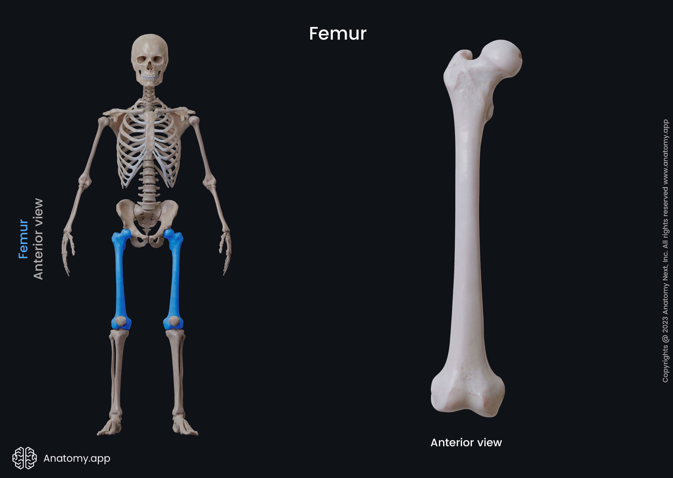
The femur is classified as a long bone, and it serves as an attachment site for numerous muscles and ligaments. Like every long bone, also the femur has three parts:
Proximal end of femur
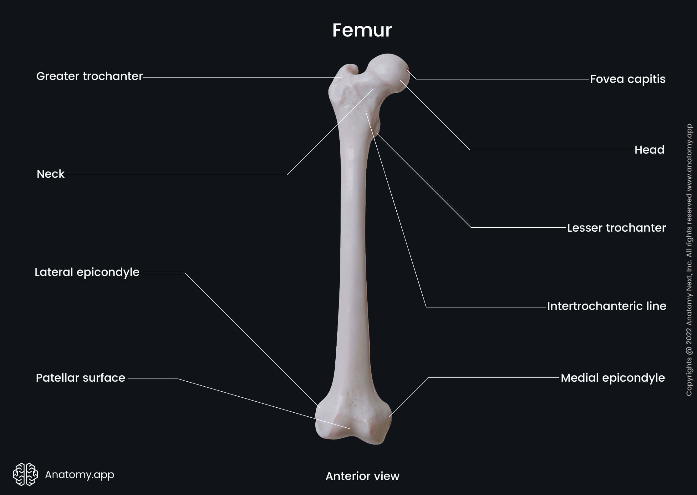
The proximal end or epiphysis is the upper end of the femur. It articulates with the acetabulum of the hip bone, forming the hip joint. The proximal end presents several landmarks, including:
- Head of the femur featuring the fovea for the ligament of the head
- Neck of the femur
- Greater trochanter with the trochanteric fossa
- Lesser trochanter
- Intertrochanteric line
- Intertrochanteric crest
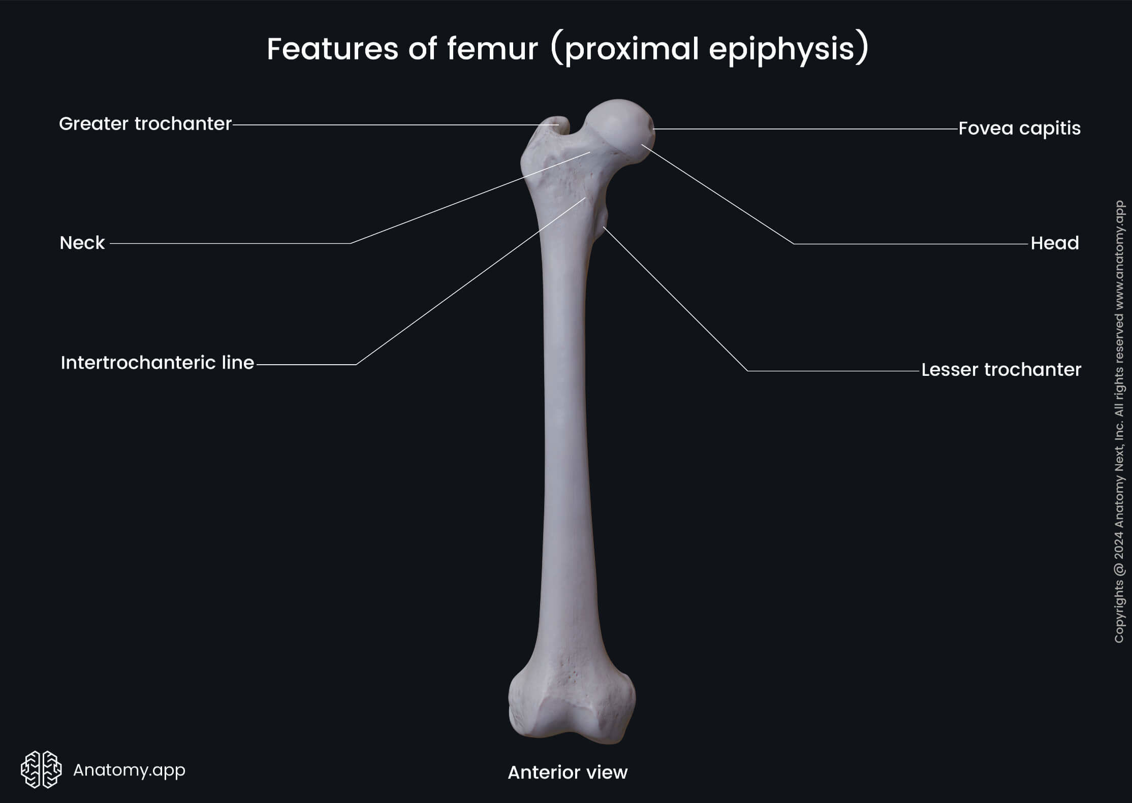
The head of the femur is the most upper aspect of the bone. It appears globular-shaped and is directed upward, medially, and slightly forward. The head articulates with the hip bone.
The femoral head presents with a slight depression called the fovea for the ligament of the head. It serves as an attachment site for the ligament of the head of femur.
The neck of the femur is a portion between the femoral head and the greater trochanter. It supports the femoral head, and it attaches to the femur at a certain angle.
The greater trochanter is a large prominence located on the superolateral aspect of the proximal end. It serves as an attachment site for the gluteus medius, gluteus minimus, and piriformis muscles.
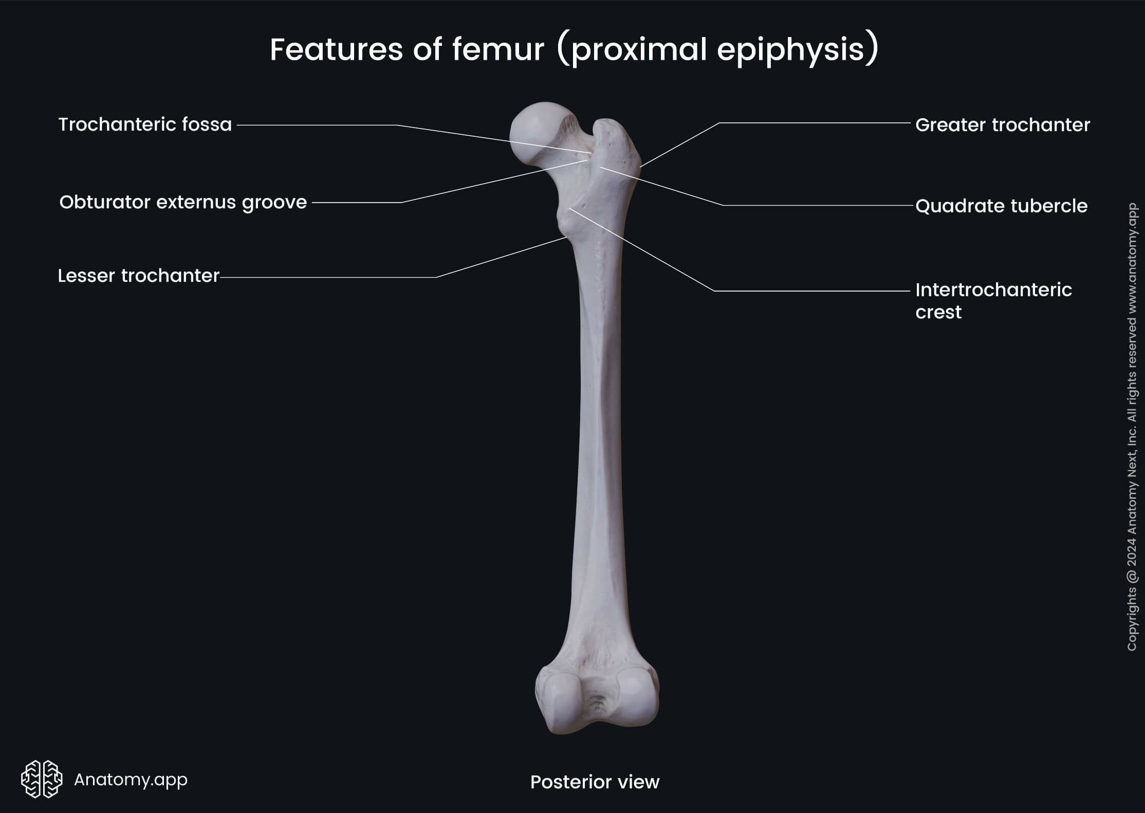
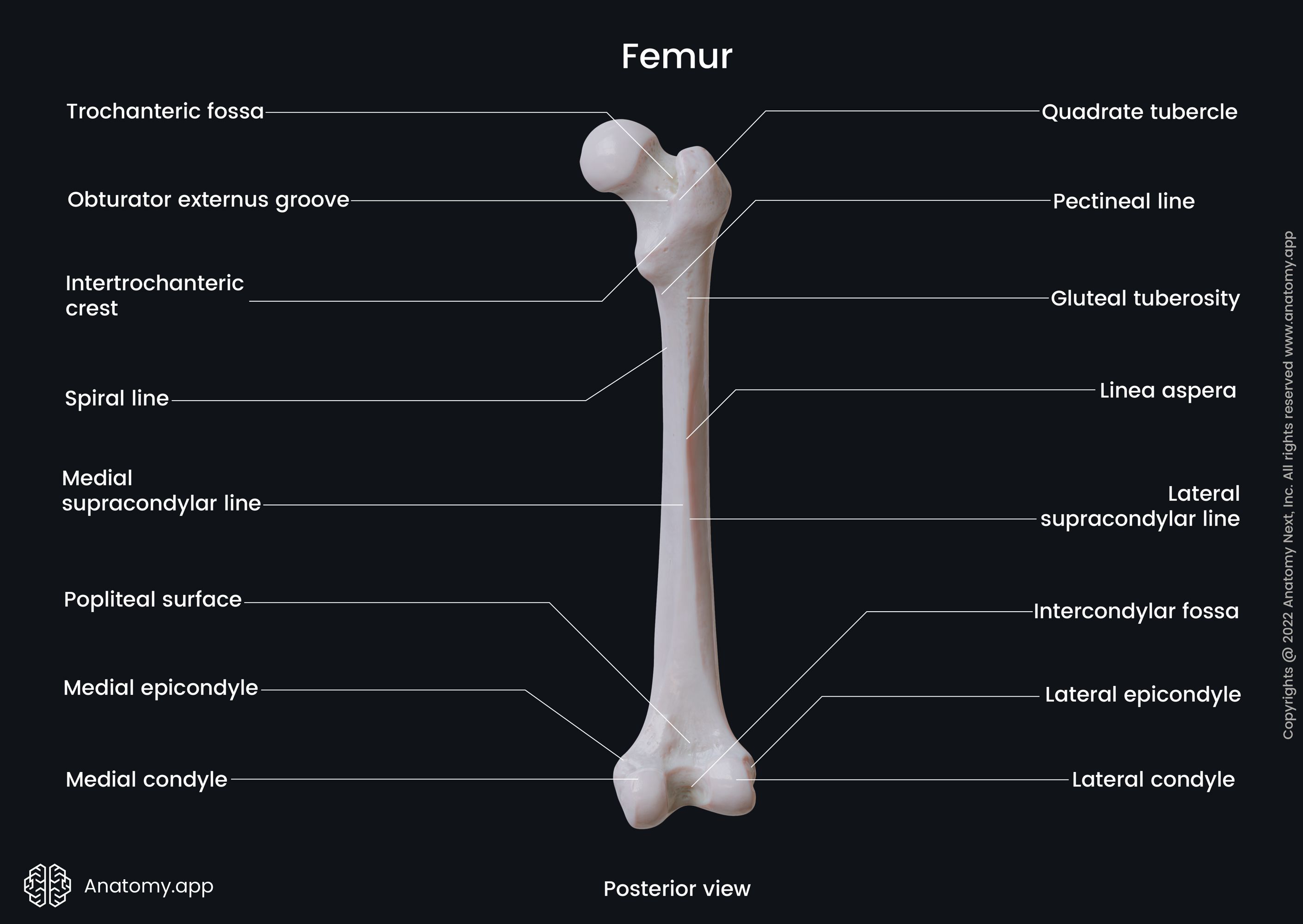
The trochanteric fossa is a depression located medial to the root of the greater trochanter. It serves as the origin site for the obturator internus, superior gemellus, and inferior gemellus muscles.
The lesser trochanter is a small prominence situated on the posteromedial aspect of the proximal end. It serves for the attachment of the iliopsoas muscle.
The intertrochanteric line is a rough anterior line between the femoral shaft and the neck of the femur. It extends from the greater trochanter to the lesser trochanter.
The intertrochanteric crest is a posterior bony ridge located between the shaft and the neck of the femur. It runs from the greater trochanter to the lesser trochanter.
Shaft of femur
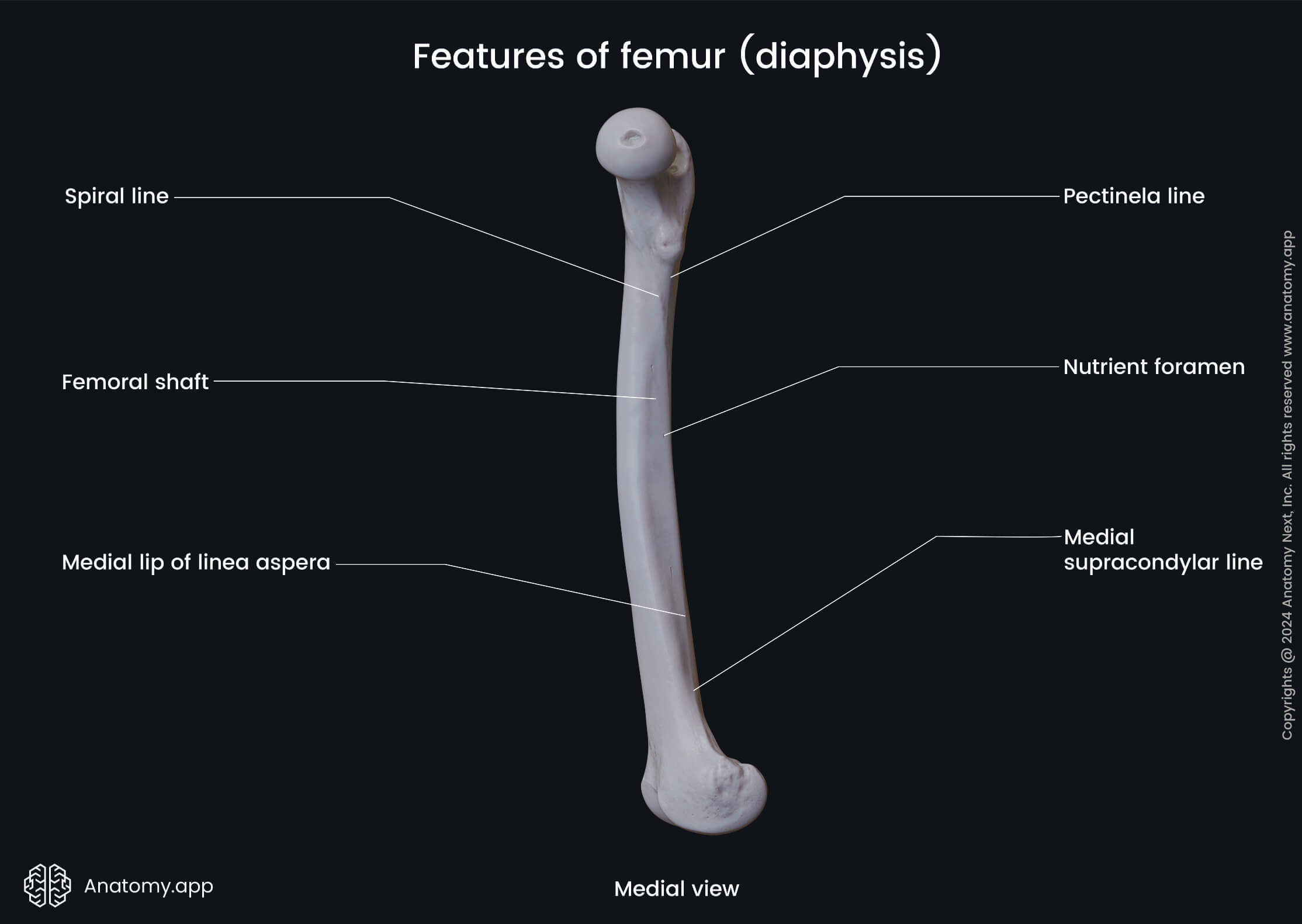
The shaft or the diaphysis is the extended middle portion of the femur. It appears cylindrical-shaped with a wide proximal part. Towards the middle, it becomes narrower, and the shaft again expands, reaching its distal end. The shaft features the following landmarks:
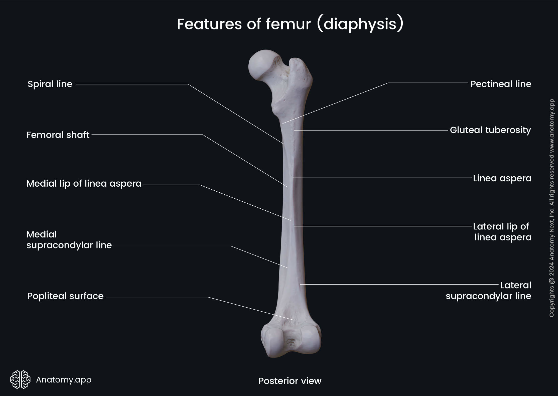
The linea aspera is a rough double line on the posterior aspect of the shaft composed of two diverging lips - medial and lateral. It serves as an attachment site for two vasti muscles (vastus medialis, vastus lateralis) and the short head of the biceps femoris. The linea aspera also is an insertion site for the adductors of the thigh, gluteus maximus and pectineus muscles.
The pectineal line is a bony ridge extending downward on the surface of the shaft from the lesser trochanter, and it nearly reaches the linea aspera. The pectineal line gives attachment to the pectineus muscle.
The gluteal tuberosity is a rough and elevated area on the shaft below the greater trochanter. It is continuous with the linea aspera. The gluteal tuberosity serves as the insertion site for the gluteus maximus muscle.
The popliteal surface is a triangular-shaped area found on the posterior aspect of the femur. It is located on the distal part of the shaft between the intercondylar and both supracondylar lines (medial and lateral). The popliteal artery lies upon the popliteal surface.
Distal end of femur
The distal end or epiphysis of the femur is the lower end of the bone. It articulates with two bones composing the lower leg - tibia and fibula - at the knee joint. The distal end features the following landmarks:
- Medial condyle containing
- Articular surface
- Medial epicondyle
- Lateral condyle featuring
- Articular surface
- Lateral epicondyle
- Patellar surface
- Intercondylar fossa
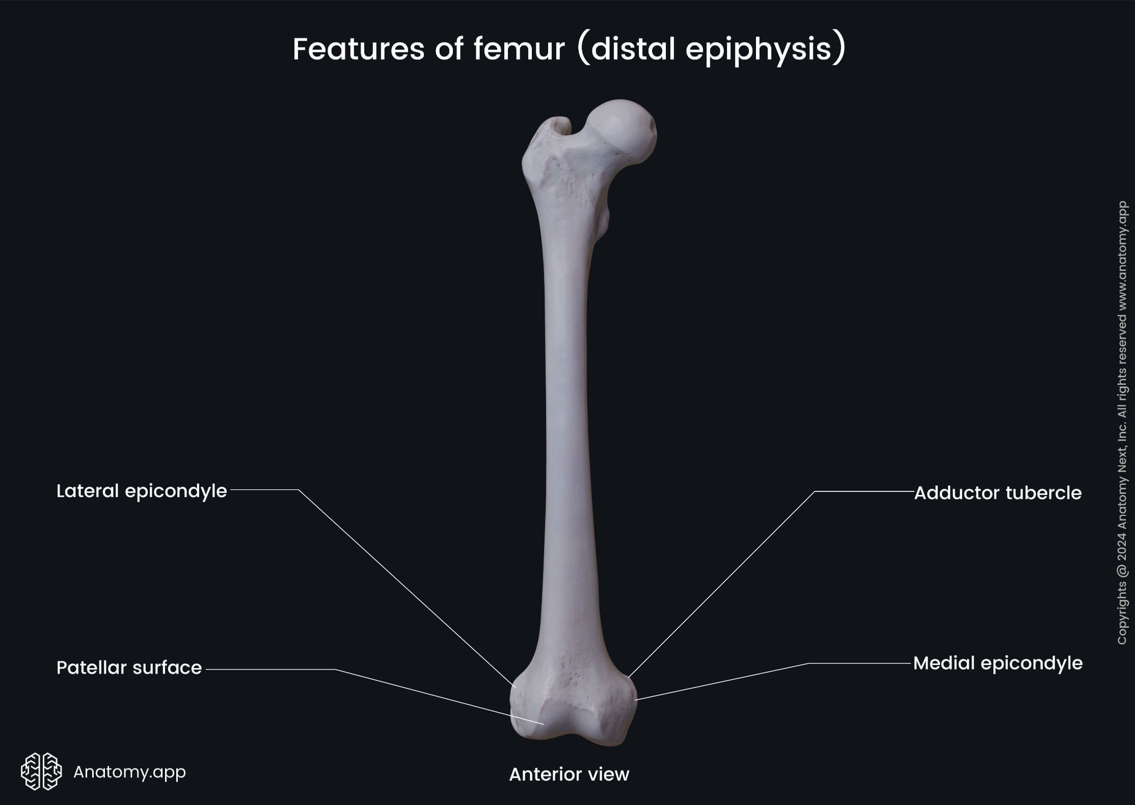
The medial condyle is a rounded projection on the medial side of the femur. It presents an articular surface for the articulation with the tibia and a medial epicondyle. The medial epicondyle is a large bony elevation on the medial aspect of the medial condyle.
The lateral condyle is a rounded projection on the lateral side of the femur. It also features an articular surface for the articulation with the tibia and a lateral epicondyle. The lateral epicondyle is a bony elevation on the lateral aspect of the lateral condyle.
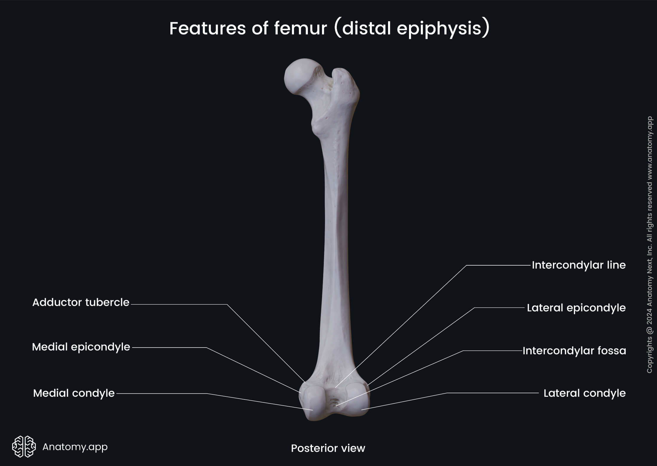
The patellar surface is an articular surface that participates in the formation of the knee joint. It is located on the anterior aspect of the distal end between both condyles. The patellar surface articulates with the articular surface of the patella.
The intercondylar fossa is a deep notch located on the posterior aspect of the distal end. It is situated between the articular surfaces of both condyles. Anteriorly the intercondylar fossa is limited by the patellar surface, while posteriorly by the intercondylar line.
Anatomy.app
Contact information
- For questions regarding business inquiries. Please contact:
- info@anatomy.app