- Anatomical terminology
- Skeletal system
- Joints
- Muscles
- Heart
- Blood vessels
- Nervous system
- Respiratory system
- Digestive system
- Lymphatic system
- Female reproductive system
- Male reproductive system
- Endocrine glands
- Eye
- Ear
Orbit
The orbit (Latin: orbita) is a paired skeletal cavity located in the bones of the skull. It is situated in the upper aspect of the face on either side of the root of the nose. The orbit accommodates and protects the eyeball and the accessory structures of the eye.
The shape of the orbit resembles a pyramidal structure with its apex directed posteriorly and the base situated anteriorly. Each orbit opens on the anterior aspect of the skull with an approximately quadrangular-shaped orbital opening.
Two margins form the orbital opening. The supraorbital margin is the upper aspect composed of the orbital part of the frontal bone. Anteromedially on the supraorbital margin lies a supraorbital foramen (sometimes called a notch), which transmits the supraorbital vessels and nerve.
In contrast, the infraorbital margin is the lower aspect formed by the orbital surface of the maxilla and zygomatic bone. The orbital surface of the maxilla presents the infraorbital notch, which opens as the infraorbital foramen on the face below the margin. It carries the infraorbital nerve and vessels.
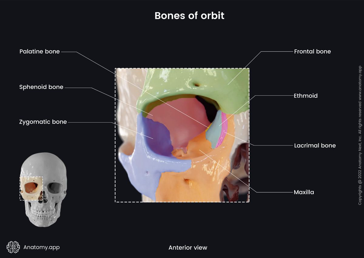
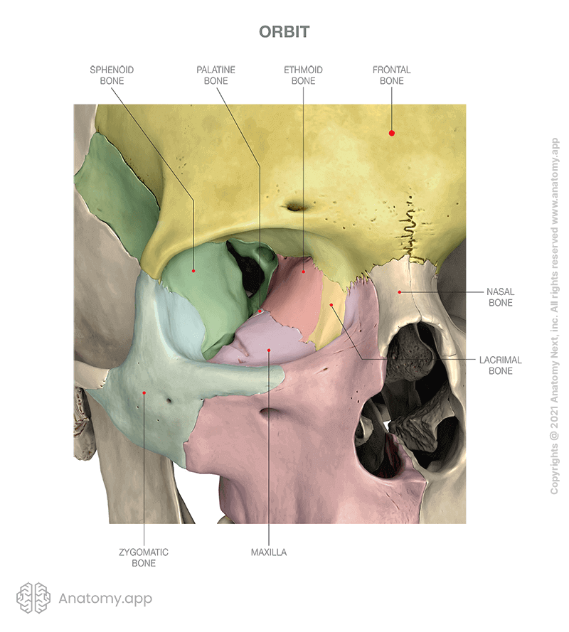
The orbit is formed by seven bones of the skull, and they include the following:
- Frontal bone - its orbital part;
- Lacrimal bone - its lateral (orbital) surface;
- Ethmoid bone - its orbital plate;
- Zygomatic bone - its frontal and maxillary processes;
- Maxilla - its orbital surface;
- Palatine bone - its orbital process;
- Sphenoid bone - greater and lesser wings and body.
Walls of orbit
The bony orbit is composed of four walls formed by several bones of the skull. The walls of the bony orbit are known as follows:
- Superior wall (roof)
- Inferior wall (floor)
- Medial wall
- Lateral wall
The roof or the superior wall anteriorly is formed by the orbital part of the frontal bone and posteriorly by the orbital surface of the lesser wing (sphenoid bone). Anterolaterally, in the roof part is situated the lacrimal fossa that houses the lacrimal gland. The floor or the inferior wall is formed by the orbital surface of the maxilla.
The medial wall of the orbit anteriorly is formed by the lacrimal bone and frontal process of the maxilla. The middle part is formed by the orbital plate of the ethmoid bone and posteriorly is the body of the sphenoid. The orbital surface of the lacrimal bone presents a fossa that lodges the nasolacrimal sac.
The lateral wall of the orbit anteriorly is formed by the frontal process of the zygomatic bone, while posteriorly by the orbital surface of the greater wing (sphenoid bone). The lateral wall of the orbit contains the zygomaticofacial and zygomaticotemporal foramina that transmit the corresponding nerves.
Contents of orbit
Both orbits contain the eyeballs and the accessory structures of the eyes, as well as their associated structures such as nerves and blood vessels. Besides mentioned structures, the orbit contains connective tissue and orbital fatty tissue that cushions the eyes and stabilizes the extra-ocular muscles. The following structures are situated within the orbit:
- Nerves and ganglia - motor nerves (oculomotor (CN III), trochlear (CN IV), abducens nerves (CN VI)), parasympathetic fibers from the oculomotor (CN III) and facial (CN VII) nerves, sympathetic fibers, sensory nerves (optic (CN II) and ophthalmic nerves (CN V1)) and a parasympathetic ganglion (ciliary ganglion);
- Blood vessels - ophthalmic artery, infraorbital branch of the maxillary artery, superior and inferior ophthalmic veins, infraorbital vein;
- Extra-ocular muscles - levator palpebrae superioris, superior and inferior oblique muscles, and four recti muscles (superior, inferior, lateral, medial);
- Lacrimal apparatus - lacrimal gland, lacrimal canaliculi, lacrimal sac and nasolacrimal duct.
Openings of orbit
The orbit is connected with other cavities and regions of the skull via several openings. These openings transmit the nerves and blood vessels, and they include the following:
- Optic canal
- Superior orbital fissure
- Inferior orbital fissure
- Anterior ethmoidal foramen
- Posterior ethmoidal foramen
- Nasolacrimal canal
- Infraorbital canal
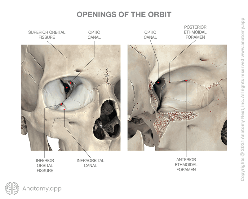
The optic canal connects the orbit with the middle cranial fossa, and it transmits the optic nerve (CN II) and ophthalmic artery. The superior orbital fissure also connects the orbit with the middle cranial fossa, but it transmits the oculomotor (CN III), trochlear (CN IV) and abducens nerves (CN VI), as well as branches of the ophthalmic nerve and ophthalmic veins.
The inferior orbital fissure connects the orbit with the pterygopalatine and infratemporal fossae. It transmits the infraorbital and zygomatic branches of the maxillary nerve (CN V2), their accompanying vessels and orbital branches arising from the pterygopalatine ganglion.
The anterior ethmoidal foramen is an opening between the orbit and the anterior cranial fossa transmitting the anterior ethmoidal nerve and its accompanying blood vessels. In contrast, the posterior ethmoidal foramen connects the orbit with the nasal cavity and conducts the posterior ethmoidal nerve and blood vessels.
The nasolacrimal canal leads to the inferior nasal meatus located in the nasal cavity. It carries the nasolacrimal duct. The infraorbital canal runs within the floor of the orbit and opens with the infraorbital foramen situated on the anterior surface of the maxilla. It transmits the infraorbital nerve, artery and veins.

Anatomy.app
Contact information
- For questions regarding business inquiries. Please contact:
- info@anatomy.app