- Anatomical terminology
- Skeletal system
- Skeleton of trunk
- Skull
- Skeleton of upper limb
- Skeleton of lower limb
- Joints
- Muscles
- Heart
- Blood vessels
- Lymphatic system
- Nervous system
- Respiratory system
- Digestive system
- Urinary system
- Female reproductive system
- Male reproductive system
- Endocrine glands
- Eye
- Ear
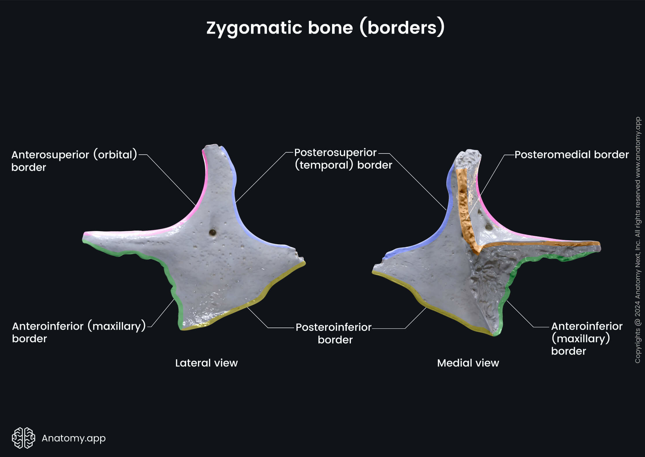
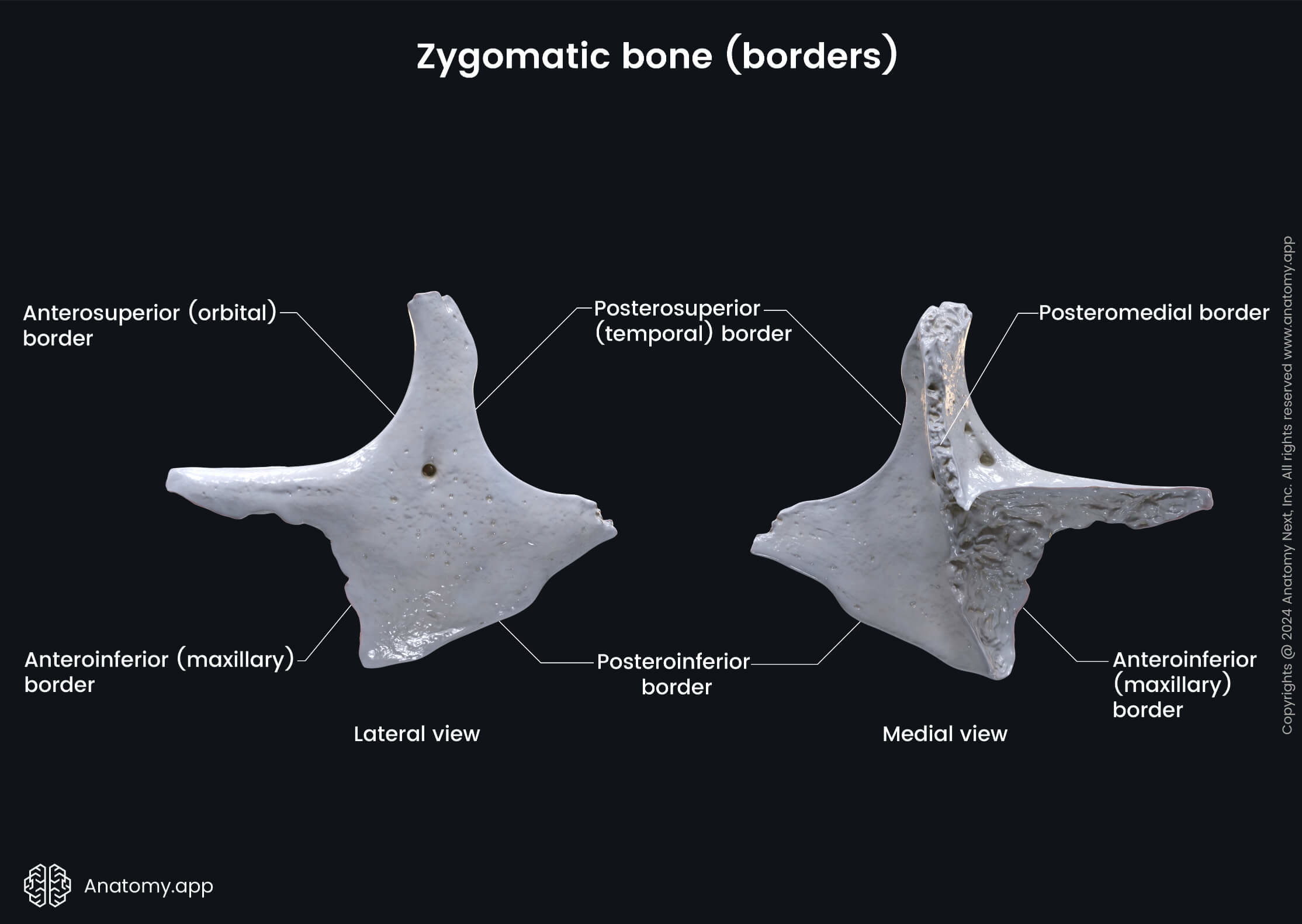
Zygomatic bone
The zygomatic bone (also known as cheek bone, malar bone, Latin: os zygomaticum) is a paired facial bone situated in the upper lateral part of the face. This bone participates in creating the prominence of the cheek.
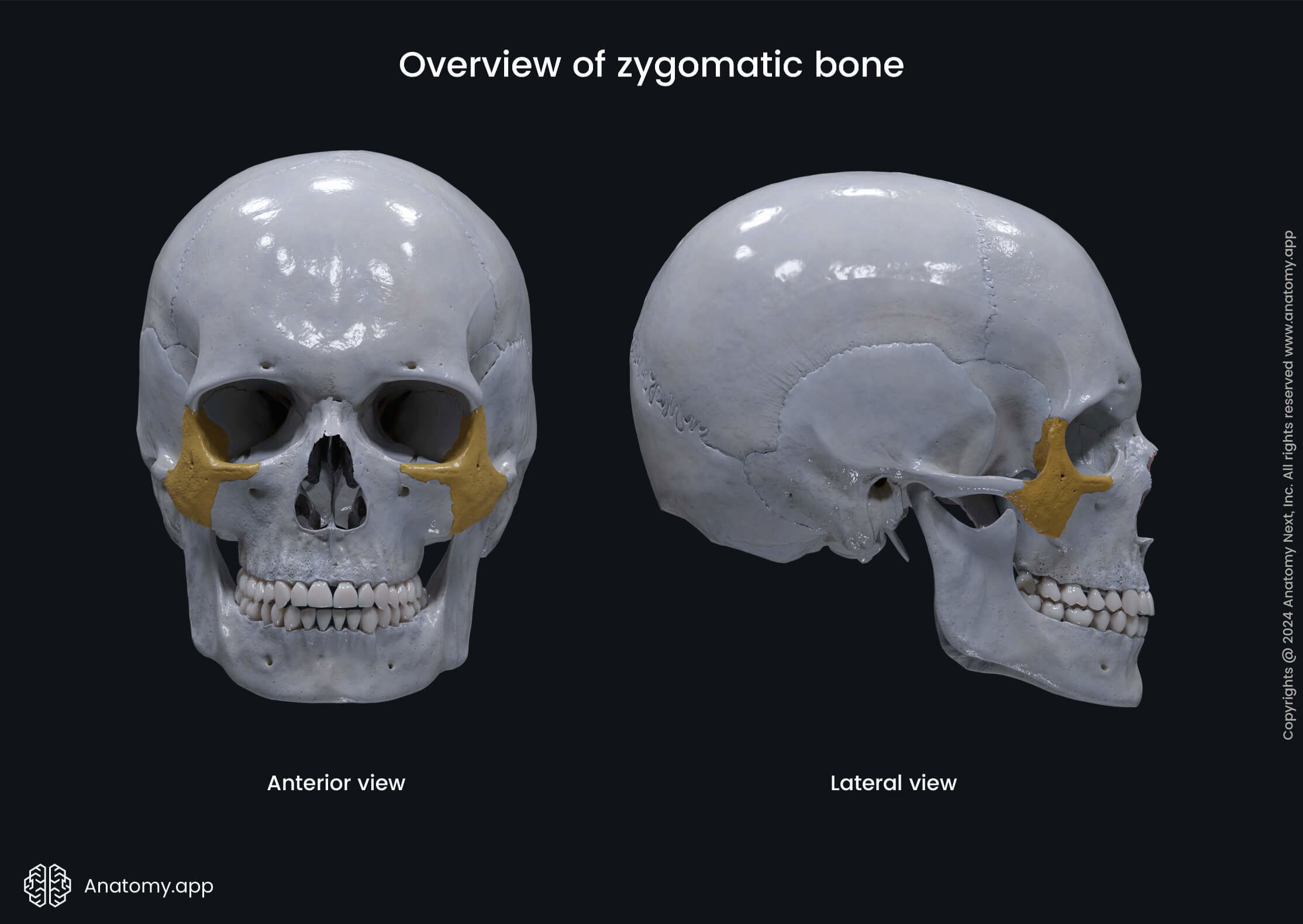
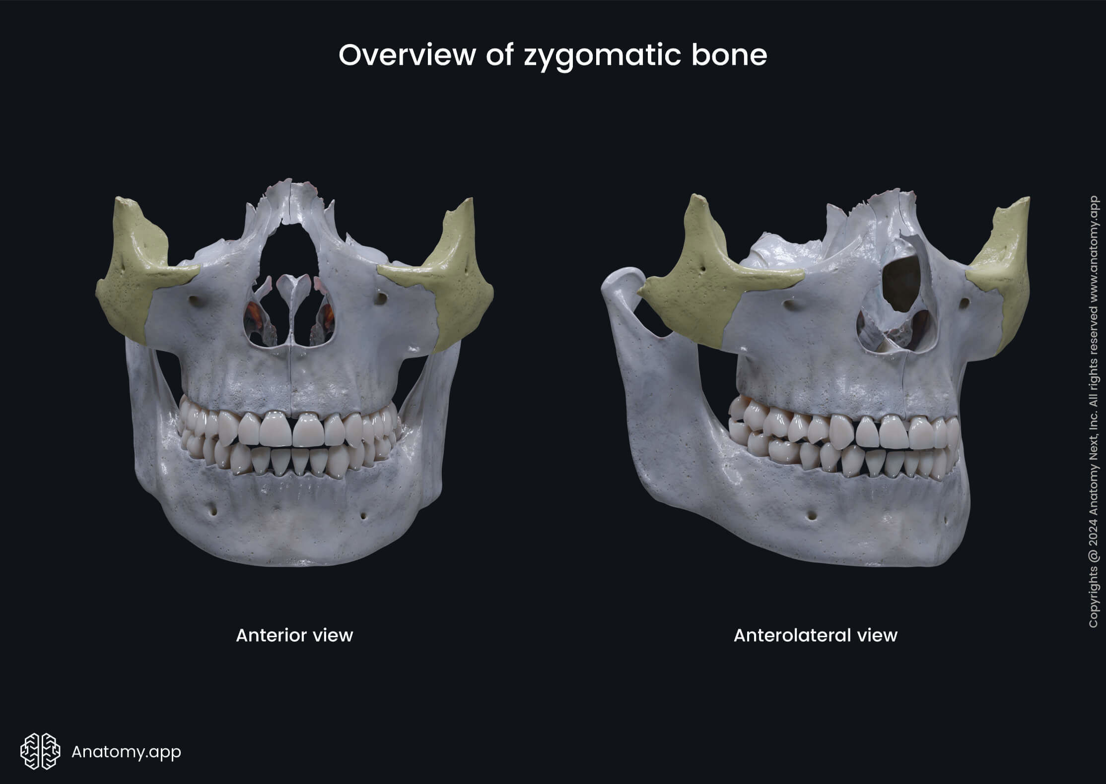

The zygomatic bone also takes part in forming the floor of the orbit, as well as the temporal fossa and infratemporal fossa. Each zygomatic bones articulates with four bones: maxilla, temporal bone, sphenoid bone, and frontal bone.
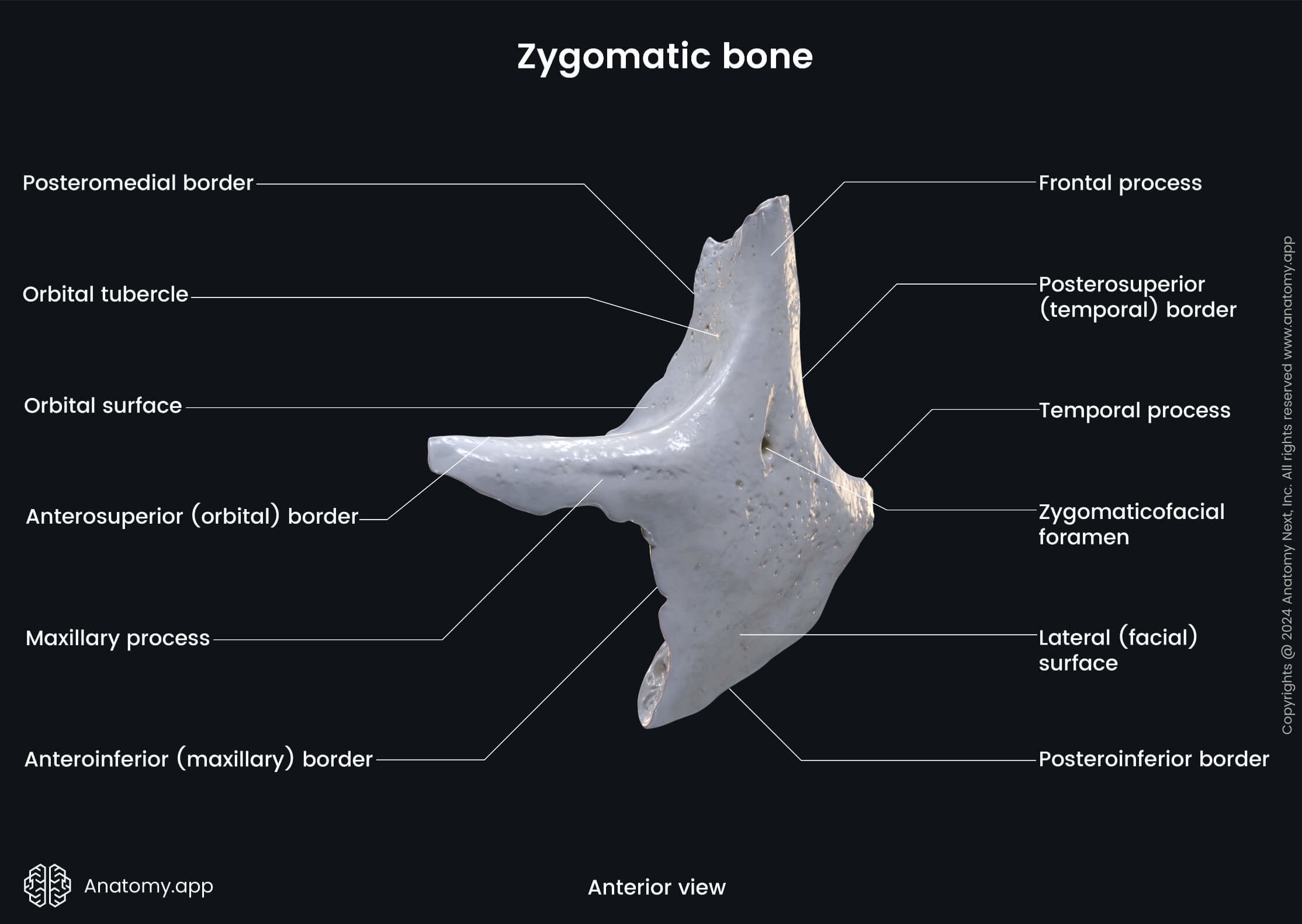
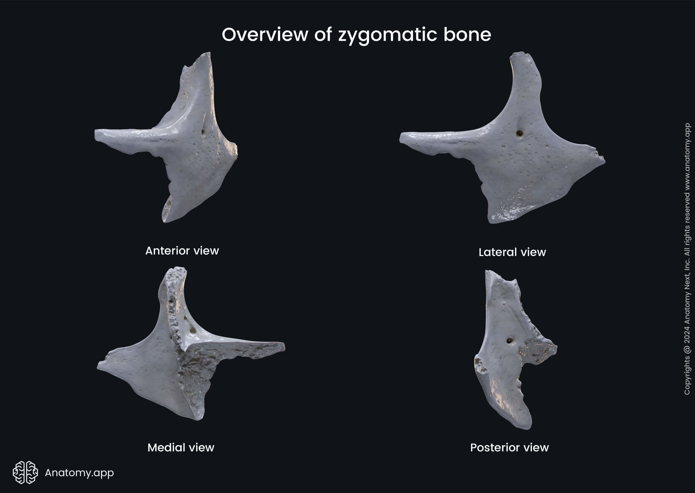
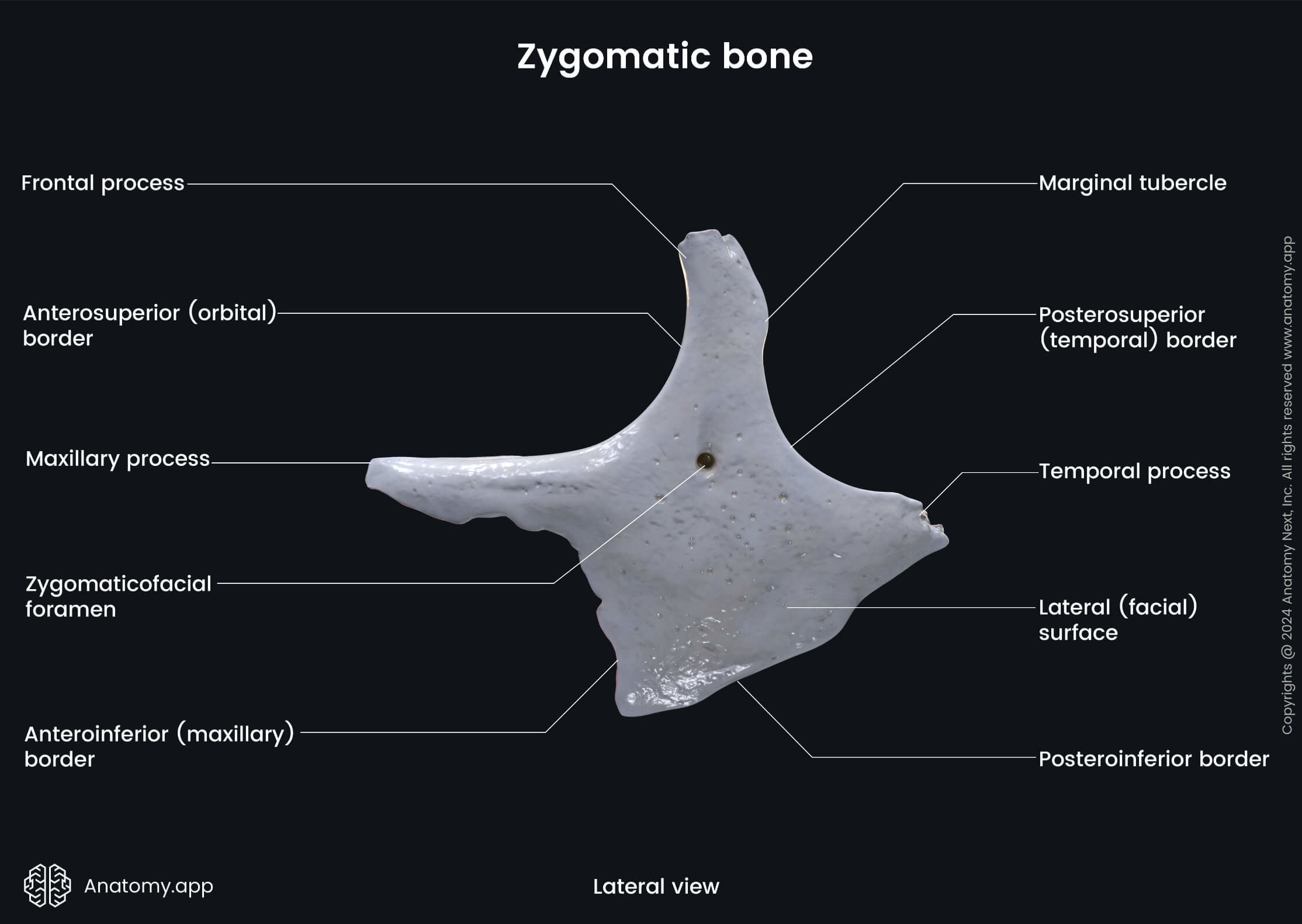
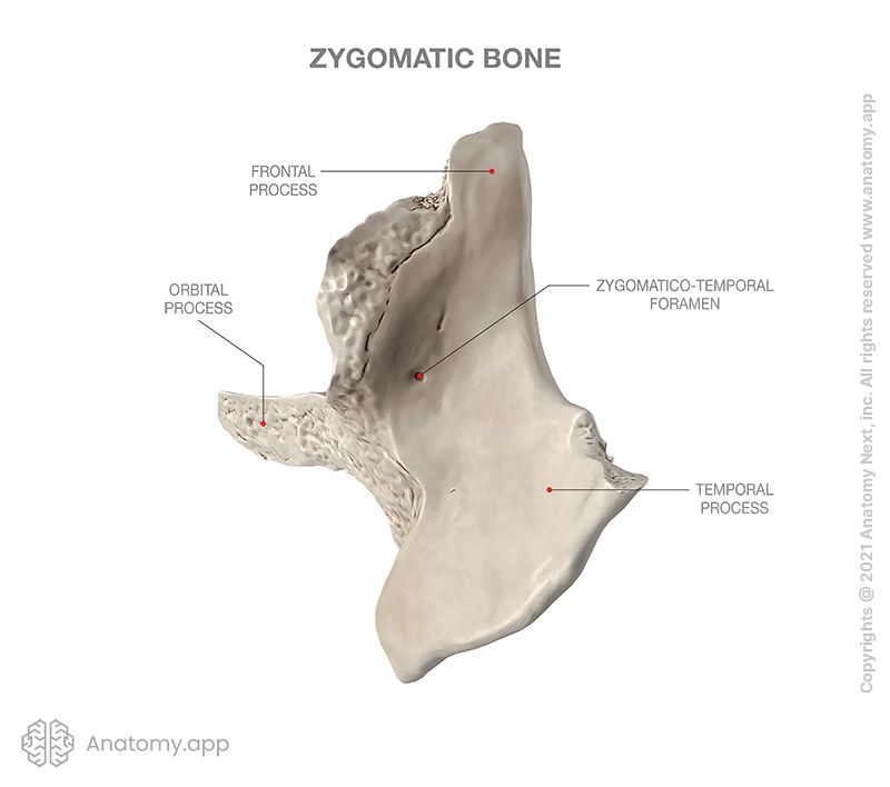
Each zygomatic bone has four margins, three surfaces (orbital, temporal, lateral or facial), and three processes (frontal, temporal, maxillary or orbital). The bone houses the zygomatic canal for the passage of the zygomatic nerve and blood vessels.
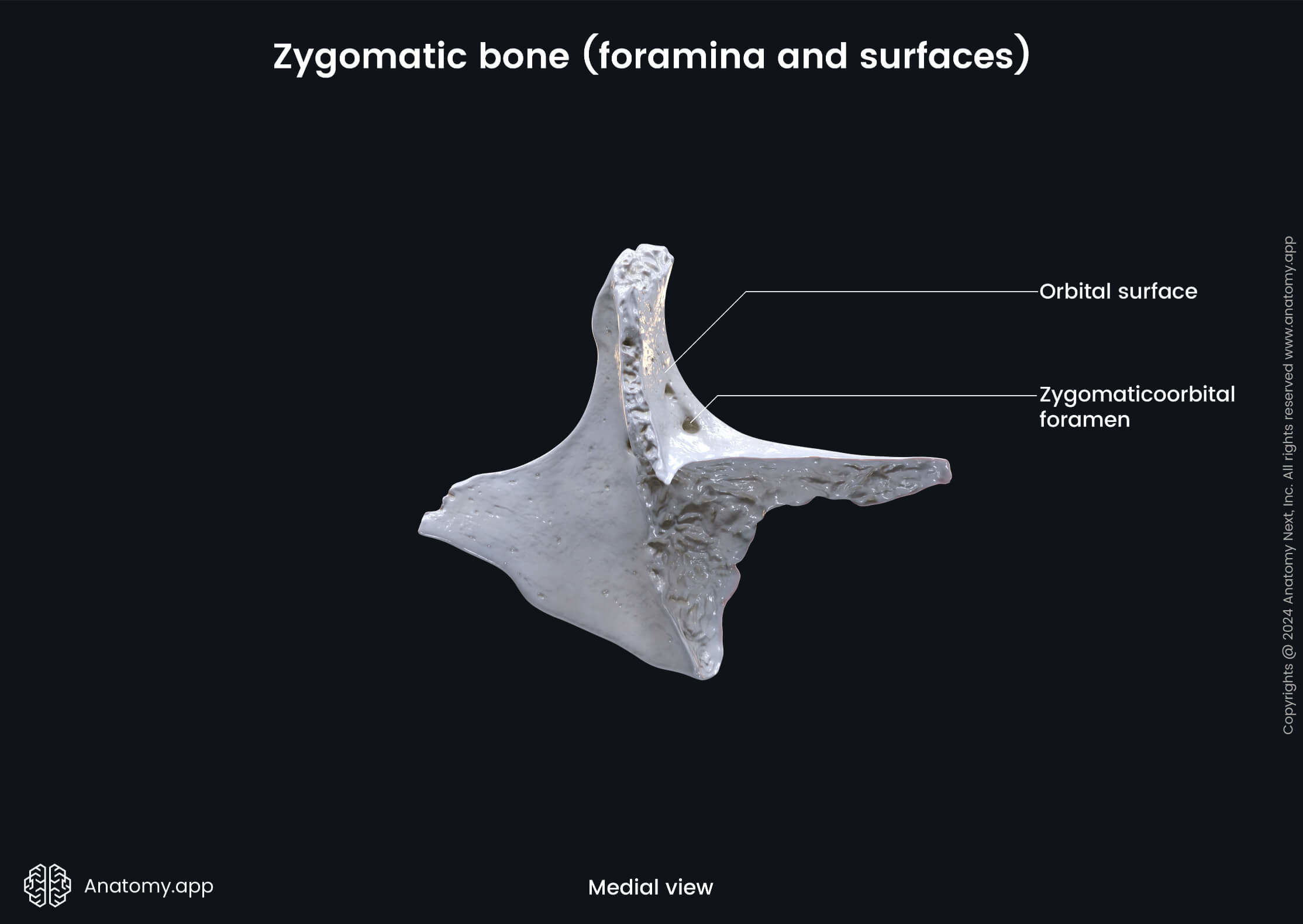
Surfaces of zygomatic bone
The orbital surface of the zygomatic bone forms part of the lateral side of the orbital floor (inferior wall of the orbit). The orbital surface presents an opening called the zygomaticoorbital foramen. The zygomaticoorbital foramen continues as the zygomatic canal leading to both zygomaticotemporal and zygomaticofacial foramina.
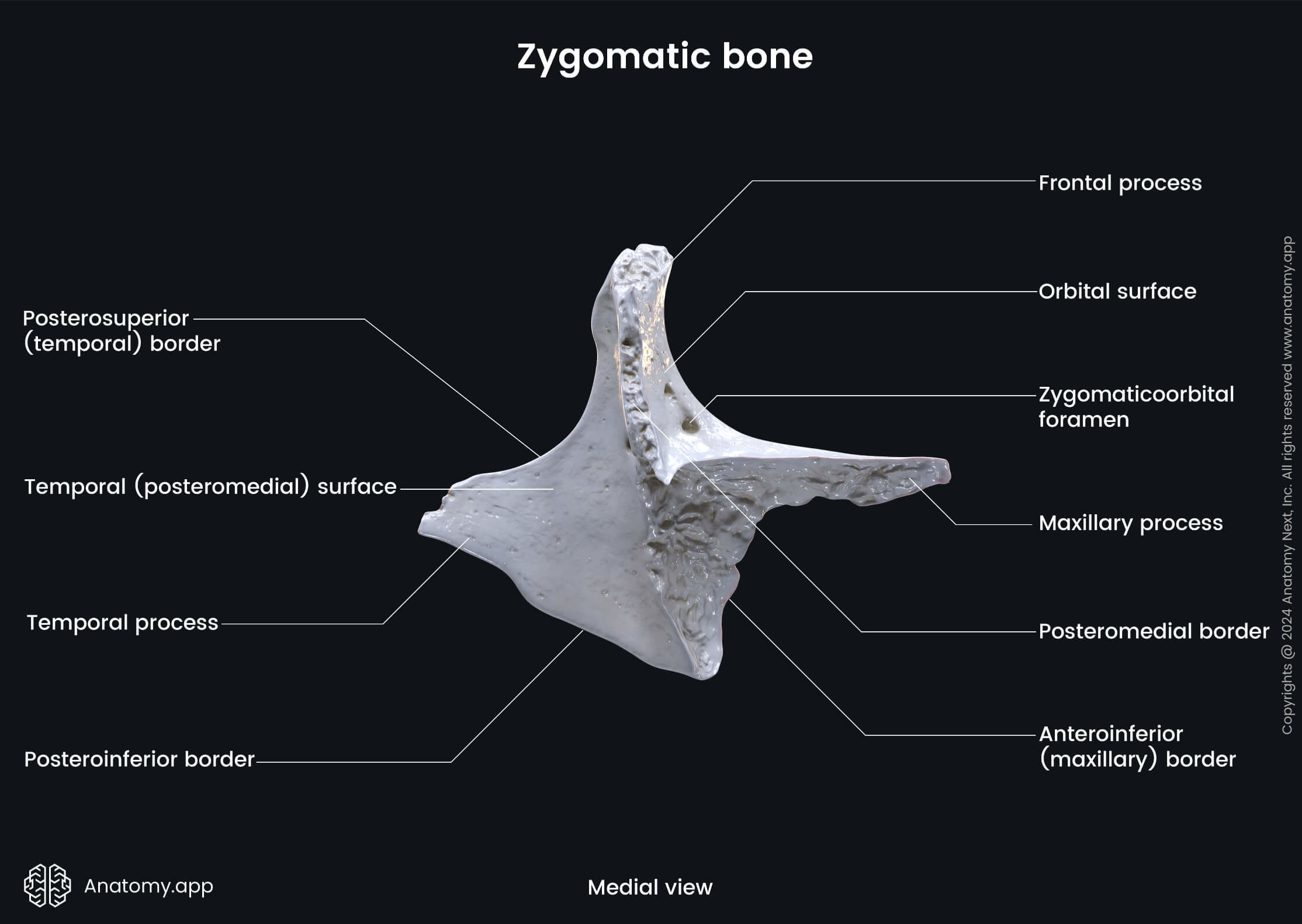
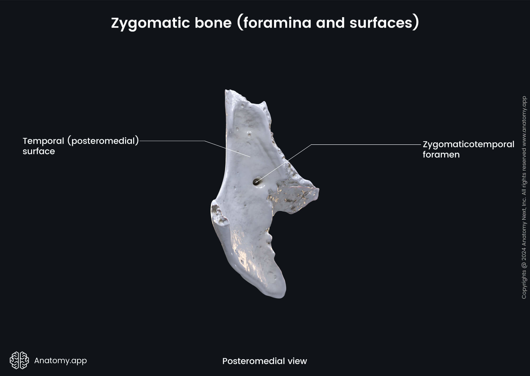
The temporal surface of the zygomatic bone is directed posteriorly and medially. It is concave and medially presents a surface for articulation with the zygomatic process of the maxilla. Laterally, it has a concave surface that forms the anterior border of the infratemporal fossa. Near the center of the temporal surface is the zygomaticotemporal foramen. It is an opening near the base of the frontal process that leads into the zygomatic canal and is a passage for the zygomaticotemporal nerve.
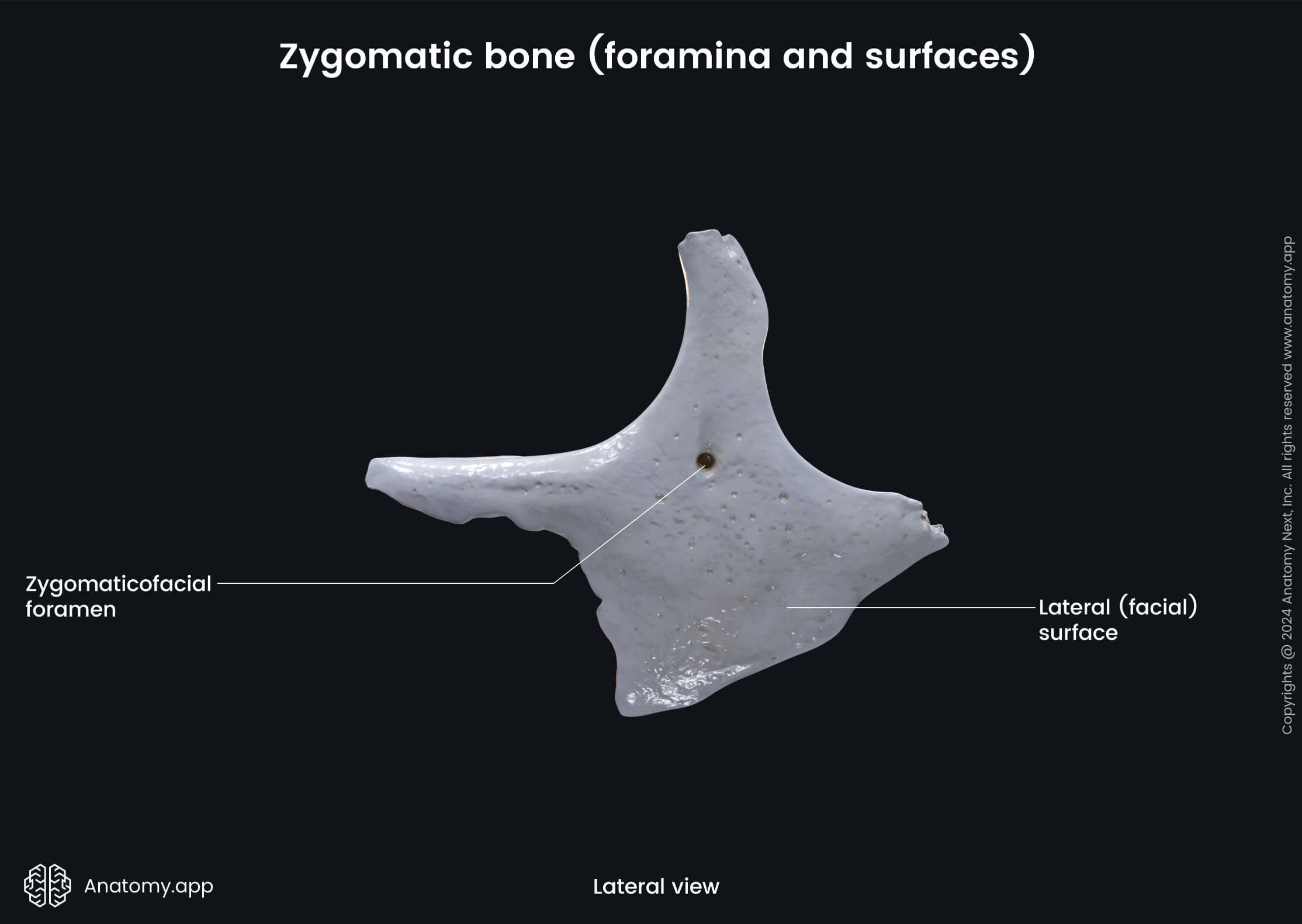
The lateral surface of the zygomatic bone is also known as the facial surface, and it is faced towards the outside. It participates in forming part of the infraorbital margin (partly formed by the zygomatic bone, partly - by the maxilla). The facial surface of the zygomatic bone features the zygomaticofacial foramen (usually, one or two openings) near its center. However, this it can sometimes be absent. This foramen leads into the zygomatic canal and is a passage for the zygomatic nerve and blood vessels.
Processes of zygomatic bone
The frontal process of the zygomatic bone is a superiorly oriented extension that unites the zygomatic bone with the zygomatic process of the frontal bone. It also participates in forming the lateral outline of the orbital opening.
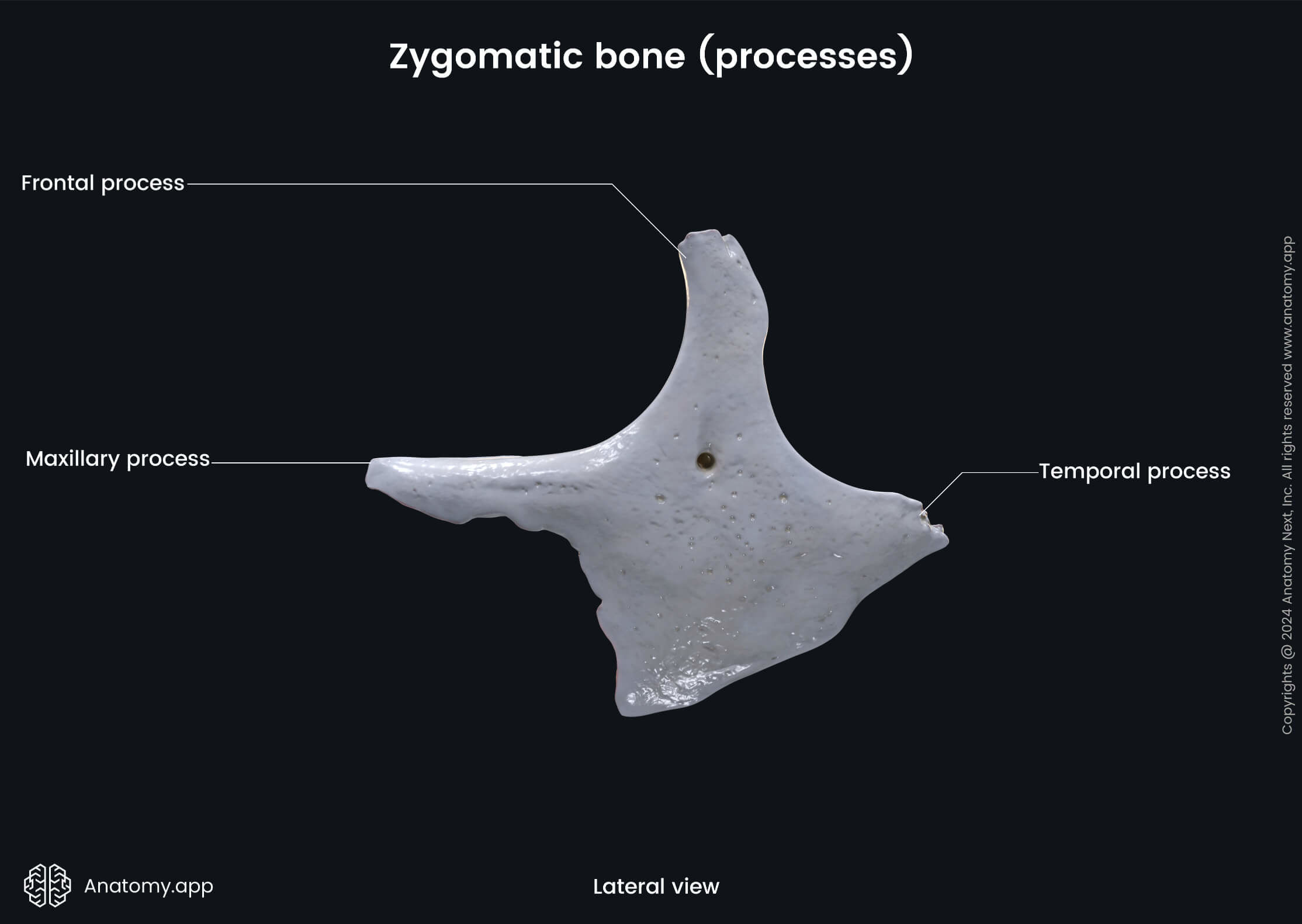
The temporal process is a posteriorly directed process that connects with the zygomatic process of the temporal bone to form the zygomatic arch. Prominent zygomatic arches appear as a facial feature that we call ''high cheekbones."
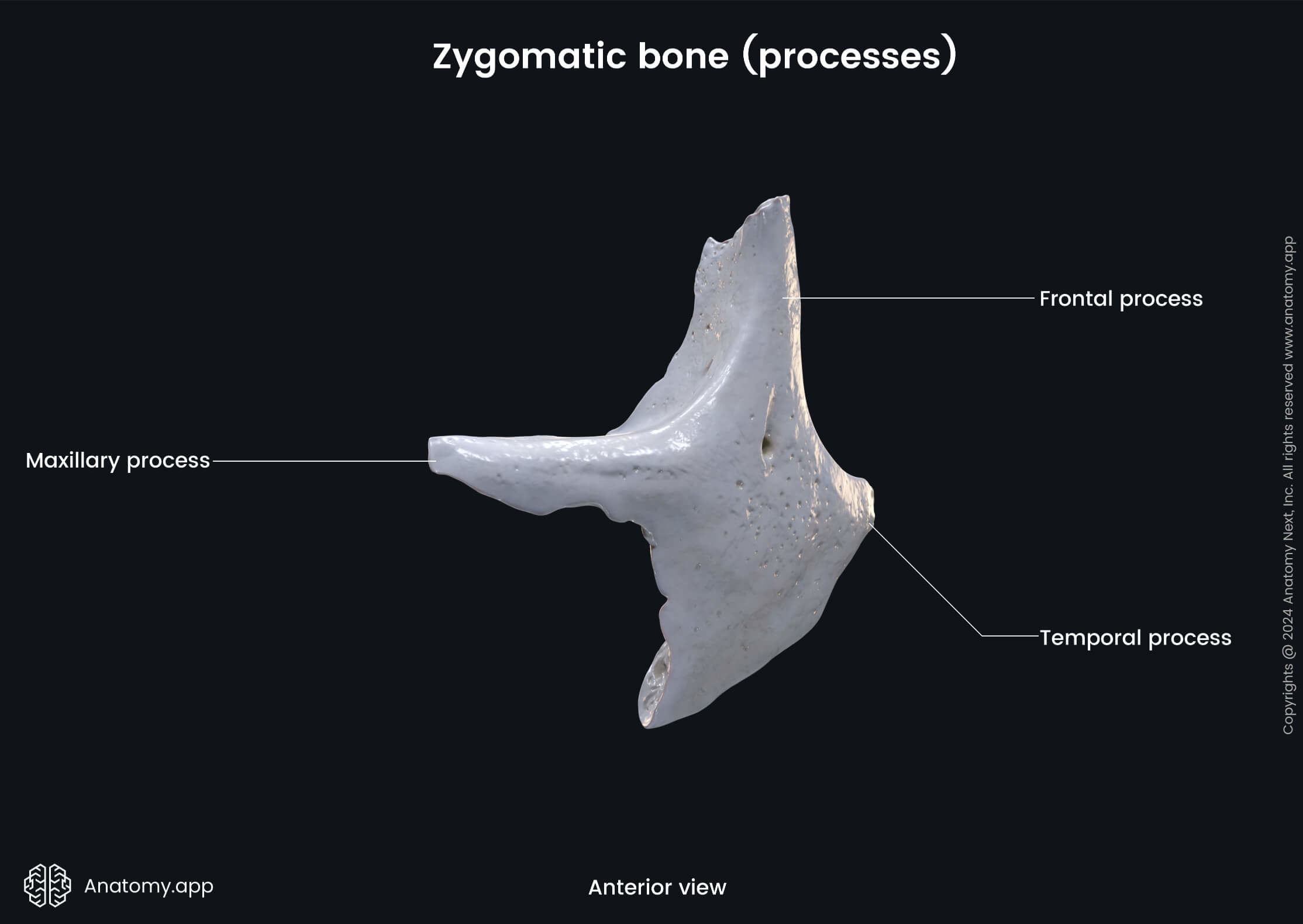
The maxillary process (sometimes called the orbital process) extends anteriorly and forms a part of the infraorbital margin and a small portion of the anterior part of the lateral orbital wall. Posterior to this process is the orbital surface of the zygomatic bone.
Zygomatic canal
The zygomatic canal is a passage within the zygomatic bone starting with one opening on the orbital surface (zygomaticoorbital foramen), and leading to openings on the temporal (zygomaticotemporal foramen) and the facial surfaces (zygomaticofacial foramen) of the zygomatic bone. The zygomatic nerve (a branch of the maxillary nerve (CN V2)) dividing into branches and the zygomatic artery and veins pass through this canal.
Anatomy.app
Contact information
- For questions regarding business inquiries. Please contact:
- info@anatomy.app