- Anatomical terminology
- Skeletal system
- Skeleton of trunk
- Skull
- Skeleton of upper limb
- Skeleton of lower limb
- Joints
- Muscles
- Heart
- Blood vessels
- Lymphatic system
- Nervous system
- Respiratory system
- Digestive system
- Urinary system
- Female reproductive system
- Male reproductive system
- Endocrine glands
- Eye
- Ear
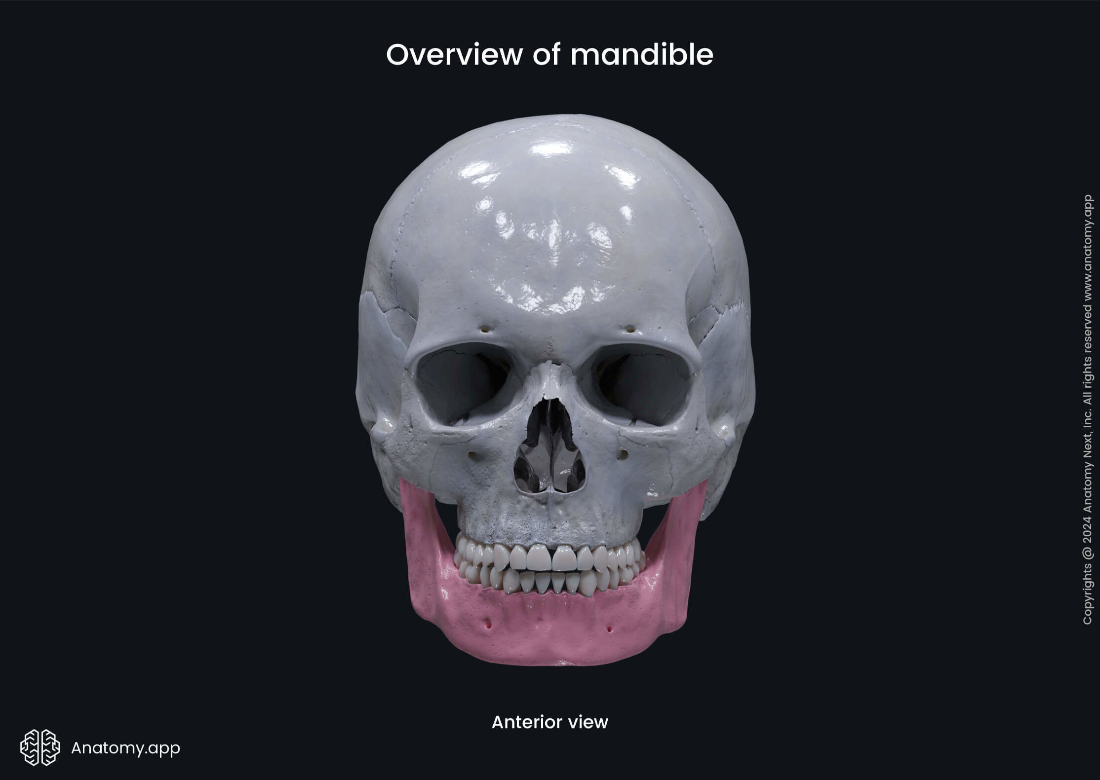
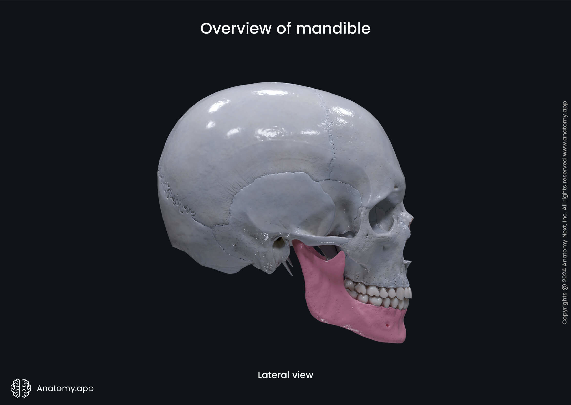
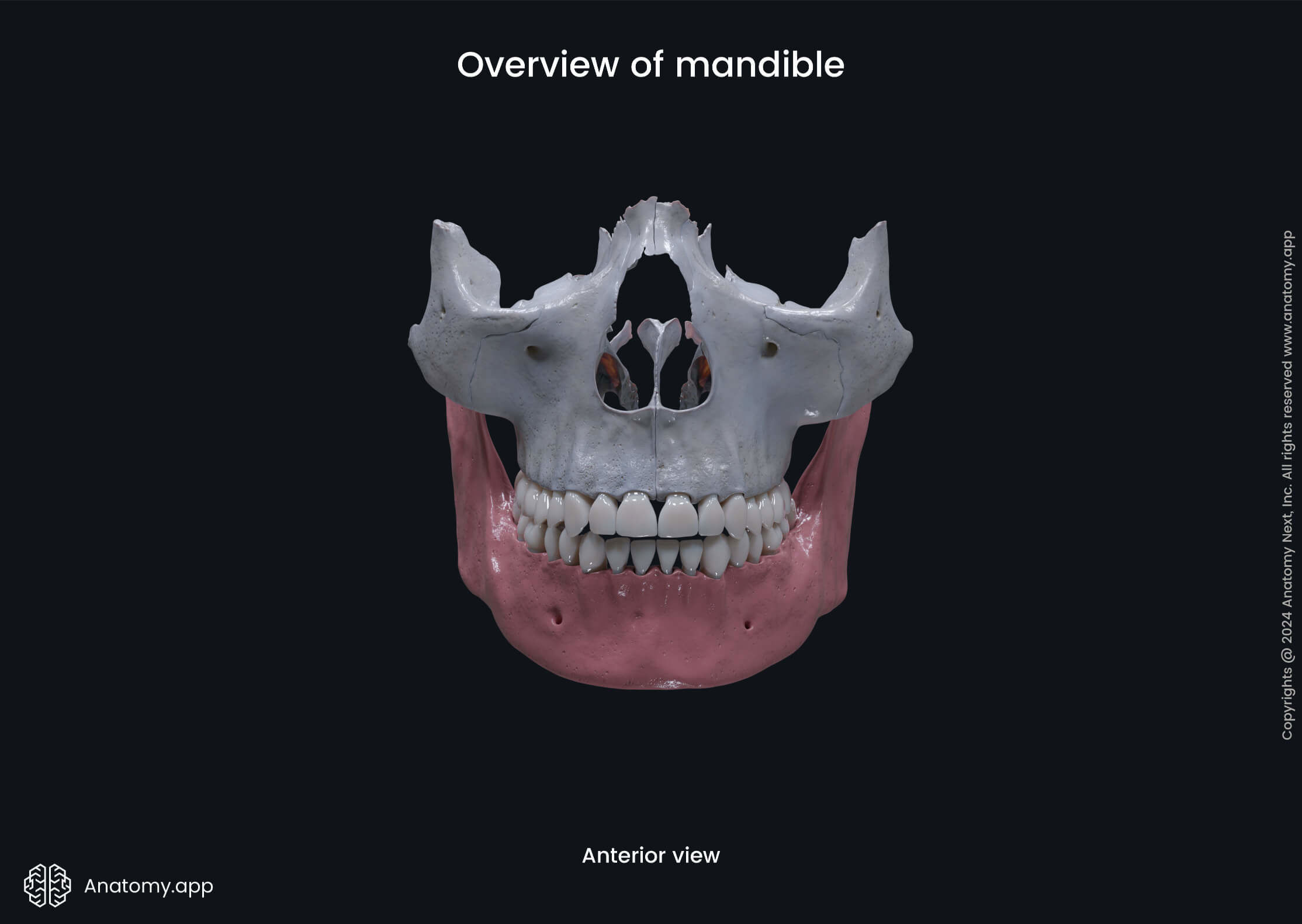
Mandible
The mandible (or lower jawbone, Latin: mandibula) is the only movable bone of the skull and the largest, strongest facial bone. It is a single bone connected to the skull by the temporomandibular joint. The mandible forms the lower jaw and houses the lower or mandibular teeth.
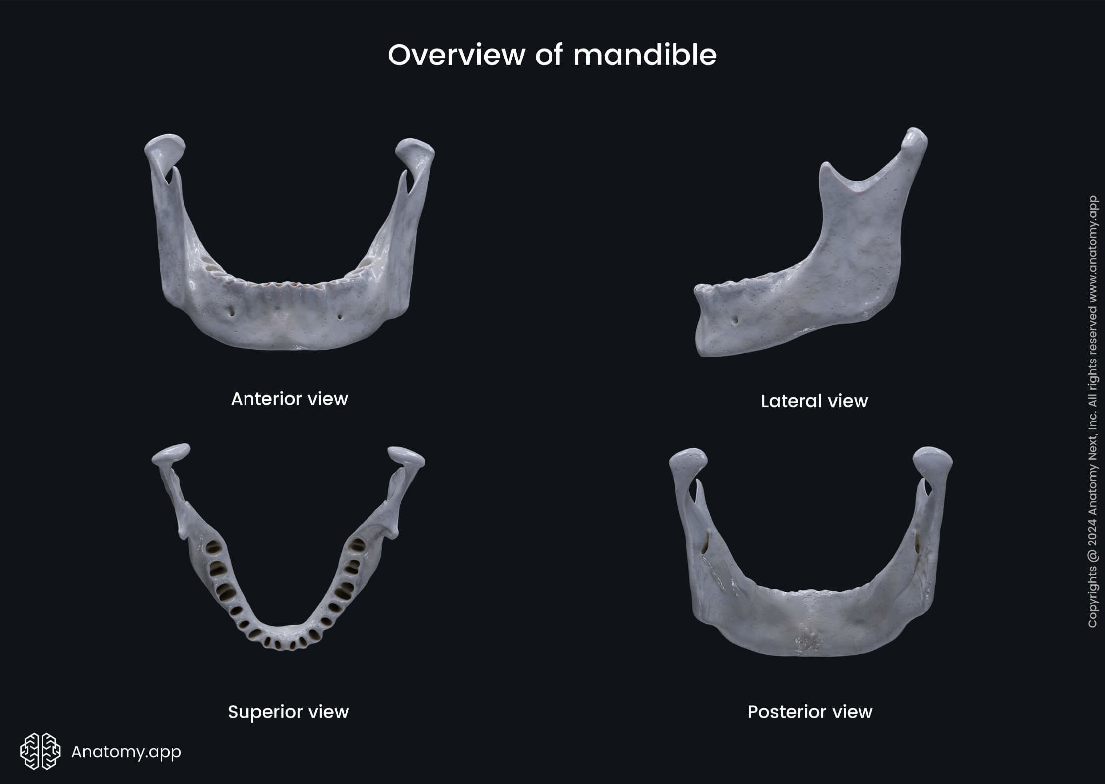
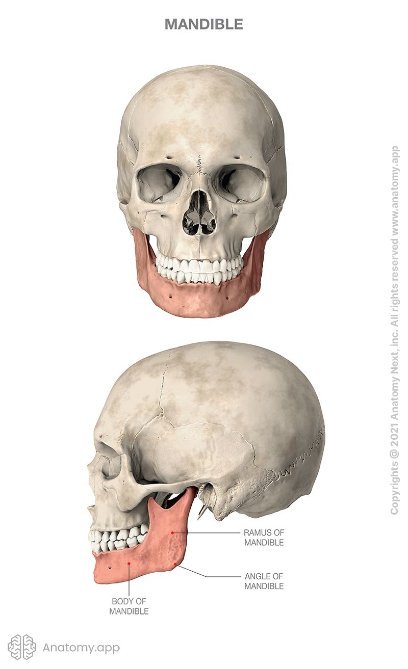
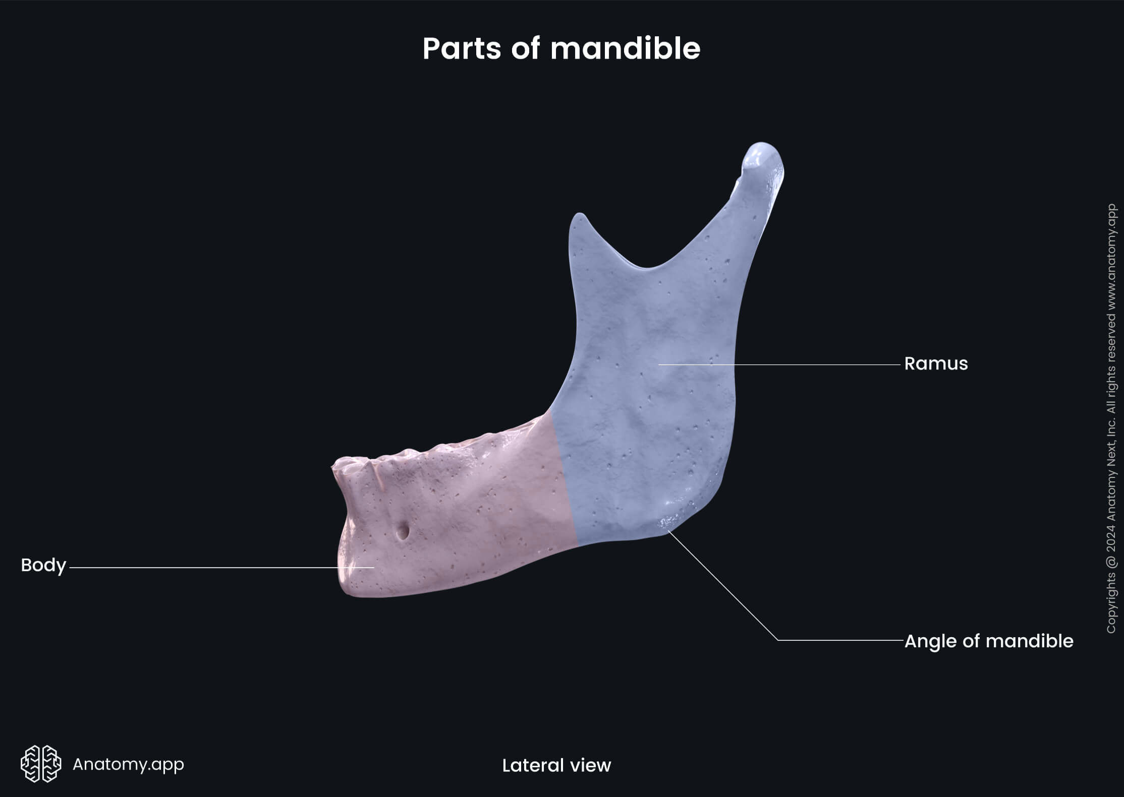
The lower jawbone has three main parts: a body and two rami of the mandible.
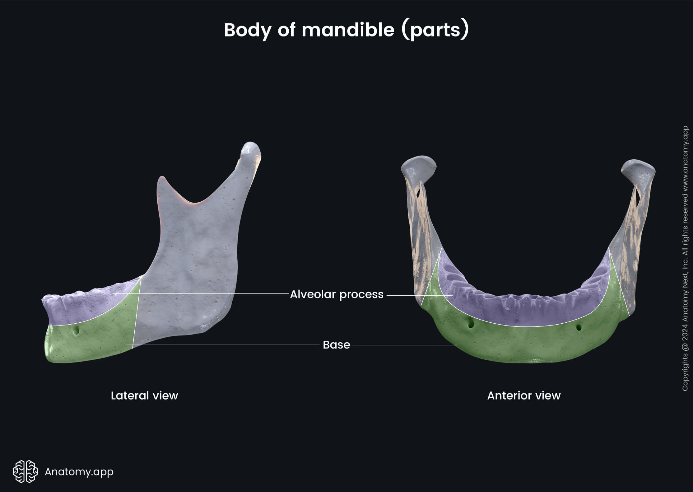
Body of mandible
The body is the anterior curved part of the mandible. The body of the mandible can be divided in two parts: the base and the alveolar part of the mandible.
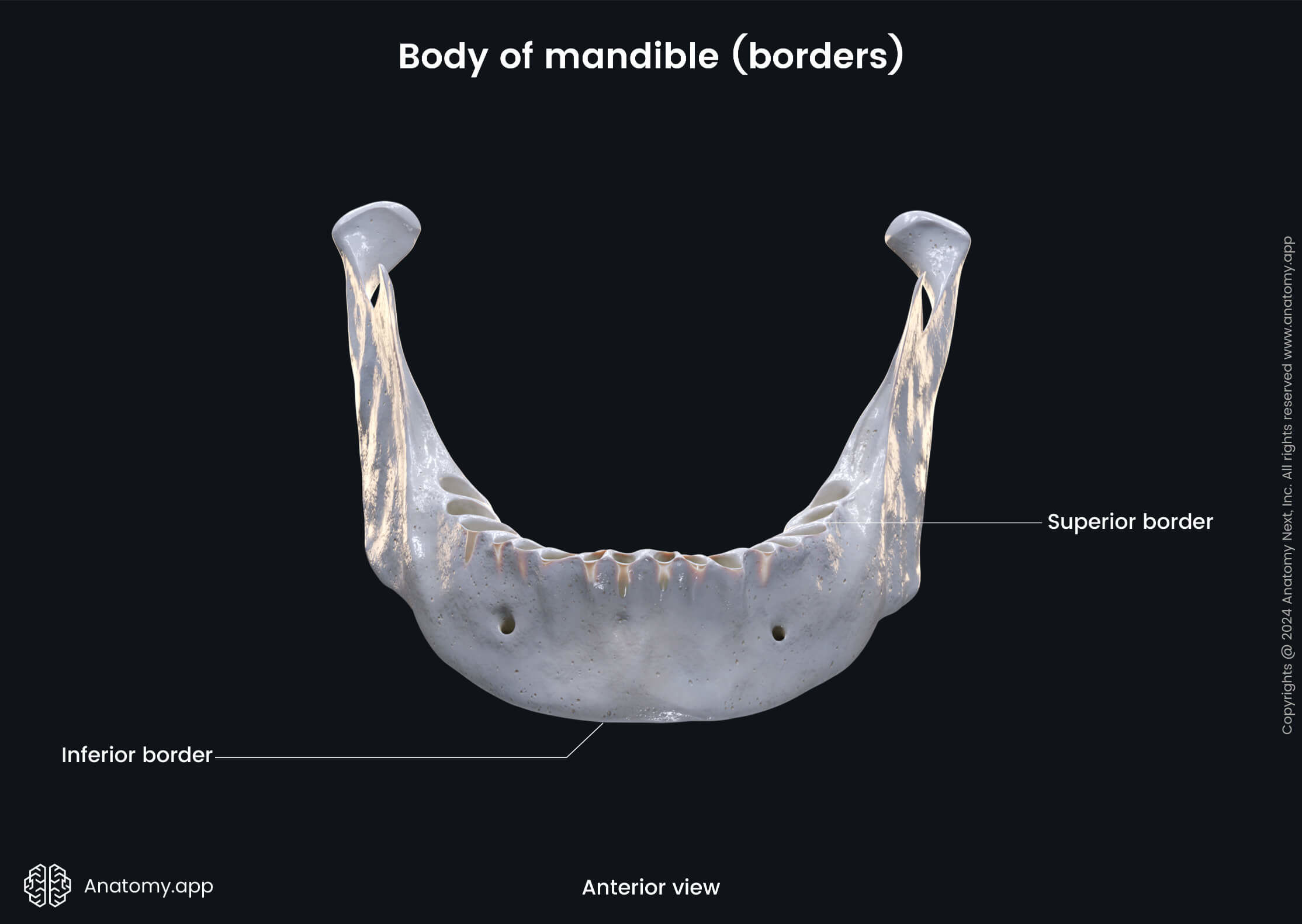
The body of the lower jawbone has two surfaces (external, internal) and two borders (superior or alveolar, and inferior).
Base of mandible
The base is the lower part of the body of the mandible, excluding the alveolar part. The base of the mandible has an external and an internal surface. The features found on the external surface of the mandibular base are:
- Mental protuberance
- Mental tubercle (2)
- Mental foramen (2)
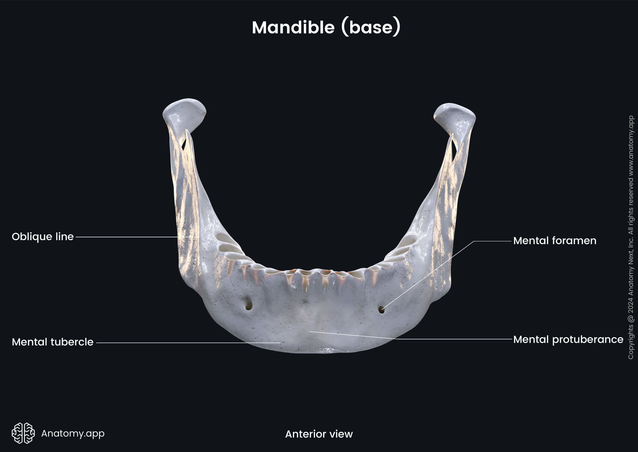
The mental protuberance is a prominence found on the outer surface of the mandible forming the chin. On either side of the mental protuberance, is a prominence known as the mental tubercle. Together the mental protuberance and mental tubercle create a landmark known as the mental trigone.
The mental foramen is an opening in the lower jawbone located below the second premolar tooth. This opening serves as the passage for the mental nerve - a nerve that arises from the inferior alveolar nerve, which itself is a branch of the mandibular nerve (CN V3).
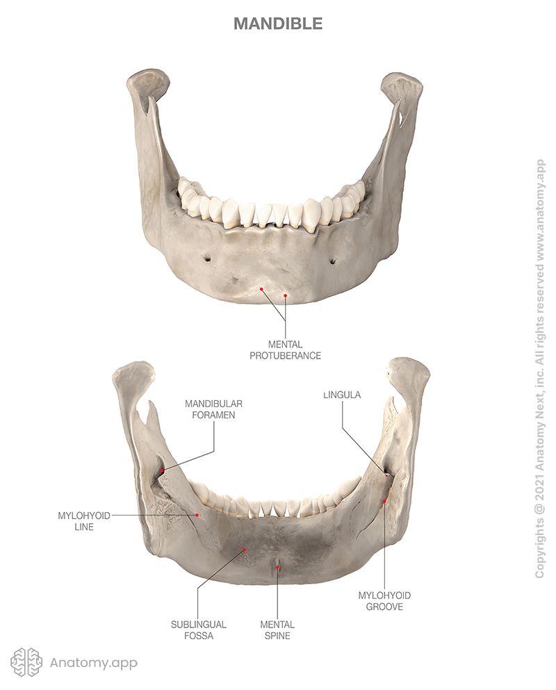
The internal surface of the mandibular base also features several anatomical landmarks, including:
- Mental spine
- Digastric fossa (2)
- Mylohyoid line (2)
- Sublingual fovea (2)
- Submandibular fovea (2)
The mental spine is a bony elevation on the inner surface of the mandible projecting toward the tongue. The mental spine is the origin site of the genioglossus and geniohyoid muscles.
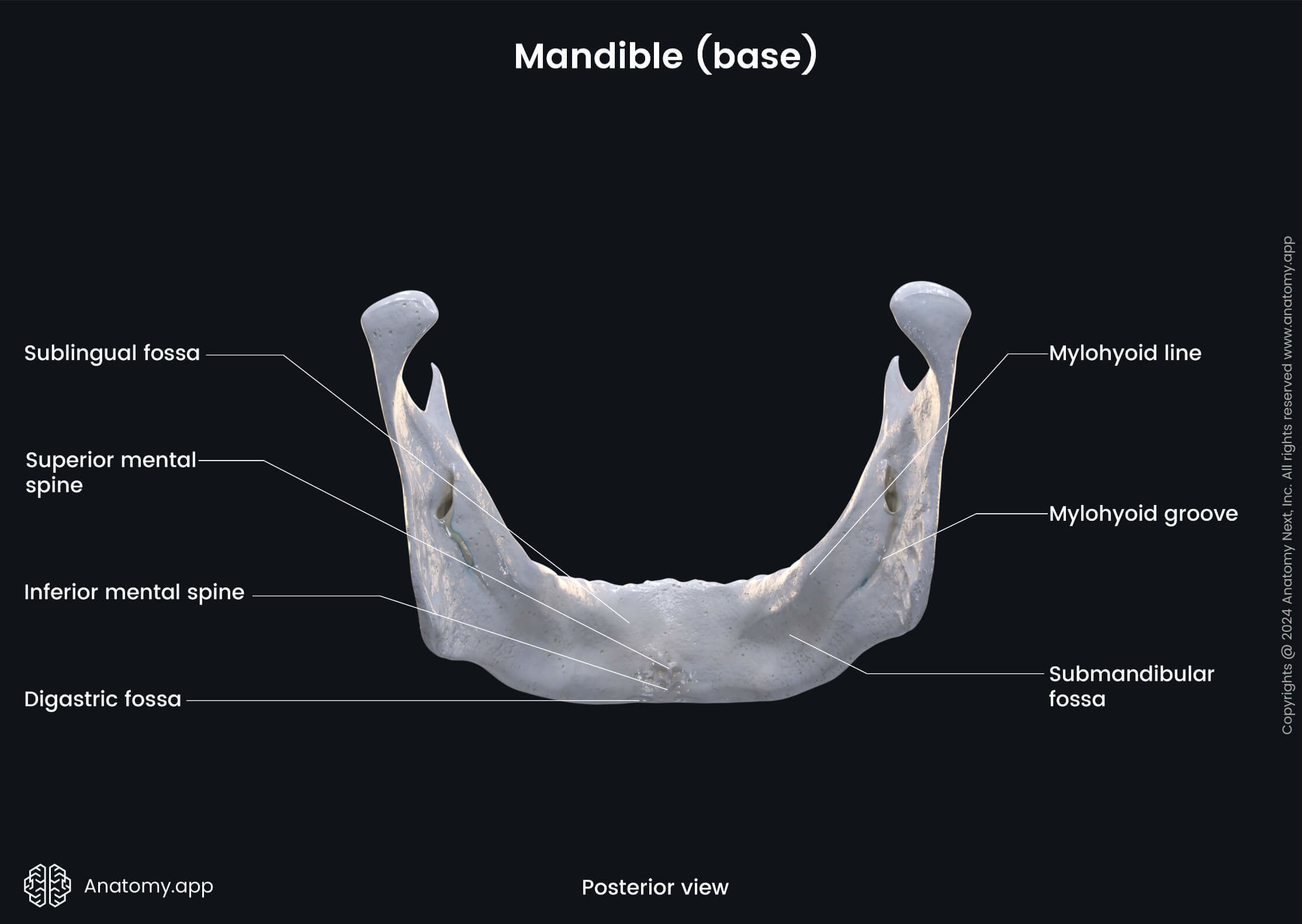
The digastric fossa is an oval depression on the lower internal surface of the mandibular body on either side of the middle line. It is an attachment site for the anterior belly of the digastric muscle.
The mylohyoid line is a paired oblique ridge extending from the posterosuperior to anteroinferior aspect of the body of the mandible. It is the origin site of the mylohyoid muscle, and its posterior part is the origin of the mylopharyngeal part of the superior pharyngeal constrictor.
The sublingual fovea is a depression on the inside of the mandible for the sublingual gland (one of the salivary glands). It is a paired structure found on the anterior half of the body of the mandible below the mylohyoid line.
The submandibular fovea is a depression on the mandible's inner surface for another salivary gland - the submandibular gland. This fovea is also a paired structure. It is found on the posterior half of the mandibular body below the mylohyoid line.
Alveolar part of mandible
The alveolar part or alveolar process of the mandible is the portion of the mandibular body that surrounds and supports the lower teeth. An alveolar process is a crested process of upper or lower jaw which houses the teeth. The curved free margin of the alveolar process is called the alveolar arch. The alveolar arch of the mandible (as the alveolar arch of the maxilla) features the following landmarks:
- Dental alveoli
- Interalveolar septa
- Interradicular septa
- Alveolar yokes
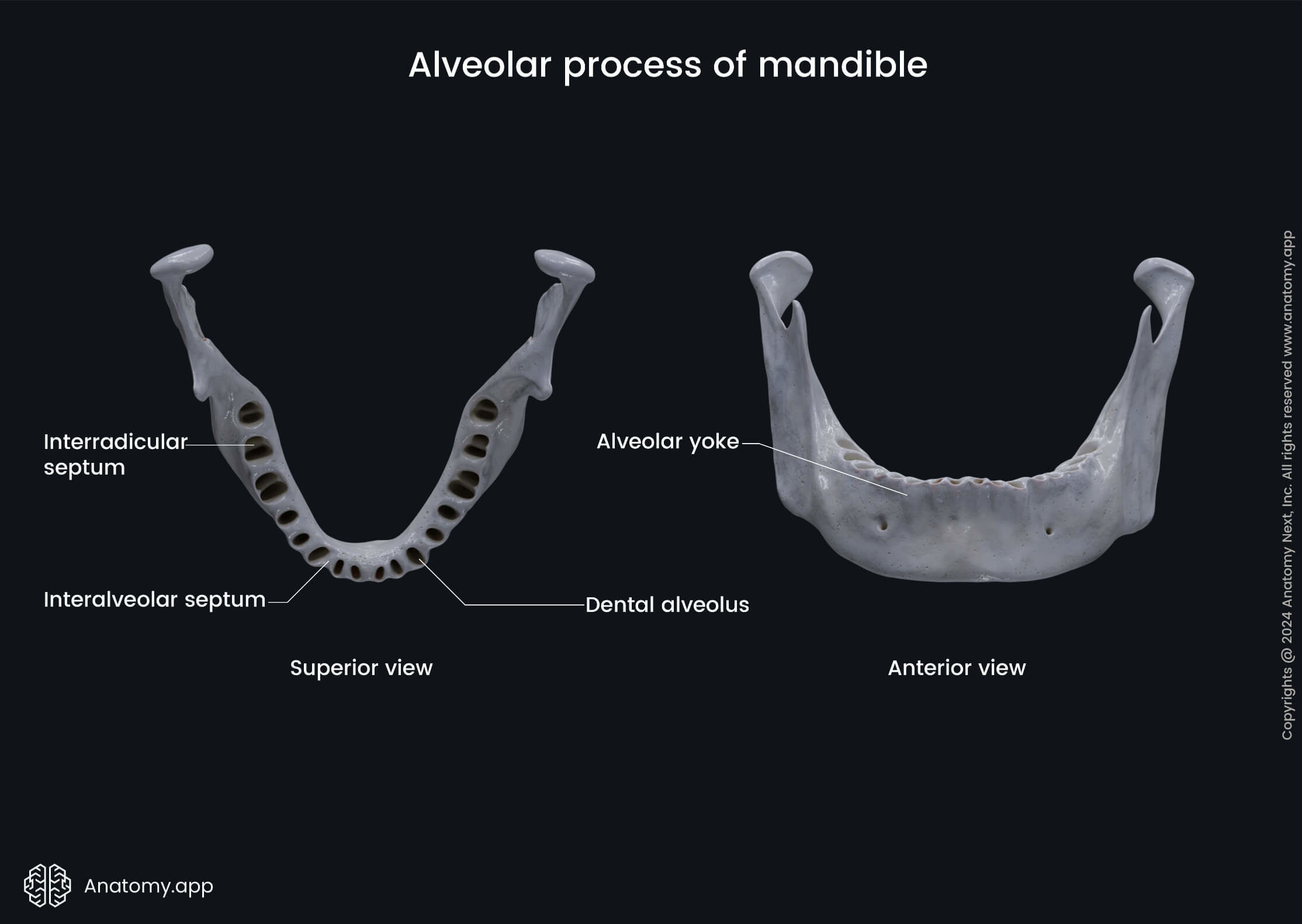
The dental alveoli are sockets in the alveolar process where the roots of the teeth lie. The dental alveoli of the mandible house the roots of the lower teeth, while the dental alveoli of the maxilla - the upper teeth.
The interalveolar septa are bony ridges between adjacent dental alveoli. But the interradicular septa are bony ridges forming compartments in dental alveoli for the roots of the teeth in both the upper and lower jaw bones.
The alveolar yokes (or juga alveolaria) are eminences on the outer surfaces of the jaw bones produced by the projections of the dental alveoli. They can be seen on alveolar processes of both the maxilla and the mandible.
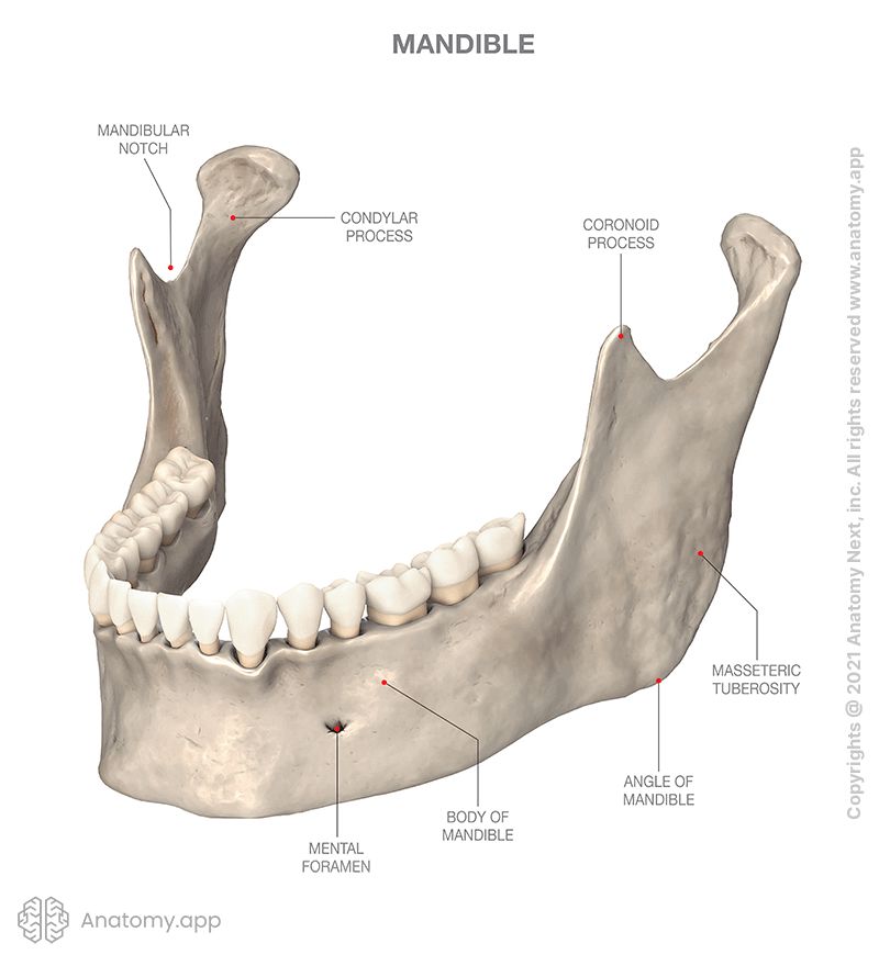
Ramus of mandible
The rami (singular: ramus) of the mandible are situated on the right and left side of the lower jawbone. Each ramus of the mandible has an internal and external structures, and features several major landmarks:
- Angle of mandible
- Condylar process, which features:
- Head of the mandible
- Neck of the mandible
- Pterygoid fovea
- Coronoid process
- Mandibular notch
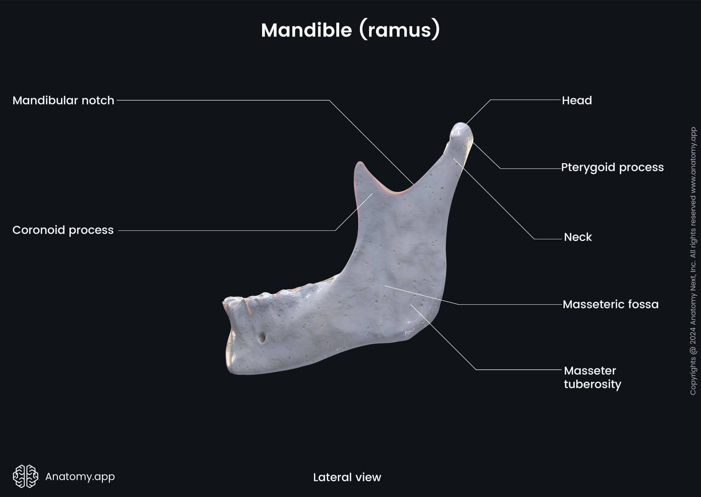
The angle of the mandible (also known as the gonial angle) is the angle seen on each side of the mandible. It is the junction between the body and ramus of the mandible.
The condylar process is an articular process of the ramus that articulates with the disk of the temporomandibular joint (TMJ). It features three structures: the head and neck of the mandible, and the pterygoid fovea.
The head of the mandible is the articular part of the condylar process. The neck of the mandible is a narrow segment of the condylar process below the mandibular head. And the pterygoid fovea is an anteromedial pit below the head of the mandible, where the lateral pterygoid muscle attaches.
The coronoid process is a muscular process located anteriorly to the condylar process of the mandible (the condylar process is thicker than the coronoid process). The temporalis muscle attaches to the coronoid process. The two processes (coronoid and condylar) of mandible are separated by a depression known as the mandibular notch.
The internal surface of the mandibular ramus features some important landmarks as well, which are:
- Inferior alveolar foramen (mandibular foramen)
- Mylohyoid groove
- Pterygoid tuberosity
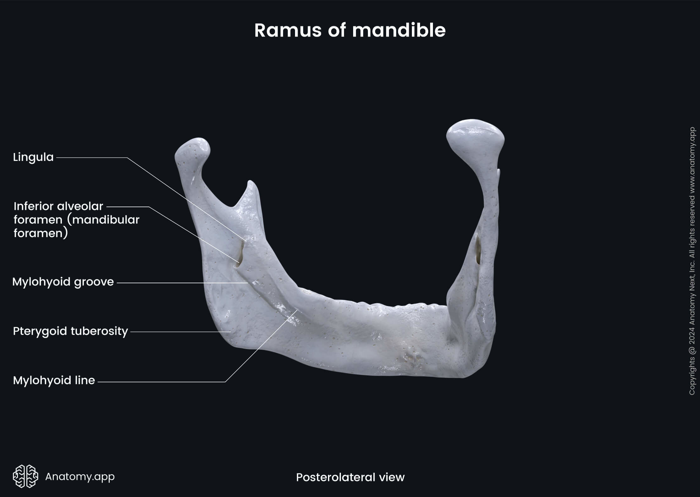
The mandibular foramen is an opening on the inner surface of the mandibular ramus leading into the mandibular canal, a bony passage in the lower jawbone that transmits the inferior alveolar artery and nerve.
The mylohyoid groove or sulcus is a groove seen on the internal surface of the mandible extending forward and downward from the mandibular foramen. This groove is the passage for the mylohyoid nerve and the mylohyoid branch of the inferior alveolar artery.
The pterygoid tuberosity is a roughened area occasionally present on the internal surface of the ramus near the angle of the mandible. The medial pterygoid muscle attaches to the pterygoid tuberosity.
The external surface of the ramus of the mandible features a roughened area known as the masseteric tuberosity. It is occasionally present near the angle of the mandible. This is an attachment site for the masseter muscle.
Anatomy.app
Contact information
- For questions regarding business inquiries. Please contact:
- info@anatomy.app