- Anatomical terminology
- Skeletal system
- Joints
- Muscles
- Heart
- Blood vessels
- Lymphatic system
- Nervous system
- Respiratory system
- Digestive system
- Urinary system
- Female reproductive system
- Male reproductive system
- Endocrine glands
- Eye
- Ear
Teeth
The teeth (Latin: dentes) are small bone-like structures that are arranged into two rows within the oral cavity. They consist of solid tissue on the outside and soft connective tissue on the inside. Teeth are positioned in the dental alveoli of the maxillae and mandible. Overall, they aid in the mechanical processing of food during eating. Therefore, they are classified as a part of the digestive system.
Sets of teeth
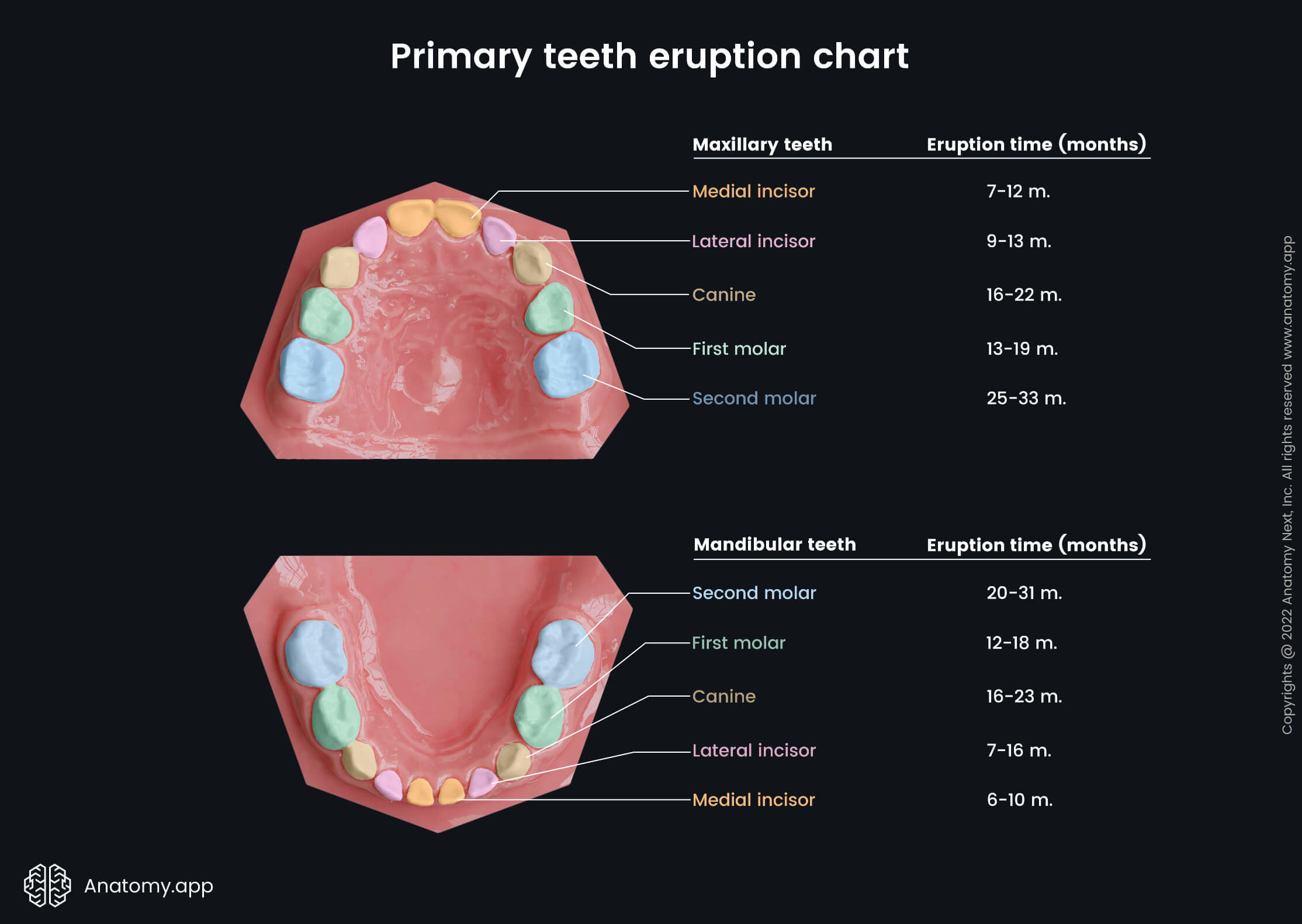
Humans have two sets of dentitions throughout their lifetime. The first set is known as primary dentition or deciduous teeth, and it is also referred to as baby or milk teeth. This dentition is slowly replaced by the secondary dentition known as the permanent teeth. The replacement happens during childhood.
Deciduous teeth are smaller in size compared to permanent teeth. Also, they have smaller roots but larger pulp chambers than permanent teeth. Overall, children have 20 milk teeth, and they start to appear at six to eight months. In contrast, the milk teeth replacement process usually begins around 5 to 7 years and continues until 12 to 14 years.
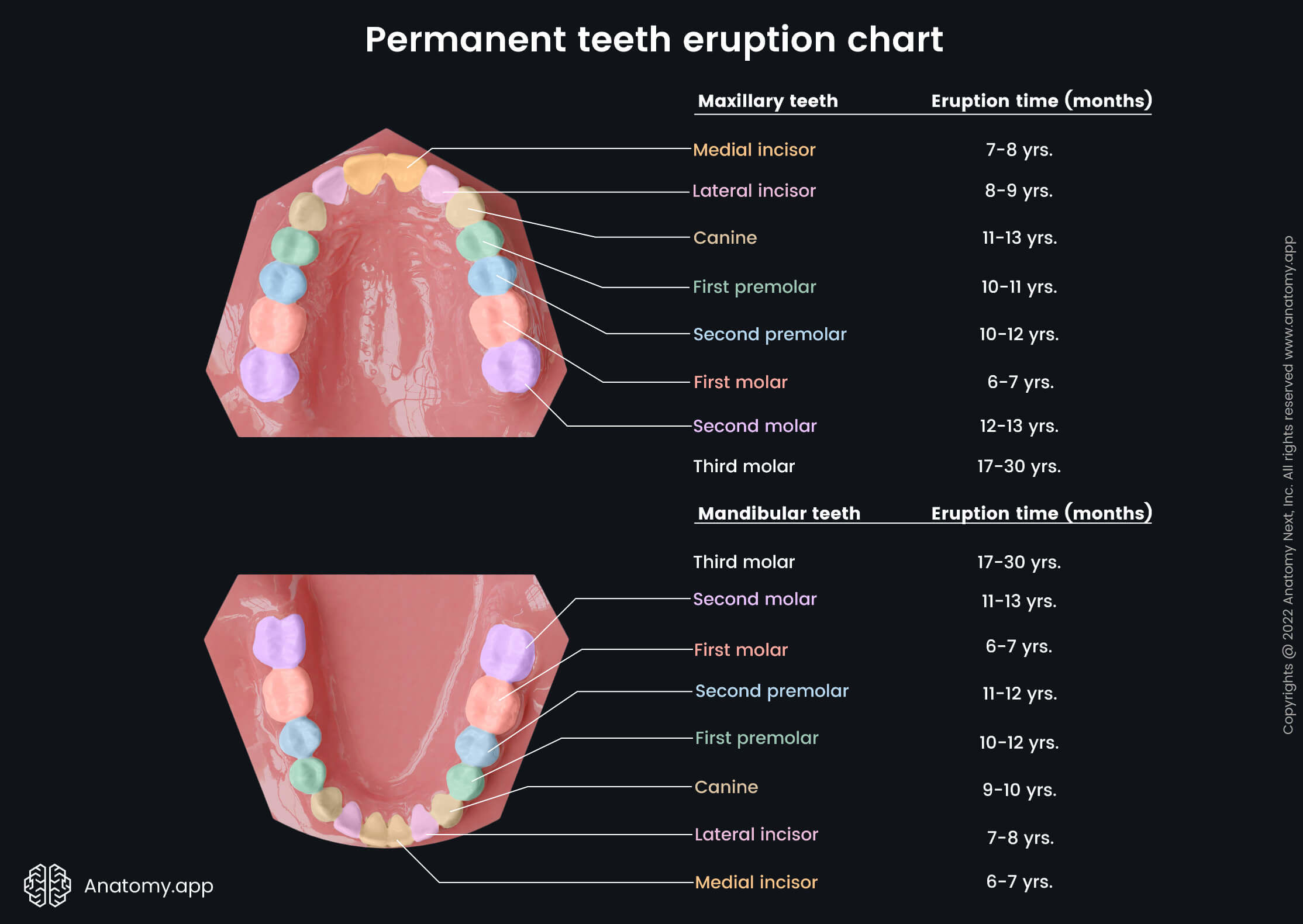
Permanent teeth are larger, and each person has 32 secondary teeth. They can be subdivided into two groups:
- The first group includes the incisor, canine, and premolar teeth. Altogether they are also known as succedaneous teeth, and these teeth replace the identical teeth of primary dentition.
- The second group includes molar teeth, which do not have counterparts in the primary dentition.
The permanent teeth increase the number of teeth from 20 to 32 as each side of the upper and lower jaw contains three molar teeth. Please note that the molar teeth of a child correspond to the premolars of an adult.
Teeth types
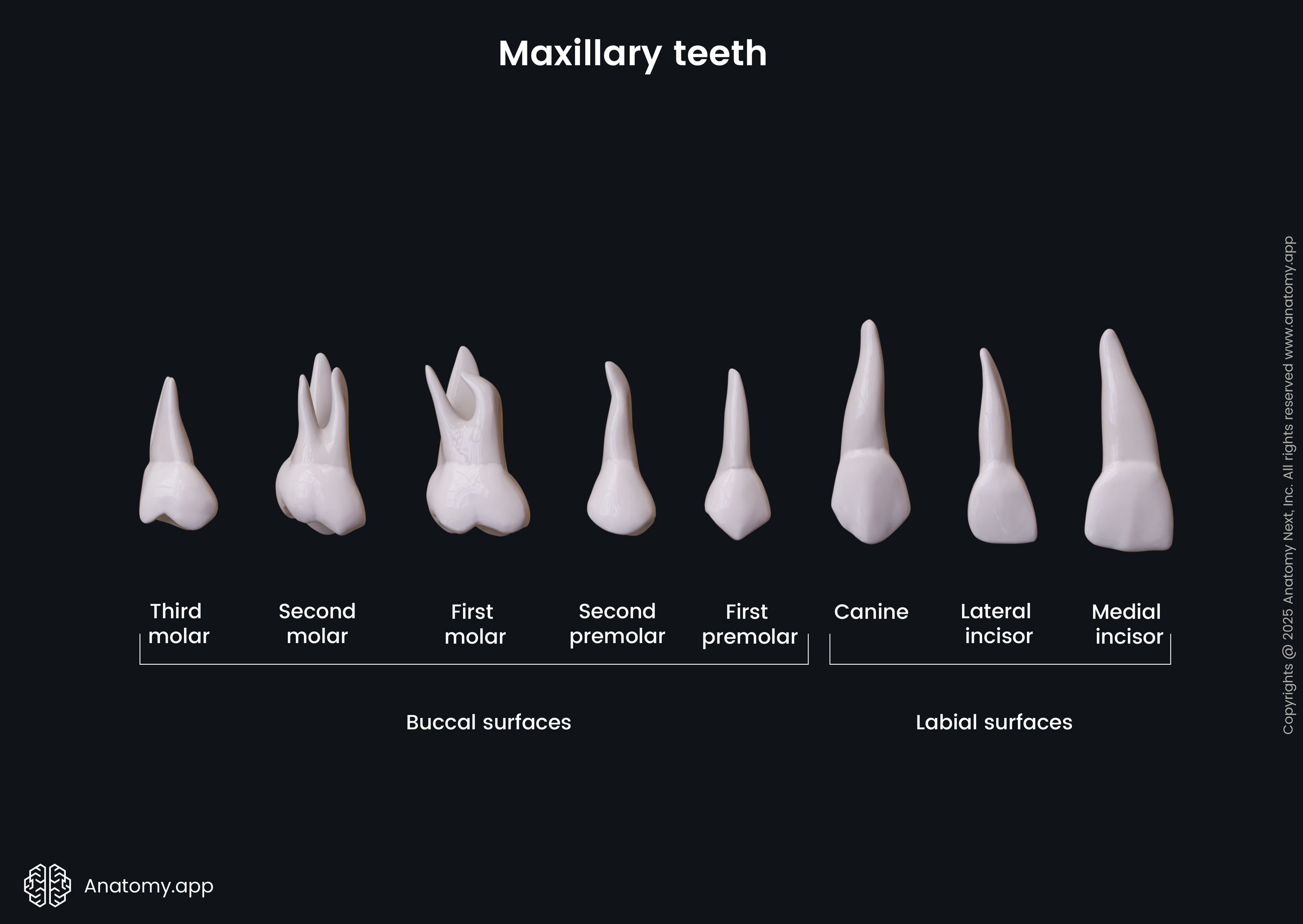
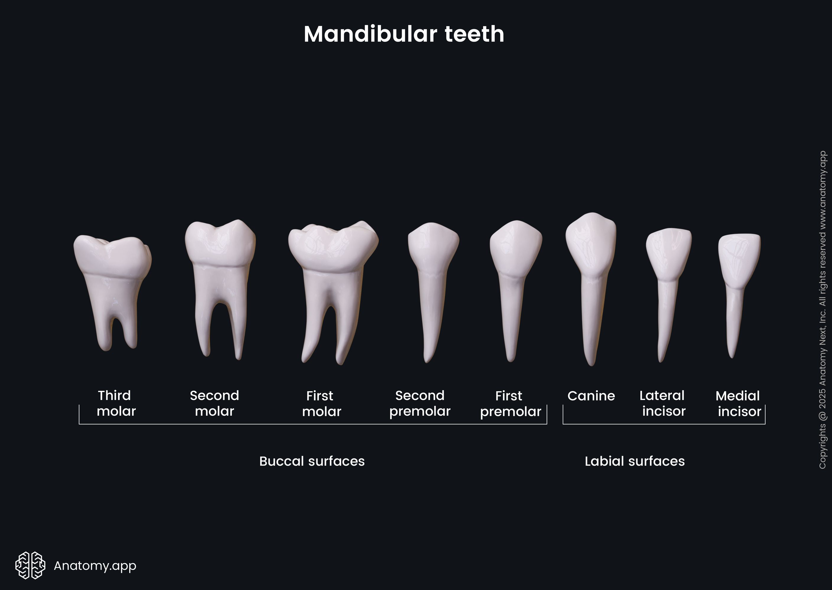
As mentioned above, adults have 32 permanent teeth. They are divided into four types based on their functions, positions in the oral cavity, number of roots, and shapes of their crowns. The oral cavity of an adult has the following four teeth types:
Additionally, the 16 upper teeth are known as maxillary teeth, while the lower 16 teeth are mandibular teeth. The maxillary and mandibular teeth of the same type are similar, but they are not identical.
Overall, the oral cavity can be subdivided into four quadrants, each having eight teeth. According to this, every quadrant contains the following eight permanent teeth:
- Medial (central) incisor
- Lateral incisor
- Canine tooth
- Two premolars
- Three molars
Incisors
Every person has eight incisors - four are positioned in the middle of each jaw. Therefore, four incisors are located in the middle of the upper jaw (maxillae), and four more are situated in the middle of the lower jaw (mandible).
Depending on how far the incisors are positioned from the midline of each jaw, they can be subdivided into medial (central) and lateral incisors. Overall, each jaw has two central (medial) incisors and two lateral incisors. Moreover, the medial incisors are the most centrally located teeth.
Incisors are used to cut, pluck, and bite food during eating. They also help to hold food within the oral cavity. These teeth are designed according to their functions, so they have chisel-shaped crowns with triangular mesial and distal surfaces. Incisors have only one long and conical root.
Canines
Canines are also known as eye teeth or cuspids. Each person has four canine teeth. Every jaw contains two canines - each is positioned between the lateral incisor and the first premolar. In every jaw between both canines are located all incisors. Also, the mandibular canines are a half-tooth closer to the midline of the jaw compared to the maxillary canines.
This teeth type has a single, long, deep root flattened and grooved on the sides. It is longer than other teeth roots. The crown of the canine has a sharpened conical shape with spear-like cutting edges. Overall, canines are used for gripping, cutting, and food tearing.
Premolars
Premolars, also known as bicuspids, are found between the canines and first molars. Each person has eight premolars - four maxillary premolars and four mandibular premolars. This type provides food crushing, grinding and grasping during eating.
Premolars usually have only one root that is bifurcated. However, the maxillary first premolar sometimes has two roots instead of one. This type has a cuboid crown that contains two cusps on its occlusal surface.
Molars
Molars are the last teeth in each jaw. Therefore, they are positioned at the back of the oral cavity. Every adult has a set of 12 molars - six teeth are found in each jaw. This teeth type is used for food grinding and crushing.
The molars have a cuboid-shaped crown and a square-shaped occlusal surface with four or five cusps. Usually, the maxillary molars have four cusps and mandibular molars present with four or five cusps. Also, the upper molars have three roots, while the lower molars have two.
The third molars on every side of the upper and lower jaw are called wisdom teeth. These are the last teeth to erupt. For some individuals, it does not happen during their lifetime.
Teeth eruption times
As mentioned, children have 20 milk teeth, and this dentition starts to appear around six to eight months. In contrast, deciduous teeth replacement usually begins around 5 to 7 years and continues until 12 to 14 years.
The dentures of the secondary teeth lie between the roots of the primary teeth until the permanent teeth are ready to replace the deciduous teeth. The last permanent teeth to appear are the last molar teeth, also known as the wisdom teeth. They do not replace any of the primary teeth, and they typically emerge between the ages of 17 and 24.
Deciduous teeth eruption times
The first deciduous teeth to appear are the medial incisors. It happens around 6 - 8 months. The medial incisors are usually followed by the lateral incisors. Overall, they can erupt anytime between 6/7 to 16 months.
The first molar teeth of the primary dentition can be visible around 12 to 15 months, while the canine teeth emerge around 16 to 23 months. And finally, the second molars appear only around 20 to 33 months.
Permanent teeth eruption times
The first permanent teeth that appear are usually the first molar teeth, and it happens around 6 - 7 years. Medial incisors replace their corresponding teeth of the primary dentition around six to eight years, while lateral incisors get replaced around 9 (7 - 9) years.
The replacement of the molar teeth of the primary dentition (premolars in adults) happens around ten years, while canines get replaced around 9 to 13 years. The second molar tooth appears only around 11 to 13 years.
And finally, the third molar teeth can emerge anytime between 17 to 30 years or even later. However, it usually happens till 24 years.
Teeth surfaces
Every tooth has several surfaces. Moreover, the names of some of the surfaces of the maxillary teeth differ from the identical surfaces of the mandibular teeth. Overall, teeth surfaces are named after the anatomical structure they face.
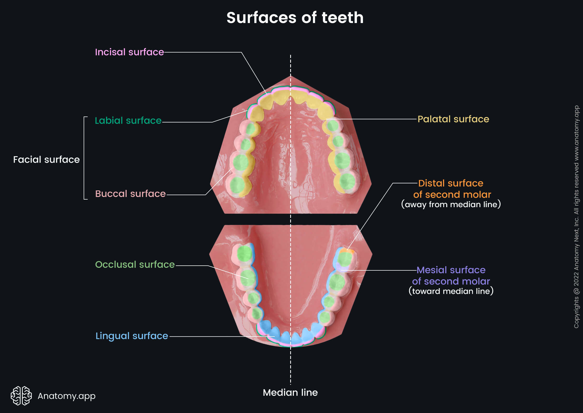
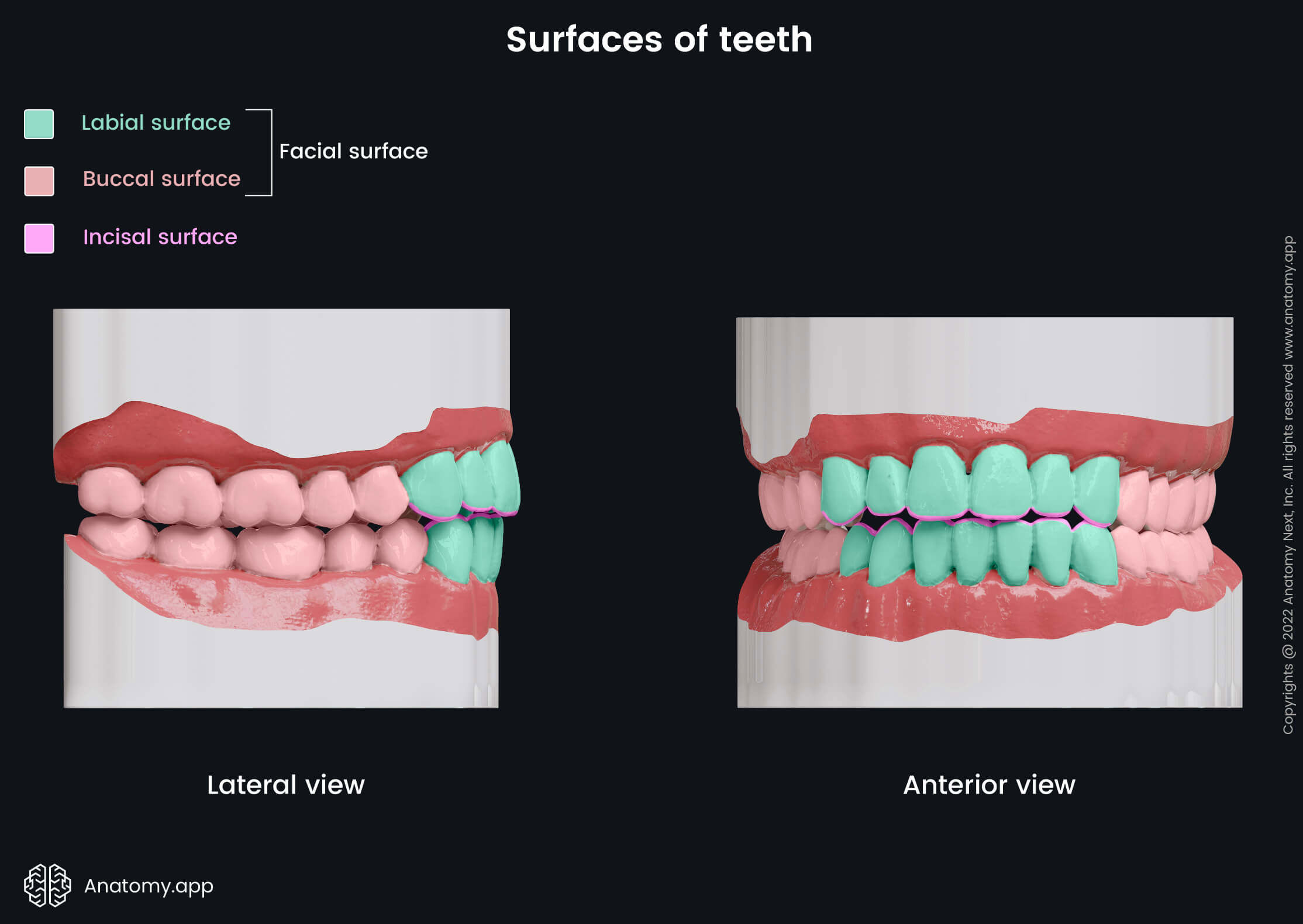
- The surface facing the lips is the labial surface. The incisors and canines are the only teeth that have this surface.
- The surface that is directed against the cheeks is the buccal surface, and it is found on the premolars and molars.
- The labial and buccal surfaces together form the facial surface.
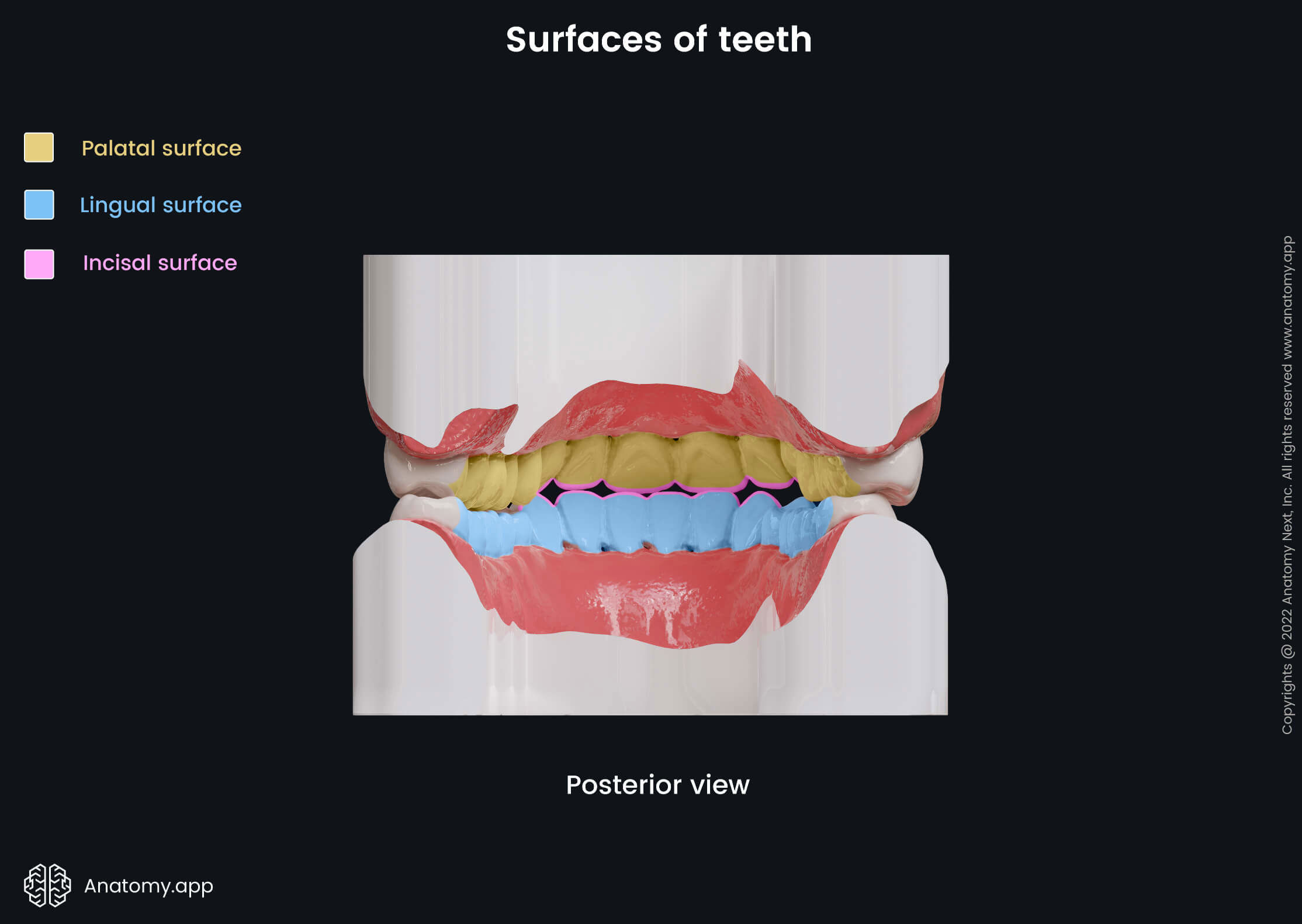
- The incisal surface is the sharp and pointed top of the incisors and canines.
- The lingual surface is the inner surface of the mandibular teeth, as it faces the tongue.
- In contrast, the palatal surface is the inner surface of the maxillary teeth, as it faces the palate.
- The mesial surface is the side of a tooth that is closest to the midline of the jaw.
- In contrast, the distal surface is the side of a tooth that is farthest from the midline of the jaw.
- The occlusal (biting) surface is found only on premolars and molars. It is the top surface that has several cusps and grooves.
Each surface has a unique design that serves a specific function. Therefore, all surfaces of each tooth are designed according to their functions. For example, canines and incisors have a single sharp top surface, serving to cut food. In contrast, premolars and molars have larger and flatter top surfaces. Moreover, these surfaces contain several cusps, grooves, fissures, and pits, as premolars and molars aid in food grinding and chewing.
Teeth parts
Each tooth consists of three parts - the crown, the neck, and the root.
- The crown is the visible part of a tooth. It is elevated above the gingiva and usually has a white or yellowish-white color depending on several factors such as the age of a person, eating and lifestyle habits. Overall, the crown is responsible for mechanical food processing during eating.
- The neck is a slightly narrow portion, and it connects the crown with the root. Also, the neck connects with the marginal gingiva, and its height varies between 0.04 - 0.06 inches (1 - 1.5 mm).
- The roots lie in the dental alveoli of the alveolar arches of the mandible and maxillae. They are fixated in the dental alveoli by a fibrous joint type called gomphosis. This joint type allows minimal or no movements. However, the amplitude of the movements can increase due to changes caused by aging, lifestyle and various disorders.
Each tooth is also fixated in its place by a specialized connective tissue structure called periodontium, which is found between the root of the tooth and bone tissue. Overall, the periodontium consists of four components - cementum, periodontal ligament, alveolar bone, and gingival tissue.
Teeth microanatomy
The primary mass of a tooth is composed of dentin, which forms the bulk of the tooth. The dentin in the crown part is covered by enamel, while the outside of the root is covered by cement. The transition between cement and enamel is the neck of the tooth, where cement thinly layers on the enamel. This site is known as the cementoenamel junction.
Cement and dentin are formed by so-called bone tissue, while enamel is a secretion of a special mineralized epithelial cell. Teeth appear very similar to bones, but they have a completely different structure and histology.
Enamel is the hardest substance in the human body, forming a durable protective layer that protects the tooth from the outside from various harmful factors. The thickness of the enamel varies, with the top part being the hardest, and the hardness gradually decreases closer to the dentin. Additionally, the enamel is the only tooth structure that can heal by itself.
The enamel of a newly erupted tooth is covered by a delicate membrane called Nasmyth’s membrane, also known as the primary enamel cuticle. This membrane is removed during chewing but typically remains intact on the sides of the crown close to the gingiva. It serves a protective role and is acid-resistant.
Every tooth has a pulp cavity or chamber that contains various blood vessels and nerves. It is located in the middle of the crown and neck. This cavity narrows towards the root and becomes the pulp or root canal in the root part of the tooth. Every root canal ends with a small opening called the root end opening or apical foramen. It allows the nerves and blood vessels to enter the root canal and further reach the pulp cavity.
Teeth functions
The teeth are primarily involved in digestion, and they aid in chewing. Overall, teeth cut, bite, grip, and grind food into smaller pieces to form a semi-solid mass called food bolus that can be easily swallowed.
However, teeth also play a significant role in articulation and speech formation. Moreover, they shape the face and support surrounding tissue, giving the person a unique and personal appearance.
Neurovascular supply of teeth
Arterial blood supply of teeth
The arterial blood supply to the teeth directly or indirectly is provided by the maxillary artery - a terminal branch of the external carotid artery.
- The maxillary incisors and canines are supplied by the anterior and middle superior alveolar arteries, which are branches of the infraorbital artery. The infraorbital artery originates from the pterygopalatine part of the maxillary artery.
- The maxillary premolars and molars receive arterial blood from the posterior superior alveolar arteries that arise from the maxillary artery when it travels through the pterygopalatine fossa.
- In contrast, all mandibular teeth are supplied by the inferior alveolar arteries and these arteries branch off the mandibular portions of the maxillary arteries.
Venous drainage
Overall, venous drainage of the teeth is provided by the veins that travel along the arteries mentioned above and share the same names.
- The maxillary teeth are drained by the anterior, middle and posterior superior alveolar veins.
- The mandibular teeth are drained by the inferior alveolar veins.
All alveolar veins drain into the pterygoid venous plexus. However, some variations are common, and sometimes they can drain in the facial vein.
Innervation
The innervation of the teeth is provided by the maxillary and mandibular divisions of the trigeminal nerves (CN V2 and CN V3).
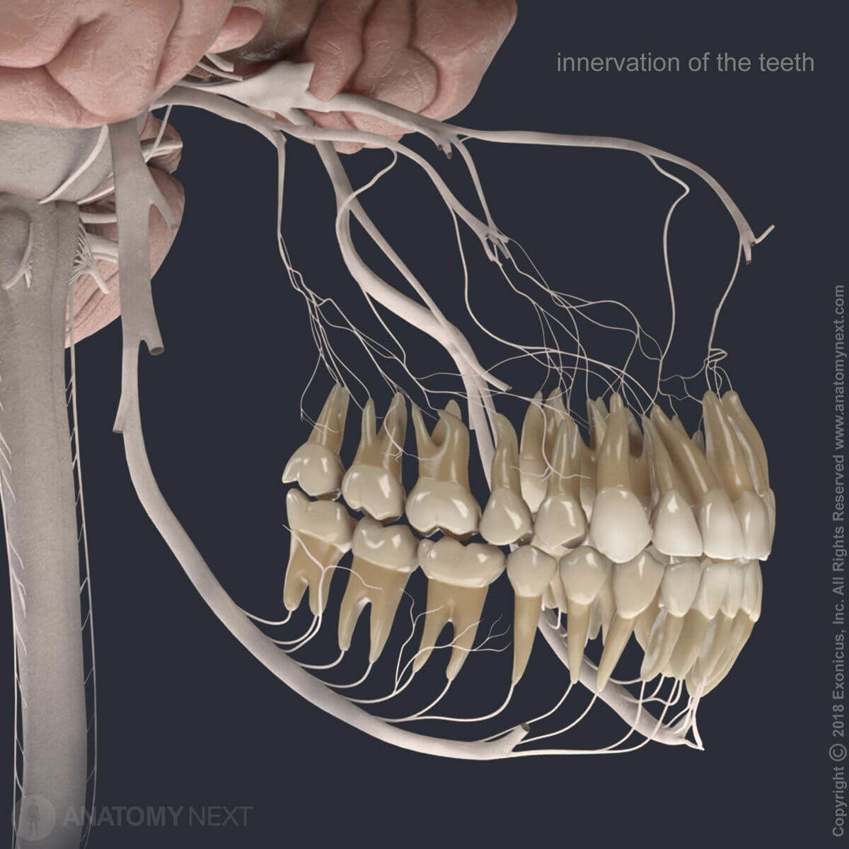
The maxillary teeth are innervated by the anterior, middle and posterior superior alveolar nerves that arise from the maxillary nerves (CN V2).
- The maxillary incisors and canines are innervated by the anterior superior alveolar nerves of the infraorbital nerves. The infraorbital nerve is a branch of the maxillary nerve.
- The maxillary premolars and the mesiobuccal (MB) roots of the first molars receive innervation from the middle superior alveolar nerves (branches of the infraorbital nerves).
- In contrast, the maxillary molar teeth (except the MB roots of the first molars) are innervated by the posterior superior alveolar nerves.
The mandibular teeth are supplied by the inferior alveolar nerves and their branches. The inferior alveolar nerve arises from the mandibular nerve (CN V3).
- The mandibular incisors and canines receive innervation from the incisive branches of the inferior alveolar nerves. Also, the incisive branches innervate the mandibular first premolars.
- The mandibular second premolars and all molars are innervated by the dental branches of the inferior alveolar nerves.
Anatomy.app
Contact information
- For questions regarding business inquiries. Please contact:
- info@anatomy.app