- Anatomical terminology
- Skeletal system
- Joints
- Muscles
-
Head muscles
- Extraocular muscles
-
Facial muscles
- Occipitofrontalis
- Corrugator supercilii
- Depressor supercilii
- Orbicularis oculi
- Malaris
- Buccinator
- Orbicularis oris
- Mentalis
- Depressor anguli oris
- Depressor labii inferioris
- Levator anguli oris
- Levator labii superioris
- Risorius
- Zygomaticus major
- Zygomaticus minor
- Levator labii superioris alaeque nasi
- Nasalis
- Procerus
- Depressor septi nasi
- Compressor narium minor
- Dilator naris anterior
- Muscles of mastication
- Neck muscles
- Muscles of upper limb
- Thoracic muscles
- Muscles of back
- Muscles of lower limb
-
Head muscles
- Heart
- Blood vessels
- Lymphatic system
- Nervous system
- Respiratory system
- Digestive system
- Urinary system
- Female reproductive system
- Male reproductive system
- Endocrine glands
- Eye
- Ear
Facial muscles
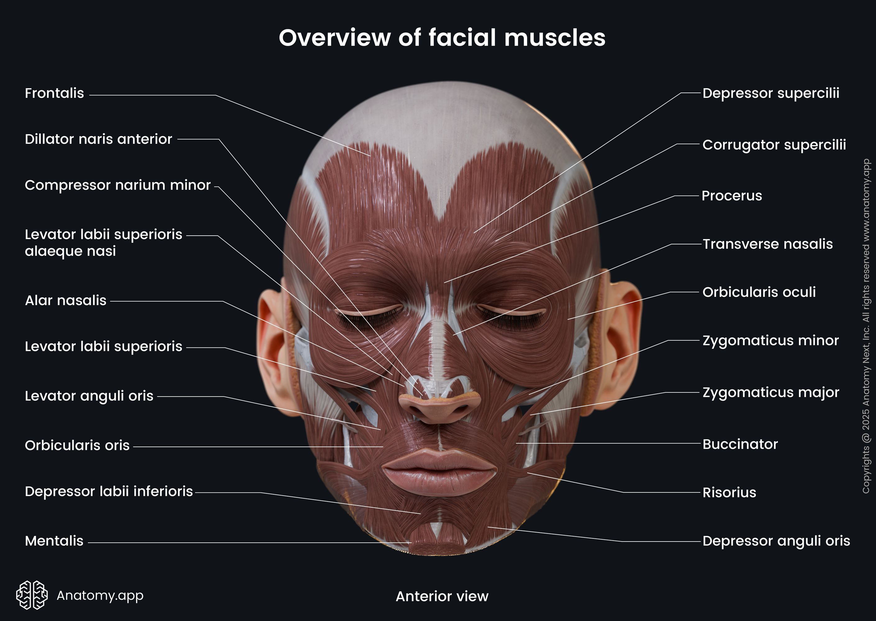
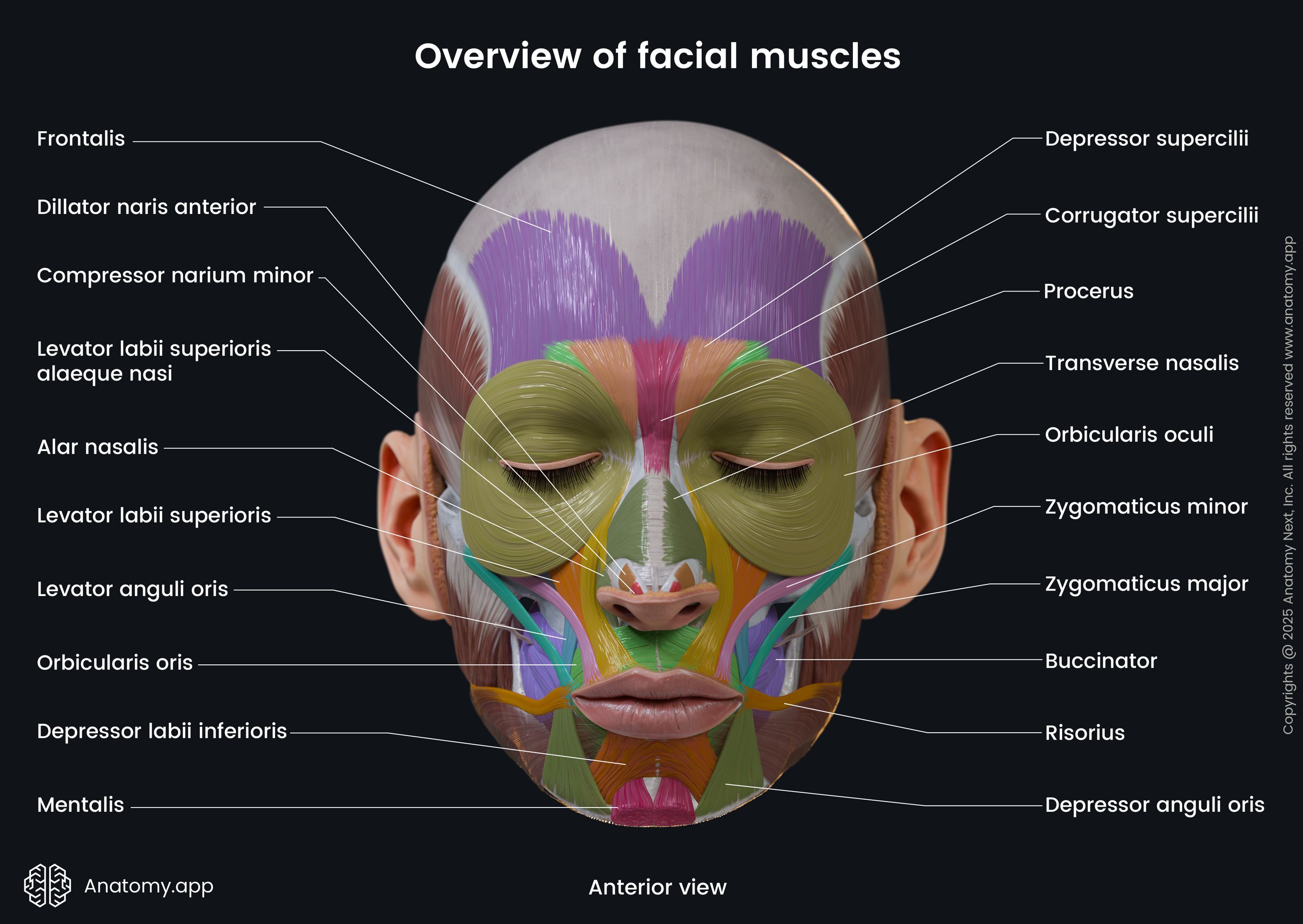
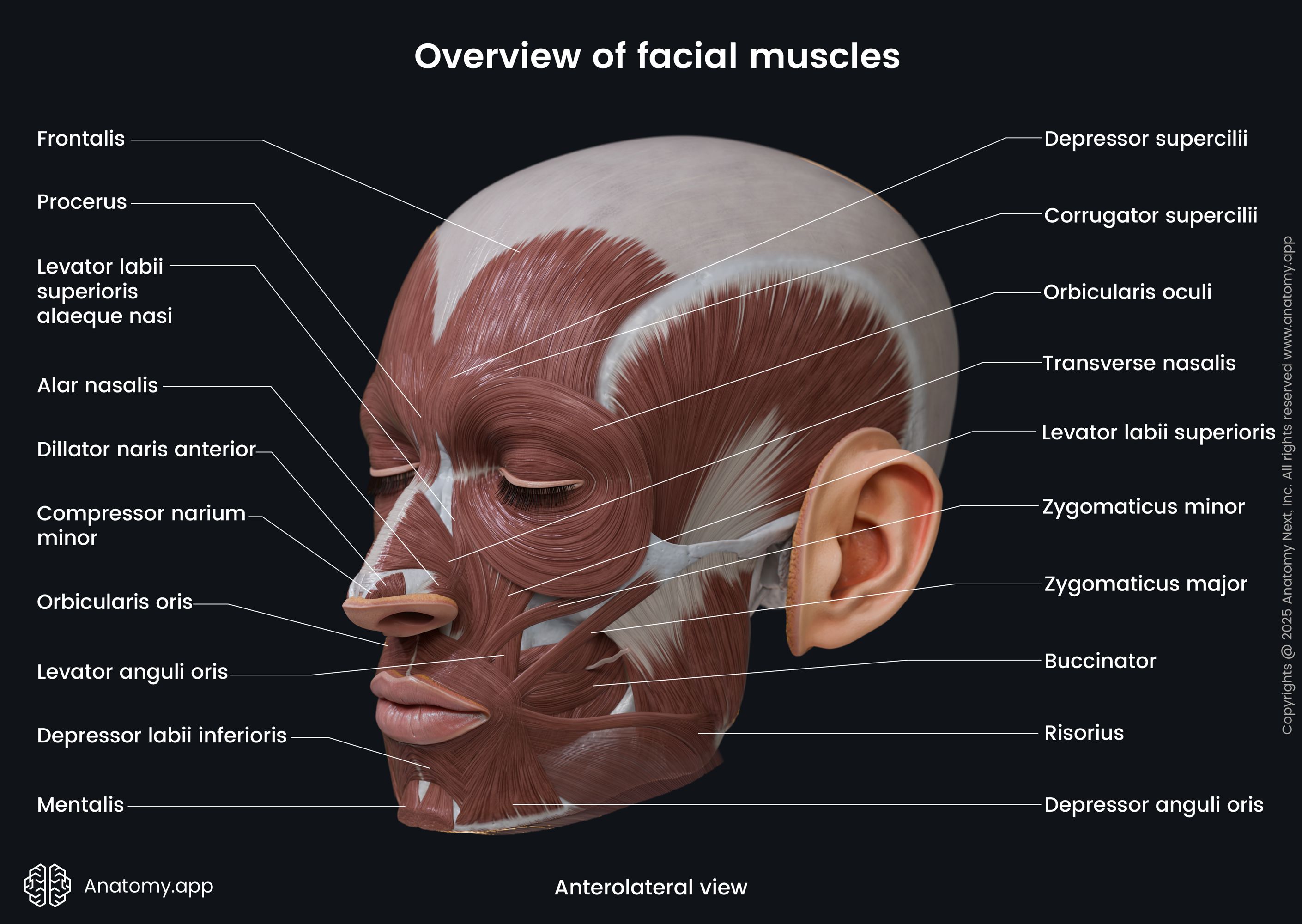
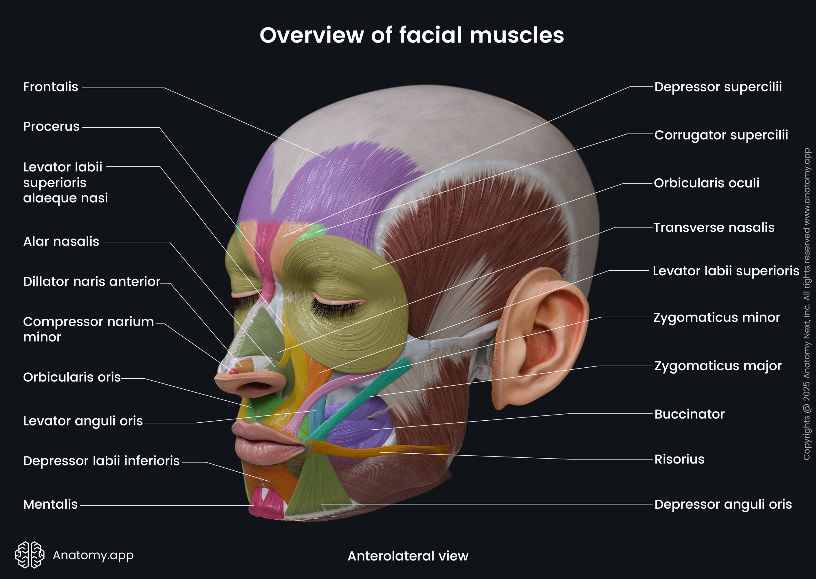
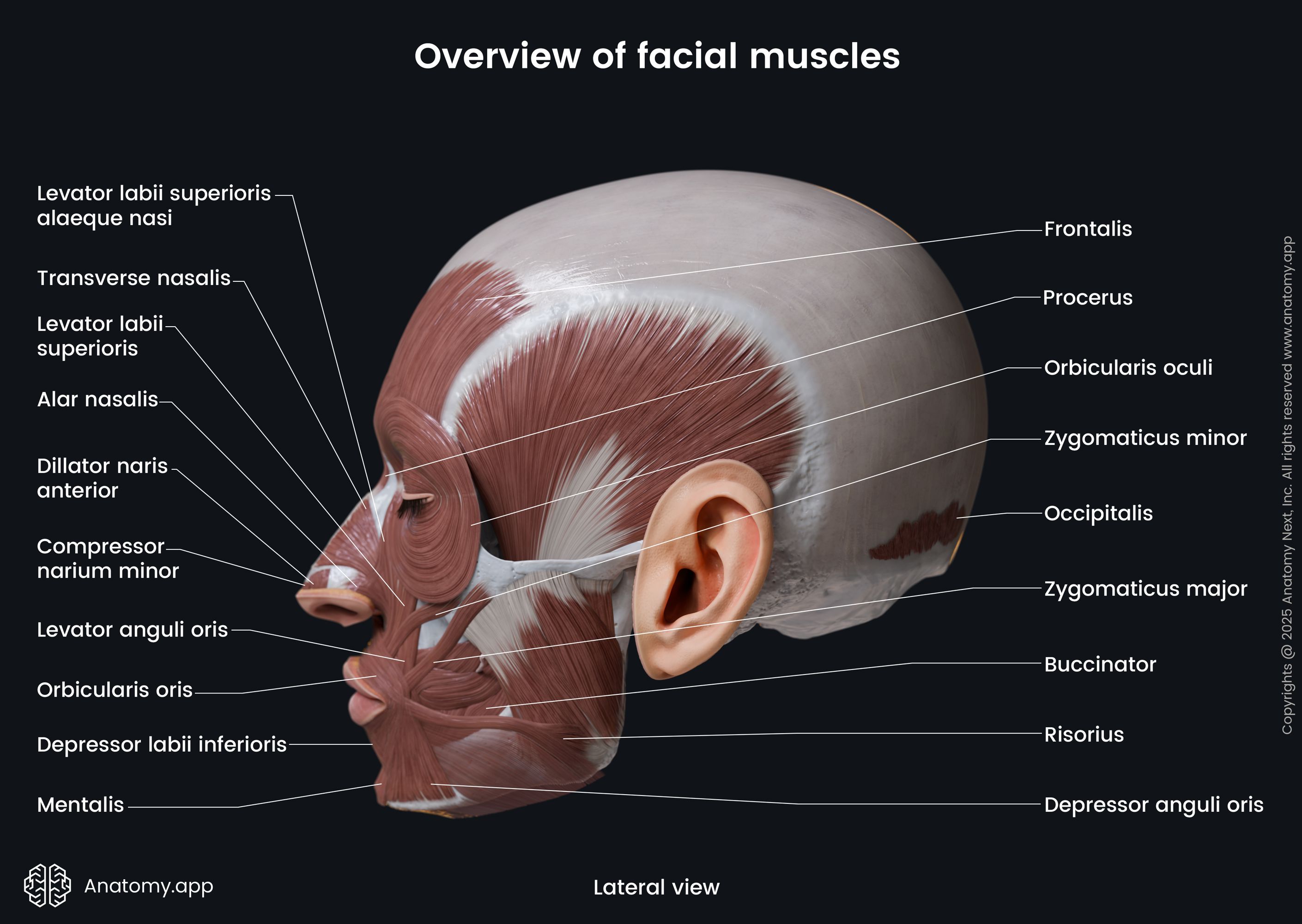
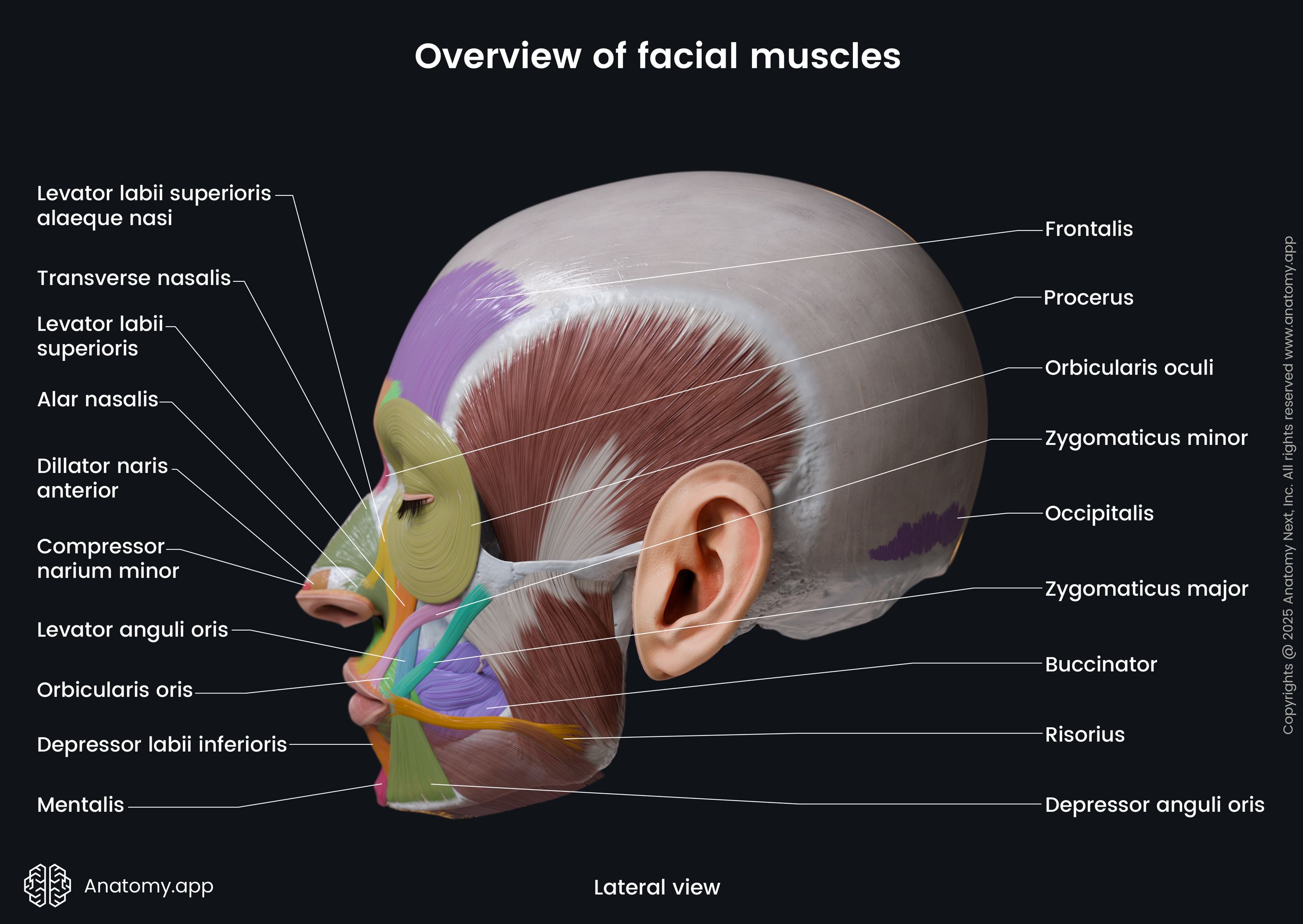
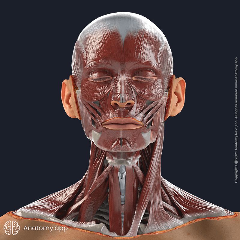
The facial muscles (Latin: musculi faciei), also called the muscles of facial expression, are the muscles around the natural orifices of the face (eyes, nose, mouth and ears) situated within the subcutaneous tissue. Usually, they originate from the bones of the facial skeleton (viscerocranium) and insert into the skin. These muscles do not have fascias, except for the buccinator. Overall, the human body has over 40 facial muscles.
Facial muscle function
The primary function of all facial muscles is to provide voluntary and involuntary movements of facial skin folds, ensuring different facial expressions. Muscles of the face act as sphincters - they take part in the closing and widening of the main orifices of the face.
Through these movements, the expression of various emotions is possible. Muscles around the eyes also have a protective function for the structures located within the orbit. In addition, muscles around the mouth participate in sound production and food consumption.
Facial muscle groups
The facial muscles can be classified in various ways. One of the ways is to divide the muscles of facial expression into five major groups, depending on their location.
1. Epicranial muscles - muscles that cover upper parts of the skull:
- Occipitofrontalis - moves the scalp and forehead backward and forward, lifts the eyebrows and forms horizontal folds in the skin of the forehead;
- Temporo-parietalis - raises the ear and temple.
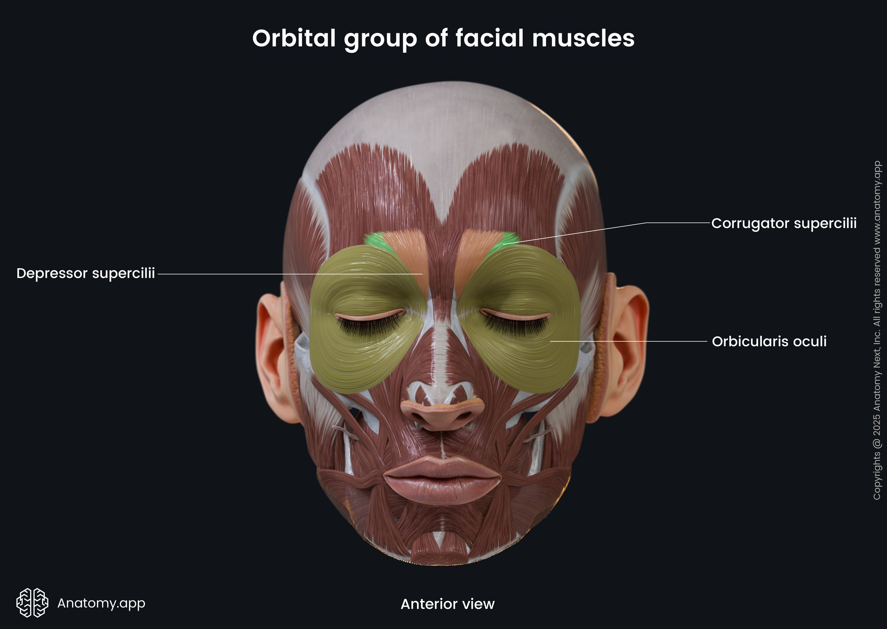
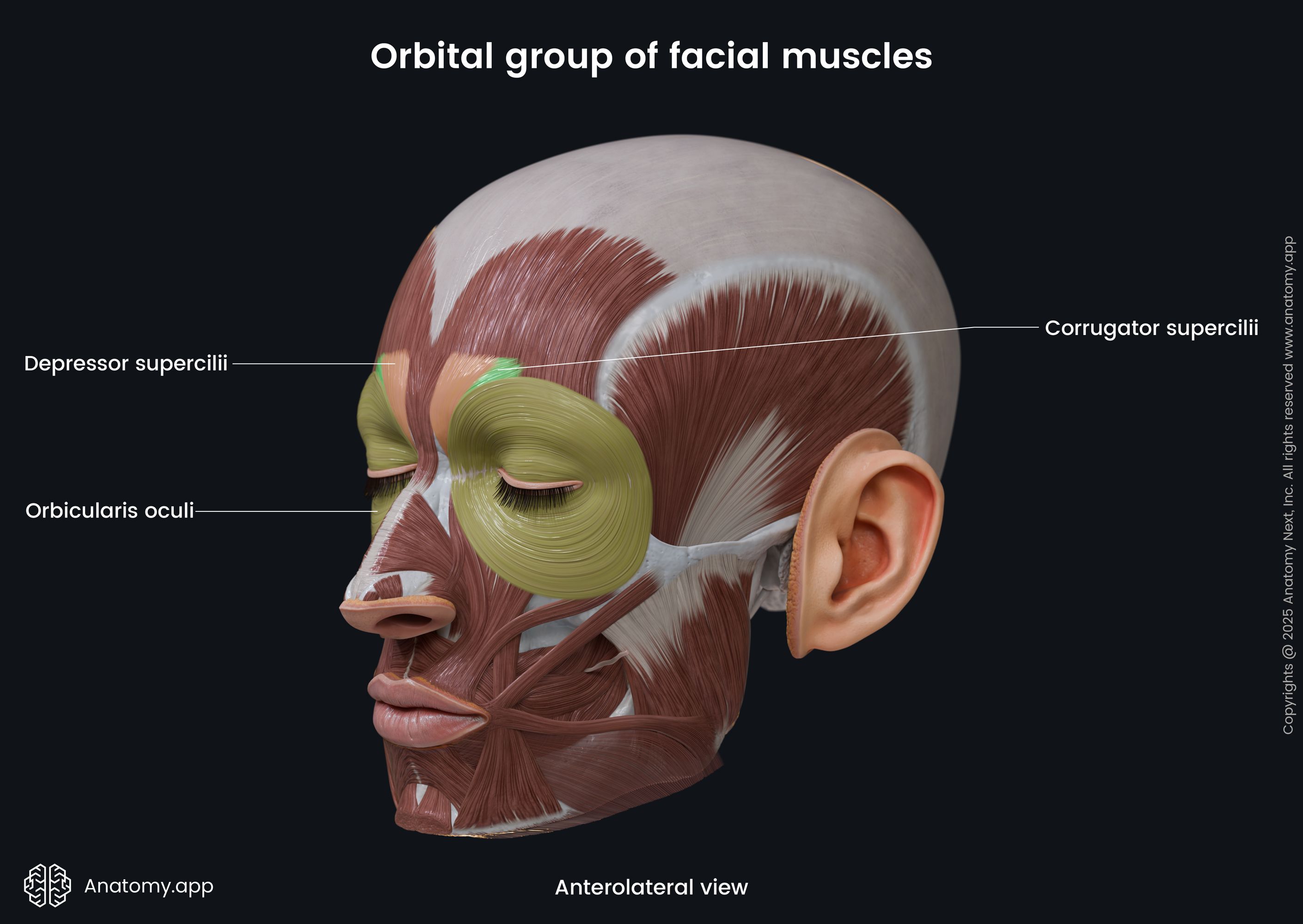
2. Circumorbital and palpebral muscles - muscles around the eyes and eyelids:
- Orbicularis oculi - closes the eye and eyelid;
- Corrugator supercilii - pulls the skin of the eyebrow downward and inferiorly, forms vertical folds in the skin of the forehead.
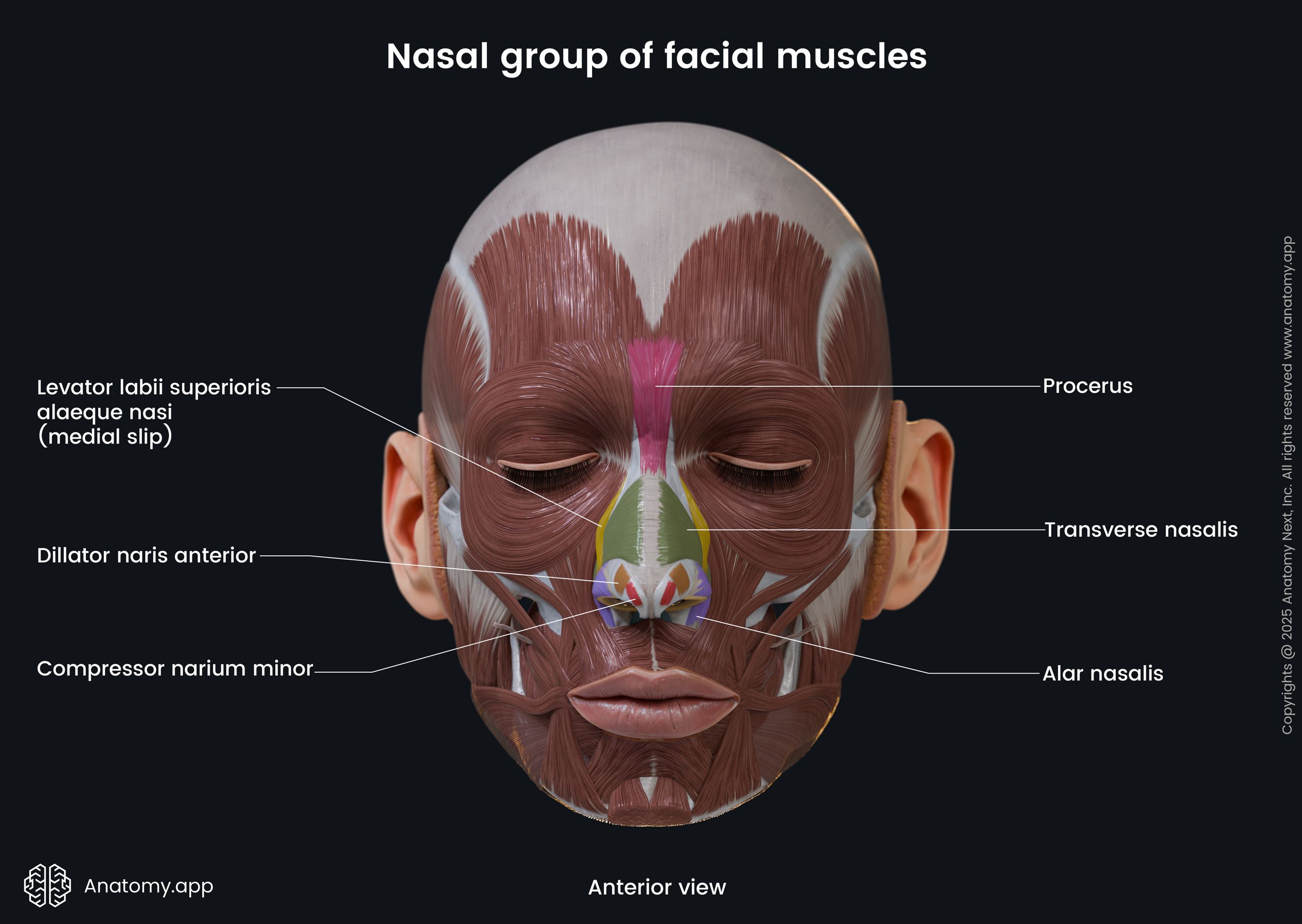
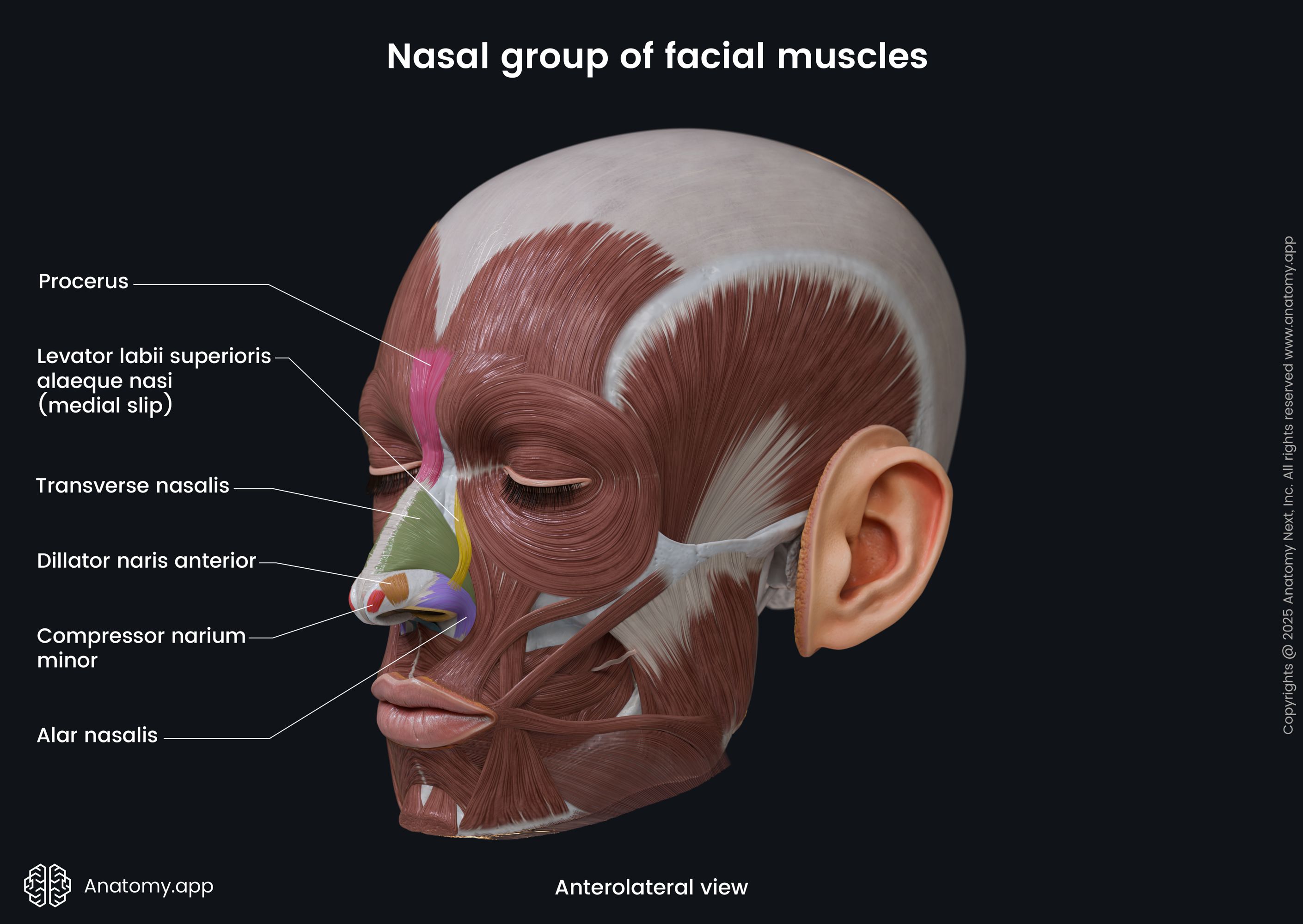
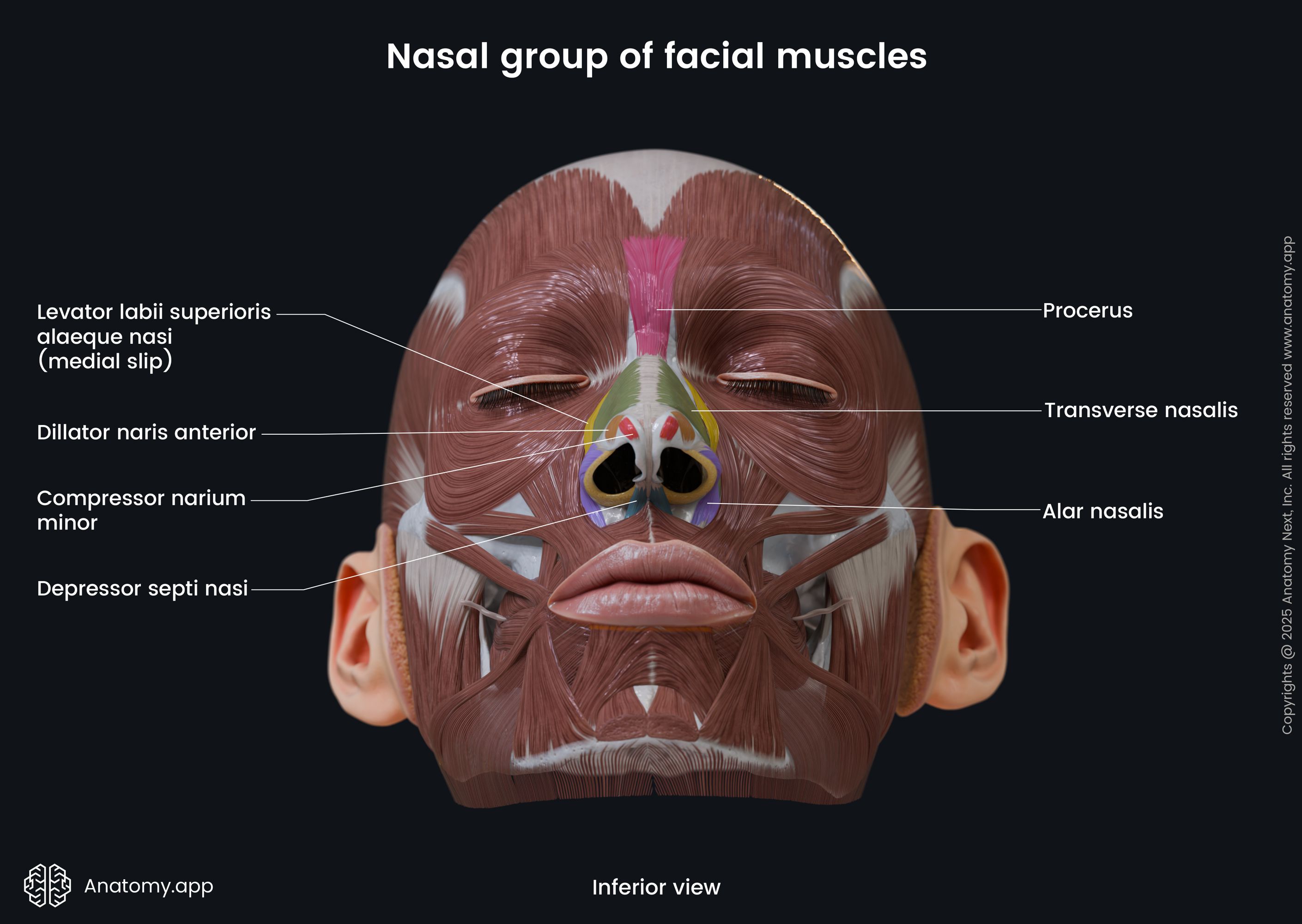
3. Nasal muscles - muscles around the nose:
- Compressor narium minor - narrows the nostril;
- Dilator naris anterior - dilates the nostril;
- Levator labii superioris alaeque nasi - widens the nostril and elevates the upper lip;
- Depressor septi nasi - pulls the nostril downward;
- Nasalis - compresses and widens the nostril;
- Procerus - depresses the medial eyebrow angle downward.
4. Buccolabial muscles - muscles around the mouth:
- Buccinator - pulls the angle of the mouth laterally, compresses the cheek;
- Depressor anguli oris - pulls the angle of the mouth downward;
- Depressor labii inferioris - pulls the lower lip downward and forward;
- Levator anguli oris - lifts the angle of the mouth upward;
- Levator labii superioris - lifts the upper lip;
- Mentalis - produces small dimples in the chin;
- Orbicularis oris - closes the mouth;
- Risorius - pulls the angle of the mouth laterally;
- Zygomaticus minor - pulls the upper lip backward, upward and outward;
- Zygomaticus major - lifts the angle of the mouth upward and to the side.
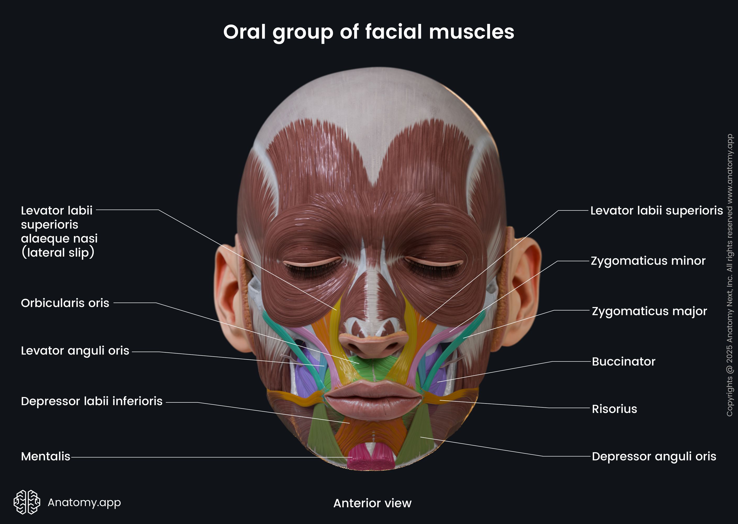
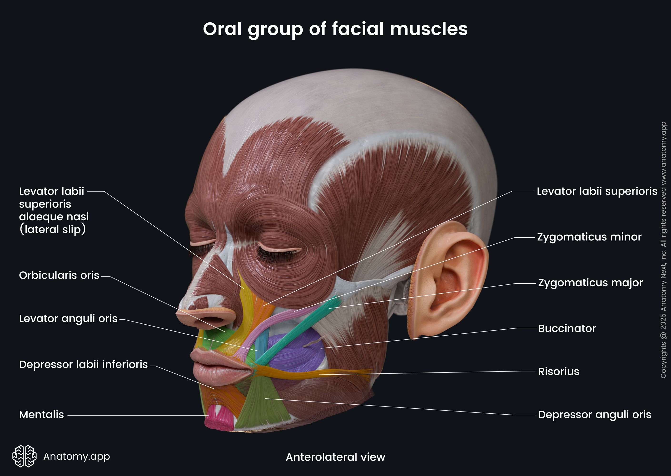
5. Auricular muscles - ear muscles that have a little functional significance. They can be subdivided into two groups - intrinsic (muscles of the auricle) and extrinsic (around the ear). Extrinsic auricular muscles are considered facial muscles, and they include:
- Auricularis anterior - draws the auricle upward and forward;
- Auricularis superior - pulls the auricle upward;
- Auricularis posterior - draws the auricle backward.
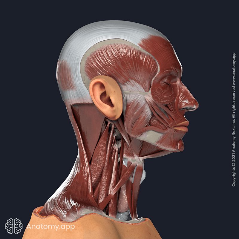
According to the level of depth and muscle origin, the muscles of facial expression can be divided into four following groups:
- First layer (most superficial) - includes the orbicularis oculi, depressor anguli oris and superficial part of the zygomaticus minor;
- Second layer (beneath the superficial one) - contains the platysma, risorius, zygomaticus major, deep part of the zygomaticus minor and levator labii superioris alaeque nasi;
- Third layer - consists of the levator labii superioris and orbicularis oris;
- Deepest layer - contains the mentalis, buccinator and levator anguli oris.
Innervation of facial muscles
All facial muscles are innervated by branches of the facial nerve (CN VII). This cranial nerve innervates only their deep parts. Therefore, the surgical incision done to the superficial part of these muscles during facial and plastic surgeries will not damage the innervation.
Every surgeon needs to remember the following muscles of facial expression - buccinator, mentalis and levator anguli oris muscles. These facial muscles located in the deepest layer are the only muscles that are innervated on their superficial parts.
NOTE: Information about blood supply, origin, insertion and function of facial muscles is provided under the description of each muscle separately.
References:
- Azizzadeh, B., & Nduka, C. (2021). Management of Post-Facial Paralysis Synkinesis. Elsevier Gezondheidszorg.
- Bolognia, J. L., Schaffer, J. V., Duncan, K. (2021). Dermatology Essentials (2nd ed.). Elsevier.
Gurtner, G. C., & Neligan, P. C. (2017). Plastic Surgery: Volume 1: Principles (4th ed.). Elsevier.
Rubin, J. P., & Neligan, P. C. (2021). Plastic Surgery: Volume 2: Aesthetic Surgery (Expert Consult - Online and Print), 3e by Richard J Warren MD FRCS(C) (2012–09-21). Saunders.
Standring, S. (2020). Gray’s Anatomy: The Anatomical Basis of Clinical Practice (42nd ed.). Elsevier.