- Anatomical terminology
- Skeletal system
- Joints
- Muscles
- Head muscles
- Neck muscles
- Muscles of upper limb
- Thoracic muscles
- Muscles of back
- Muscles of lower limb
- Heart
- Blood vessels
- Lymphatic system
- Nervous system
- Respiratory system
- Digestive system
- Urinary system
- Female reproductive system
- Male reproductive system
- Endocrine glands
- Eye
- Ear
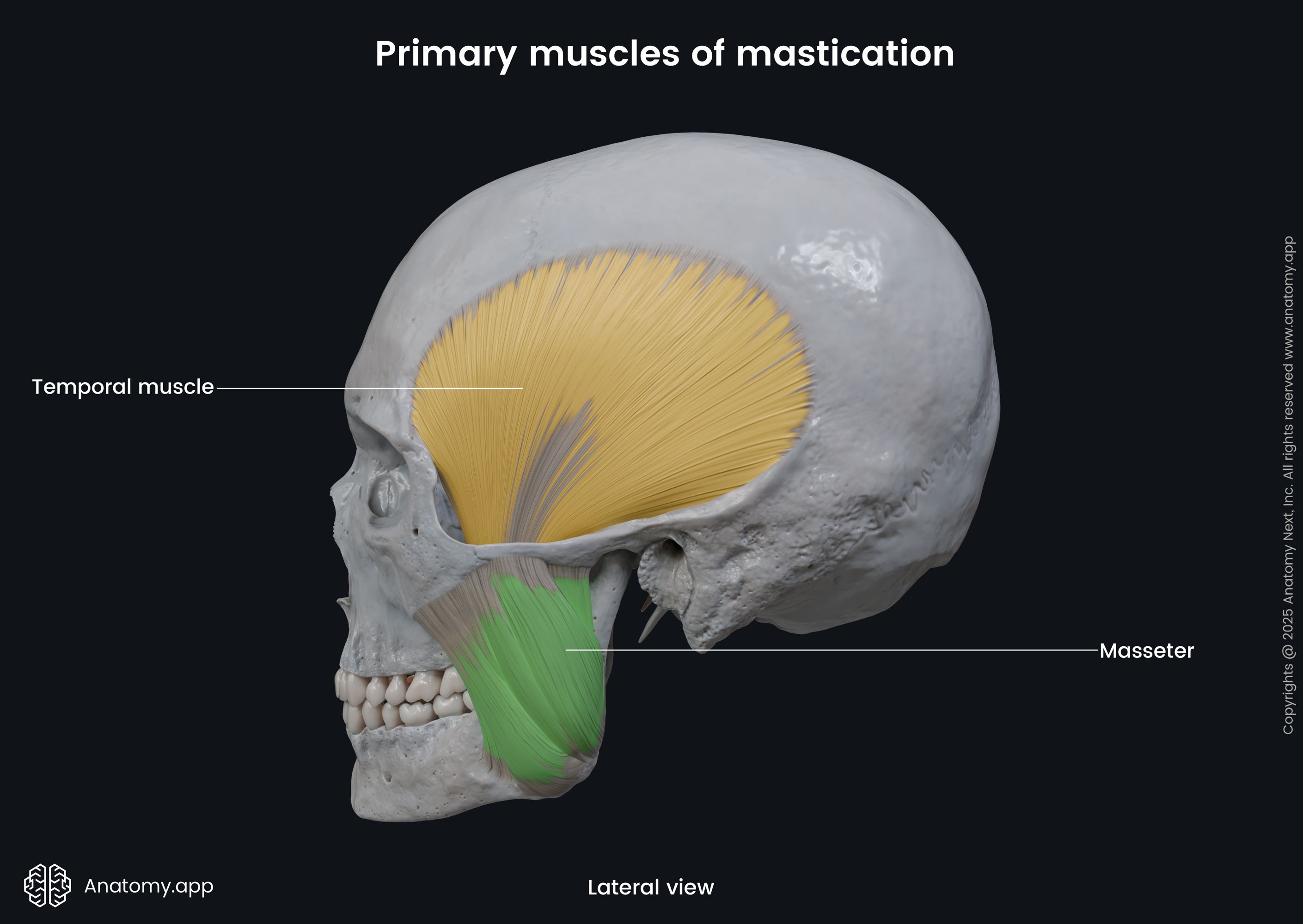
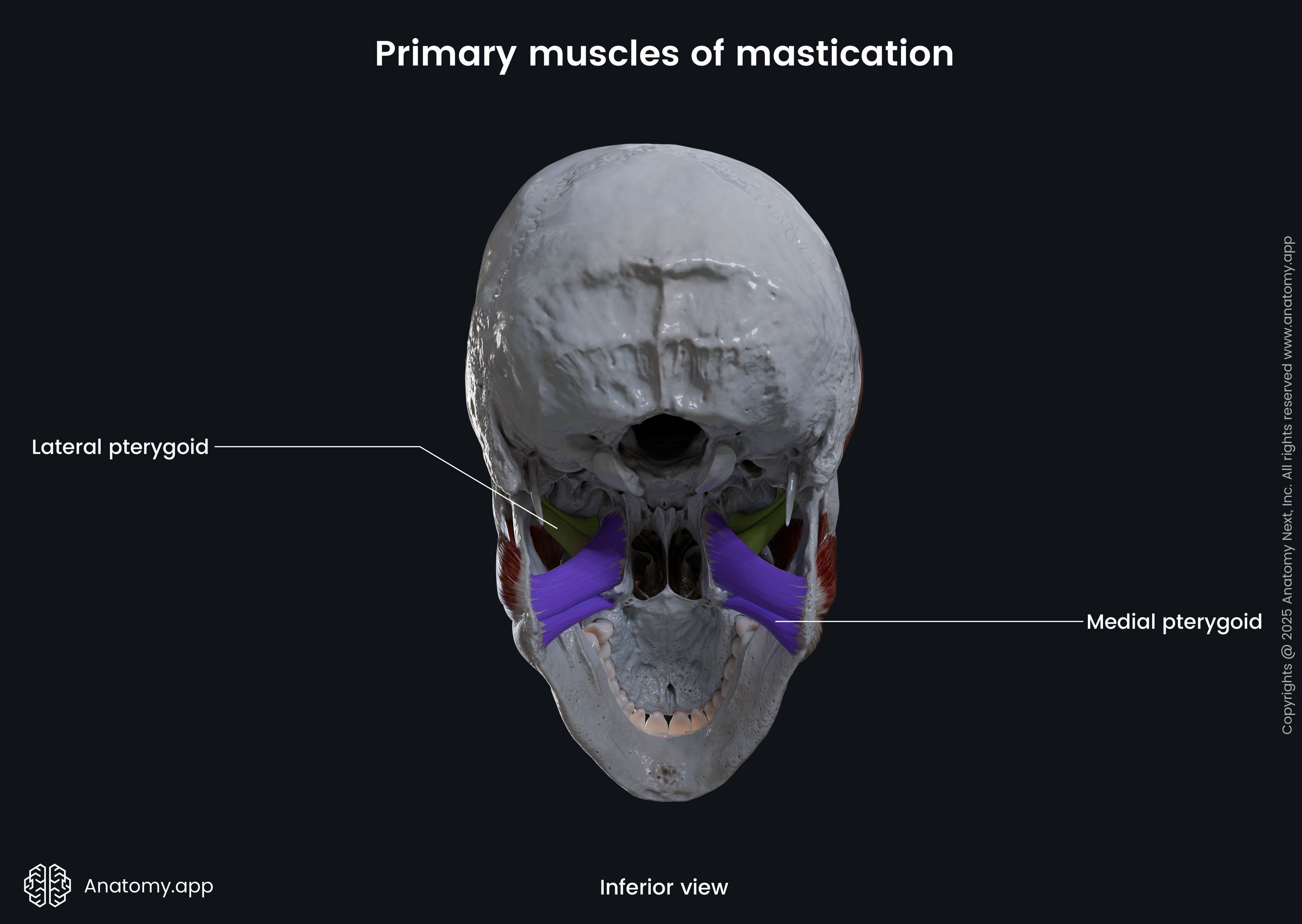
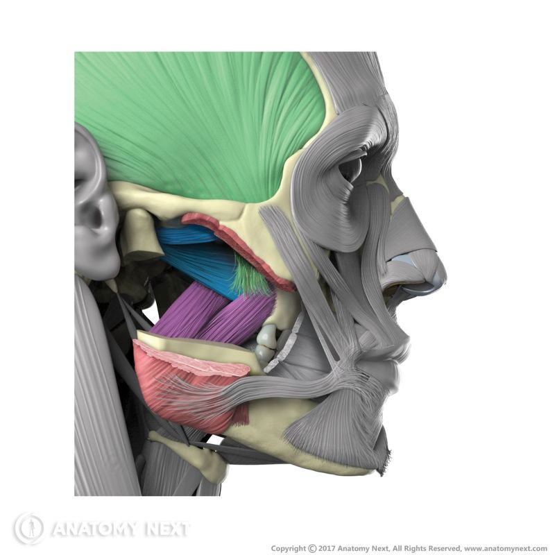
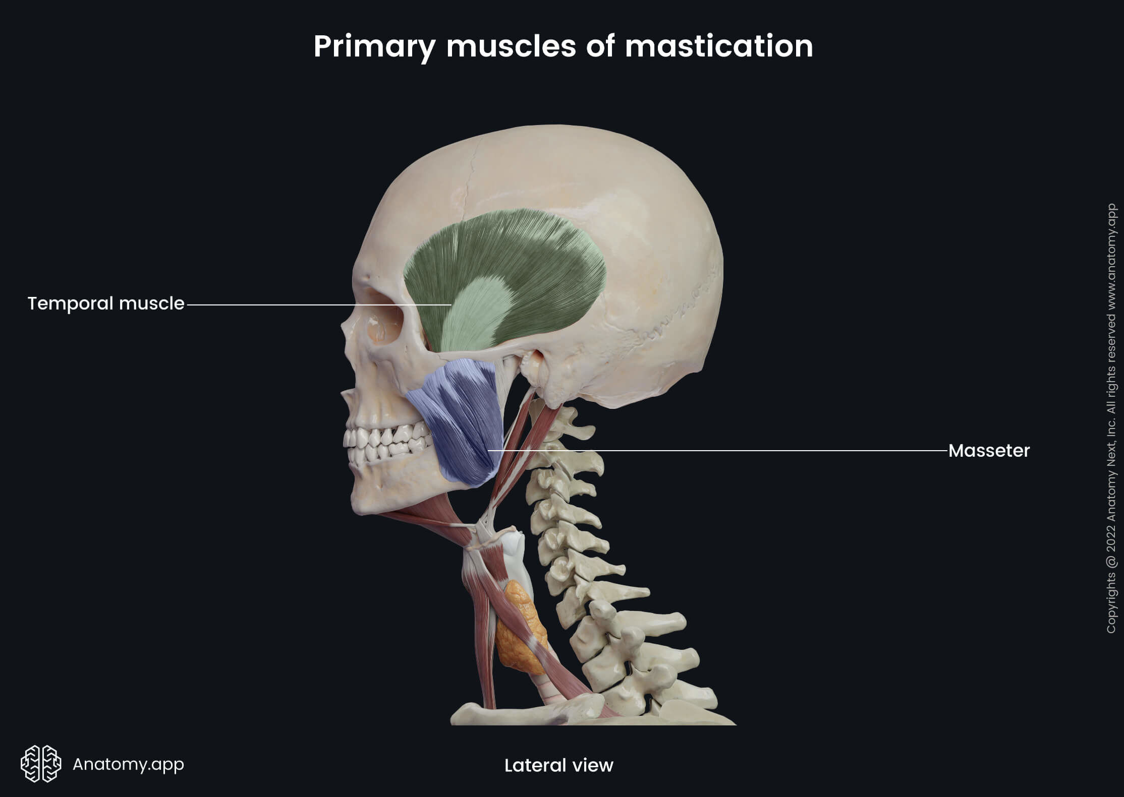
Muscles of mastication
The muscles of mastication (Latin: musculi masticatorii) or masticatory muscles are a group of paired muscles responsible for the movements of the lower jaw at the temporomandibular joints (TMJs). Therefore, these muscles are positioned within the head and neck around the mentioned joint.
The muscles of mastication primarily act during chewing. However, besides the movements associated with eating, they also move the mandible during speaking. Overall, they open and close the mouth. They elevate, depress, protrude (move forward), retract (pull backward) the mandible and provide side-to-side movements.
The masticatory muscles can be subdivided into two groups - primary (main) and secondary (accessory) muscles of mastication. All primary masticatory muscles originate from the bones of the skull and insert onto various features of the mandible.
Besides the classification that will be reviewed in this article, all muscles of mastication can also be grouped according to the functions they provide:
- Muscles that elevate the mandible - temporalis, masseter, medial pterygoid and lateral pterygoid (superior head);
- Muscles that depress the lower jaw - lateral pterygoid (inferior head), digastric (anterior belly), mylohyoid and geniohyoid;
- Muscles that protrude the mandible - masseter, medial pterygoid, lateral pterygoid (inferior head), digastric and temporalis;
- Muscles that retract the lower jaw - temporalis, digastric and lateral pterygoid (inferior head);
- Muscles that provide side-to-side movements - masseter, temporalis, medial pterygoid and lateral pterygoid.
Primary (main) muscles of mastication
The primary muscles of mastication include four paired muscles that primarily act during eating, enabling such functions as chewing. Each side of the head has the following primary muscles of mastication:
All main muscles close the mouth by elevating the mandible, except for the lateral pterygoid. It opens the mouth by depressing and moving the mandible forward. Overall, these muscles originate from various bones of the skull and insert onto the posterior half of the mandible.
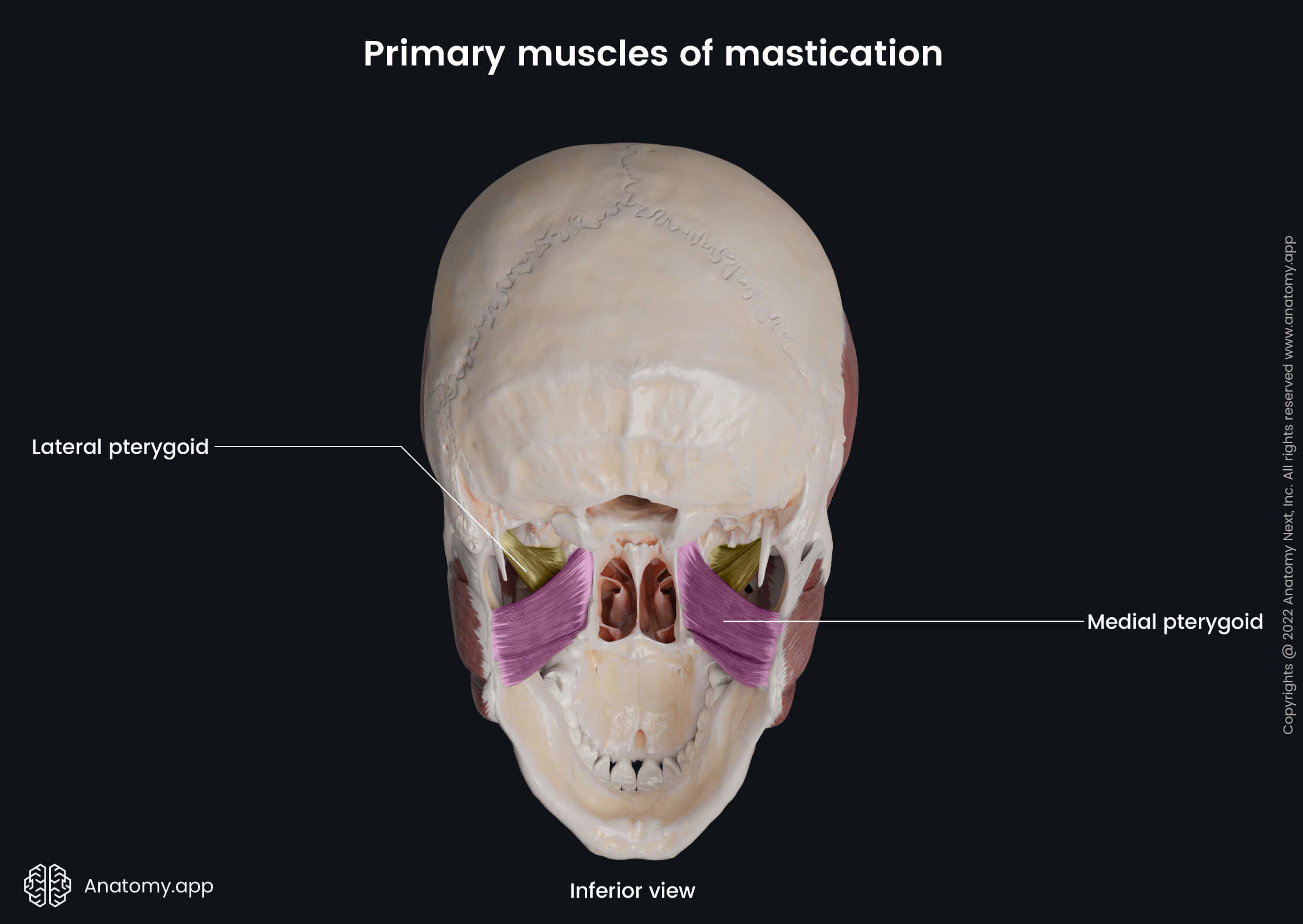
All primary muscles have a common innervation source. They all receive innervation from the branches of the mandibular division (CN V3) of the trigeminal nerve (CN V). And finally, another shared feature of the masticatory muscles is that they are supplied by the branches of the maxillary artery.
Accessory muscles of mastication
The accessory muscles of mastication do not serve as muscles that primarily act during chewing, and they include some facial and neck muscles :
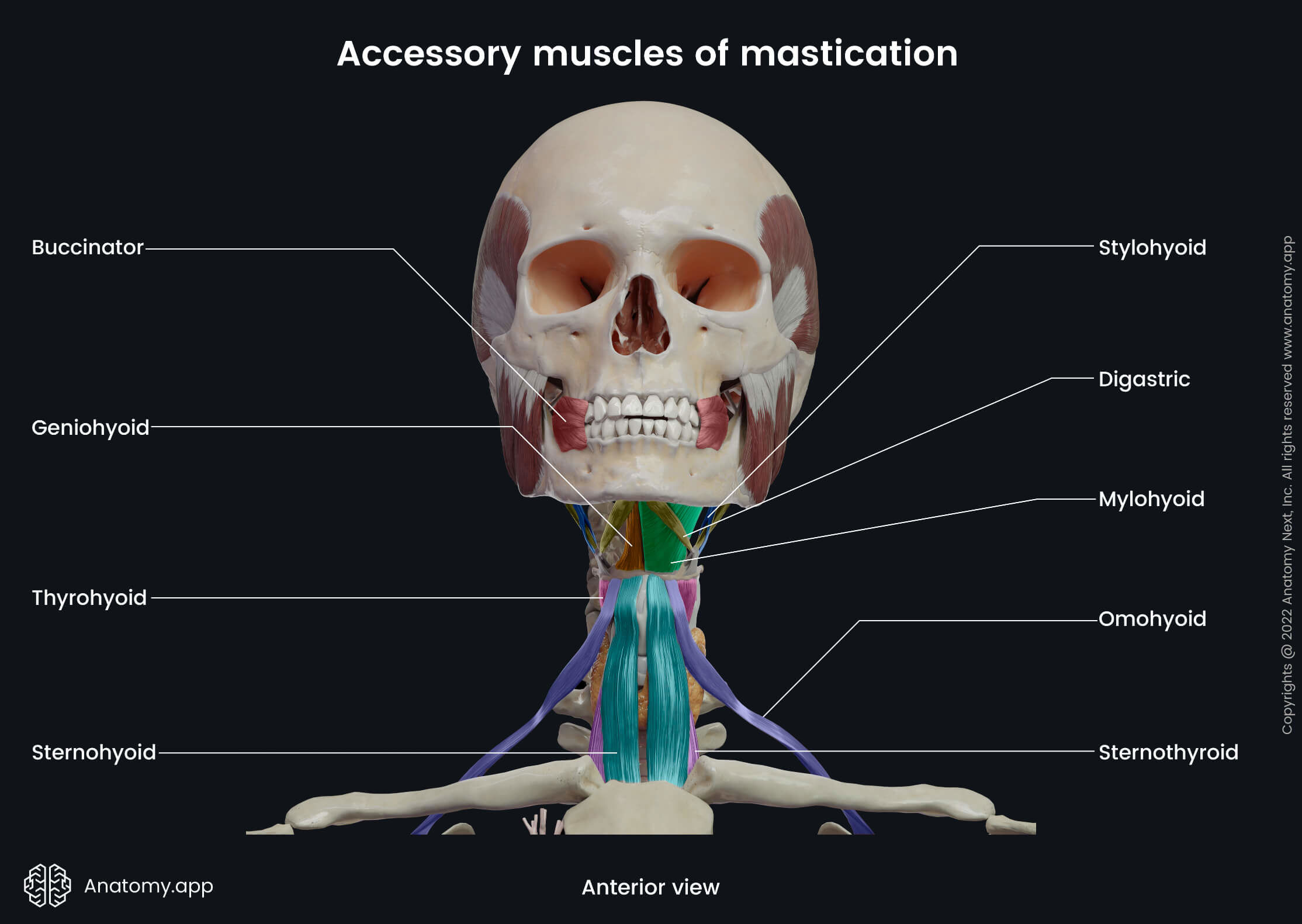

The buccinator is primarily a facial muscle that participates in forming the anterior part of the cheek and the lateral wall of the oral vestibule. The digastric, stylohyoid, mylohyoid and geniohyoid are located above the hyoid bone and are classified as the suprahyoid neck muscles. The sternohyoid, thyrohyoid, sternothyroid and omohyoid are positioned below the hyoid bone and belong to the infrahyoid neck muscles.
Anatomy.app
Contact information
- For questions regarding business inquiries. Please contact:
- info@anatomy.app