- Anatomical terminology
- Skeletal system
- Joints
- Muscles
- Heart
- Blood vessels
- Lymphatic system
- Nervous system
- Respiratory system
- Digestive system
- Urinary system
- Female reproductive system
- Male reproductive system
- Endocrine glands
- Eye
- Ear
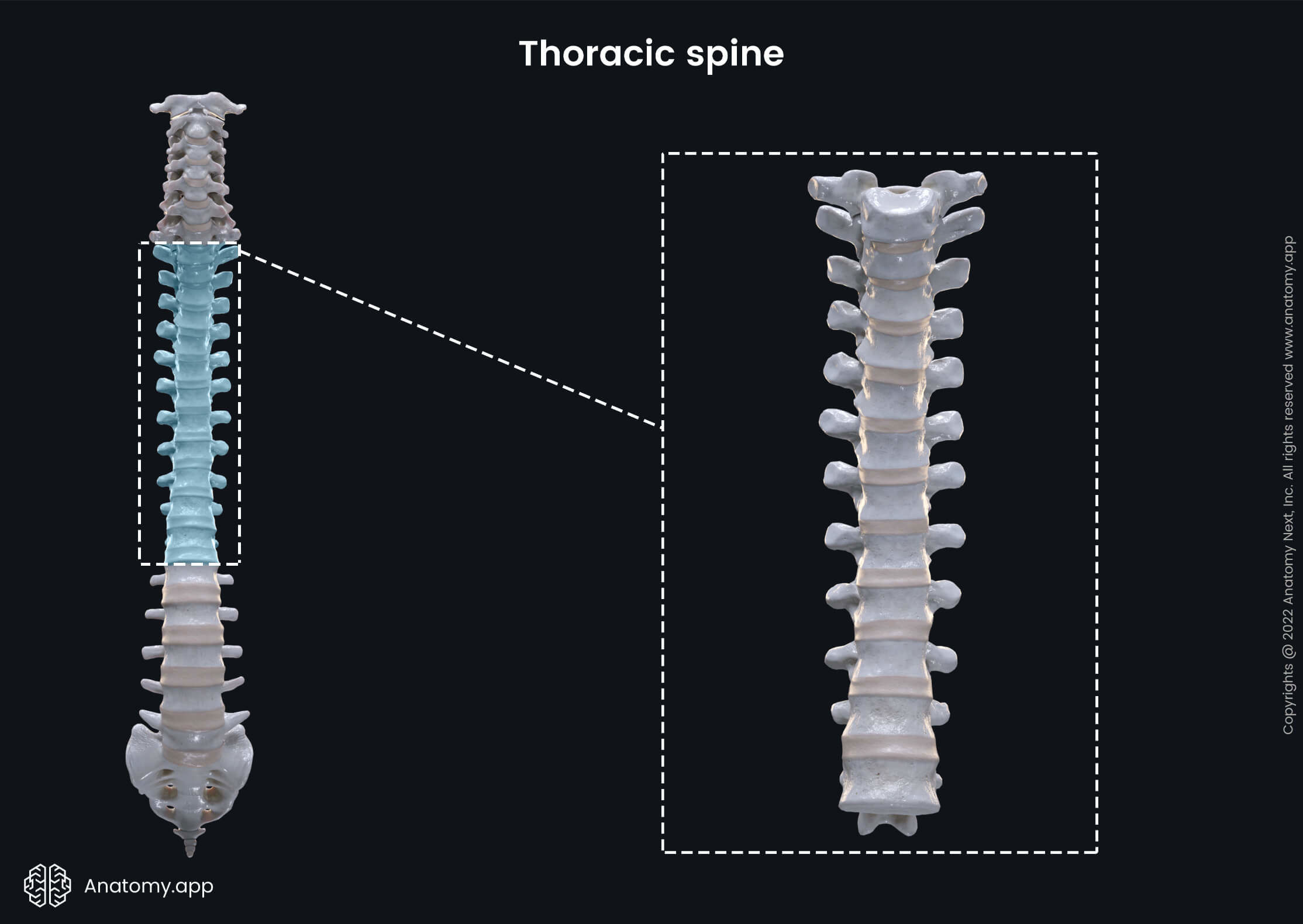
Thoracic vertebrae
The thoracic vertebrae (Latin: vertebrae thoracicae) are twelve vertebrae (T1 - T12) forming the middle or the thoracic part of the spine. These vertebrae are characterized by their articulation with the ribs. The thoracic vertebrae participate in forming the rib cage.
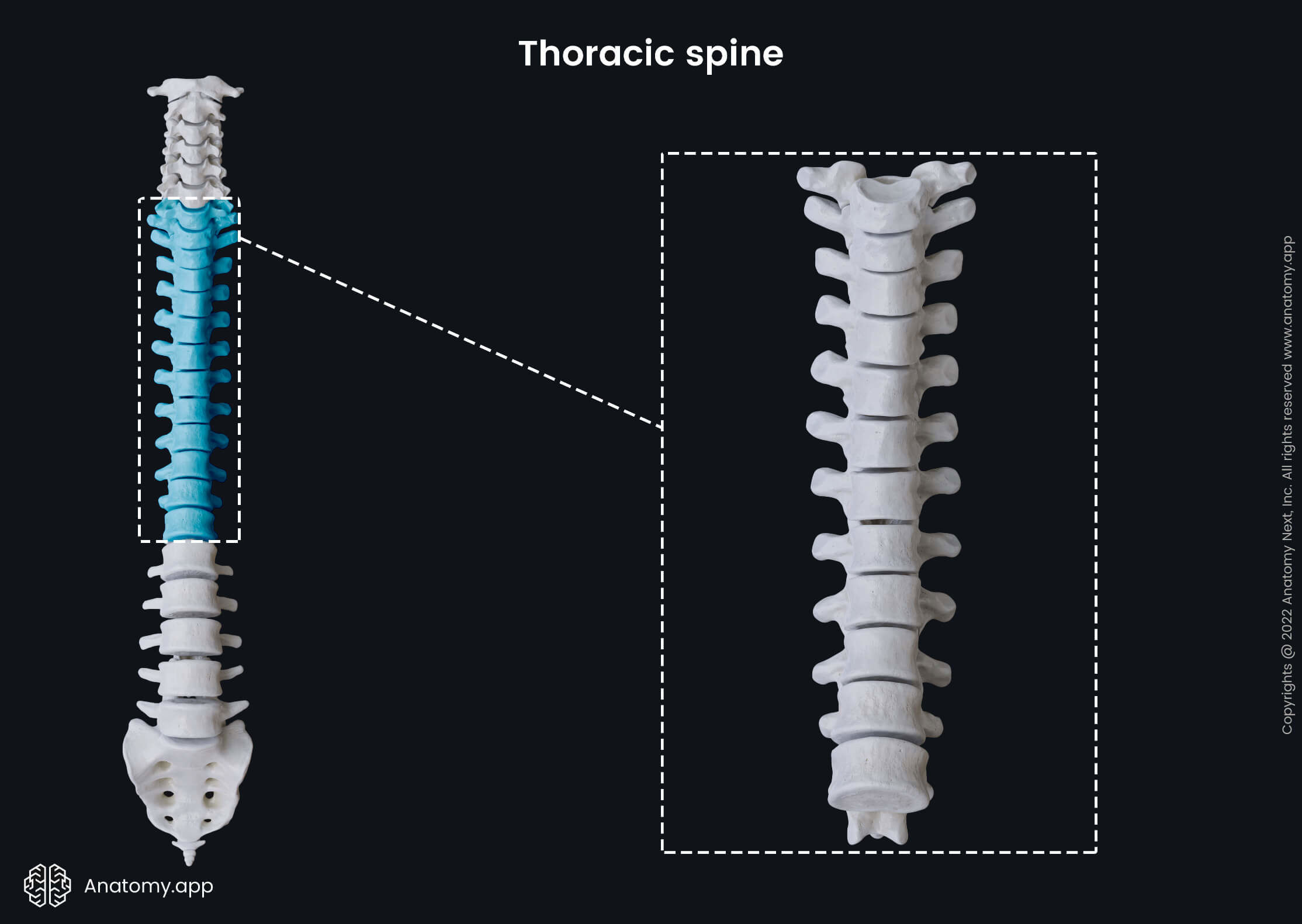
The typical characteristic features of the thoracic vertebrae are the costal or rib facets on their vertebral bodies and transverse processes. Like cervical vertebrae, thoracic vertebrae are also subdivided into typical and atypical vertebrae depending on the presence and shape of the costal facets.
Within the thorax, the thoracic vertebrae form several joints. The vertebral bodies of the thoracic vertebrae articulate with the ribs forming the costovertebral joints, while the articulations between the ribs and transverse processes of the thoracic vertebrae form the costotransverse joints.
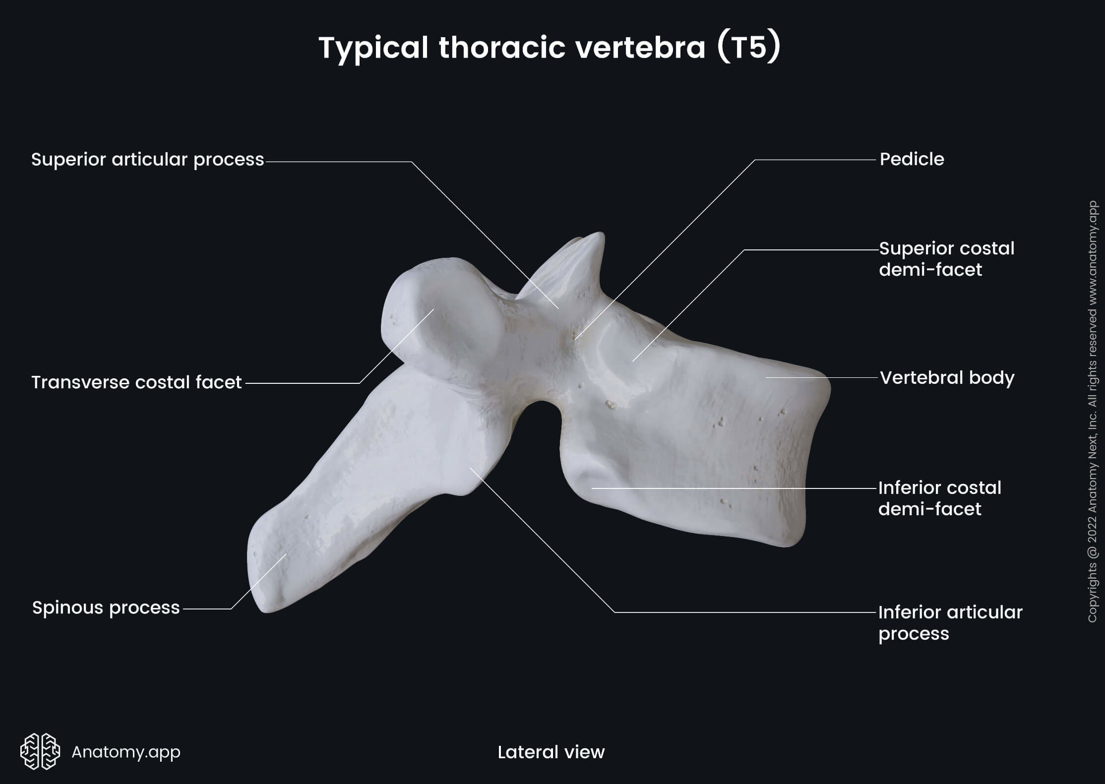
Typical thoracic vertebrae
The typical thoracic vertebrae are considered to be the second through ninth thoracic vertebrae (T2 - T9). These vertebrae present the following landmarks:
- Vertebral body
- Superior and inferior costal demi-facets (2)
- Lamina (2)
- Pedicle (2)
- Vertebral foramen
- Intervertebral foramen (2)
- Transverse process (2)
- Transverse costal facet (2)
- Spinous process
- Superior articular process (2)
- Inferior articular process (2)
The vertebral body of a typical thoracic vertebra appears heart-shaped when viewed from above, and it is usually as broad in the anteroposterior as in the transverse direction. The vertebral body on either side presents two partial facets - superior and inferior costal demi-facets - for articulation with the head of its corresponding rib and the rib below.
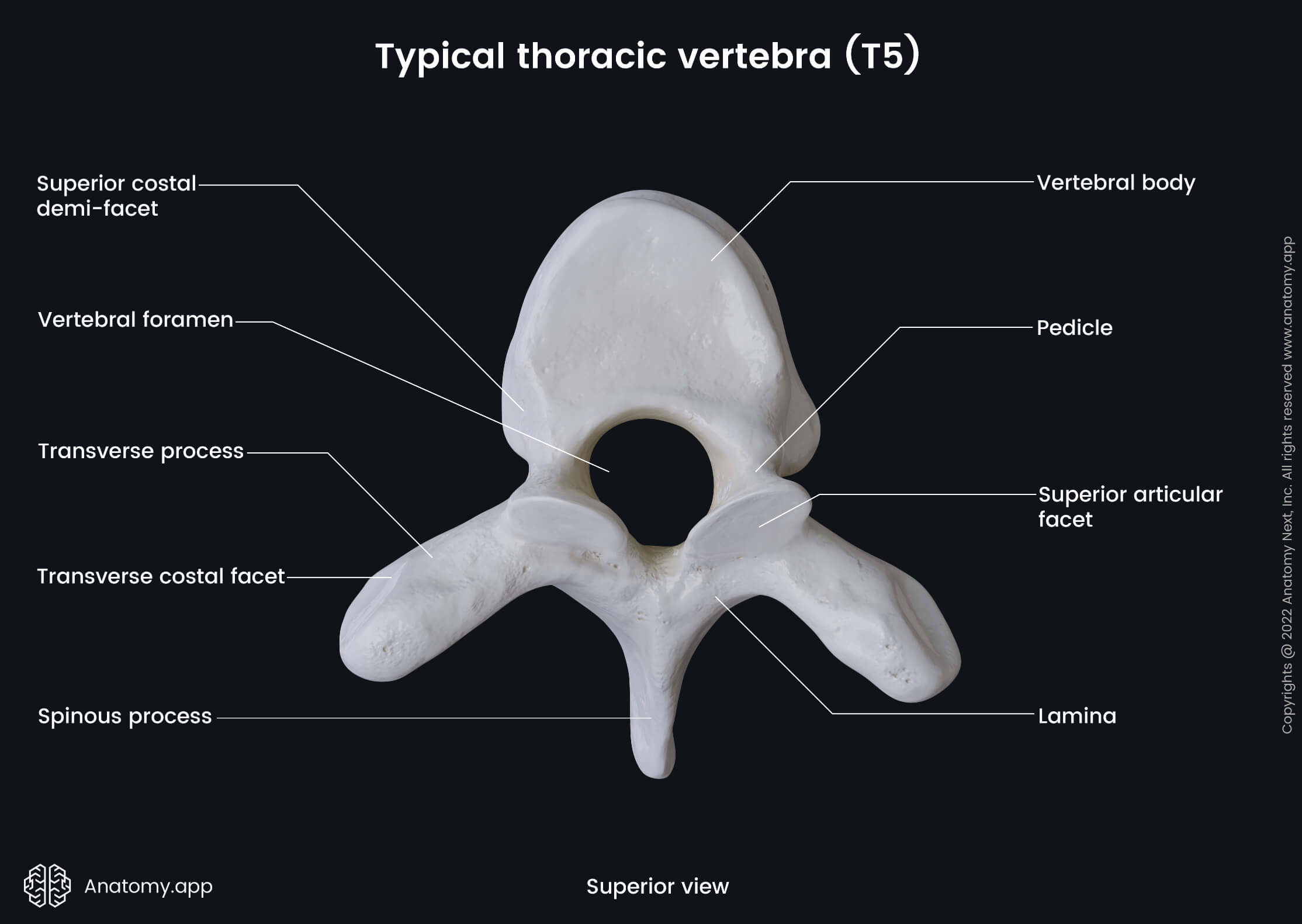
The laminae are broad and thick. They overlap the laminae of a subjacent vertebra like tiles of a roof and connect with pedicles to surround and provide protection to the spinal cord. The pedicles of the thoracic vertebrae are directed backward and slightly upward.
The typical thoracic vertebra has a circular vertebral foramen - a large opening located posterior to the body forming the spinal canal. The intervertebral foramen are two small and circular openings on either side of the vertebra in each intervertebral level.
The transverse processes of each thoracic vertebrae present a transverse costal facet that articulates with the tubercle of its corresponding rib. The spinous process appears long and triangular-shaped in coronal cross-section studies. It projects obliquely downward, arising from the laminae.
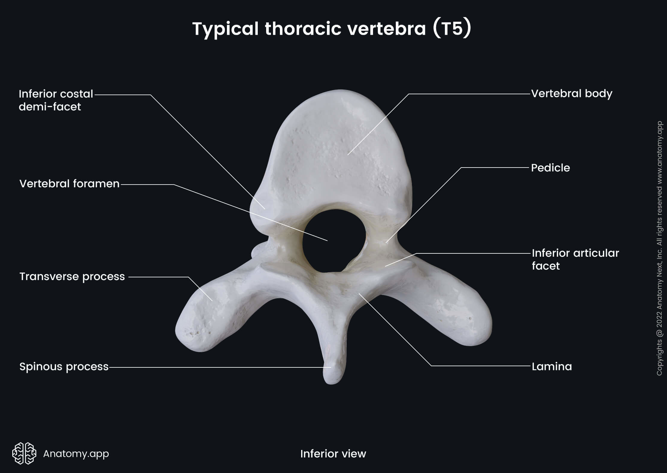
The superior articular processes are two thin bony plates projecting upward from the junctions of the pedicles and laminae. The inferior articular processes are two projections going downward from the vertebral bodies also from the junction sites of pedicles and laminae.
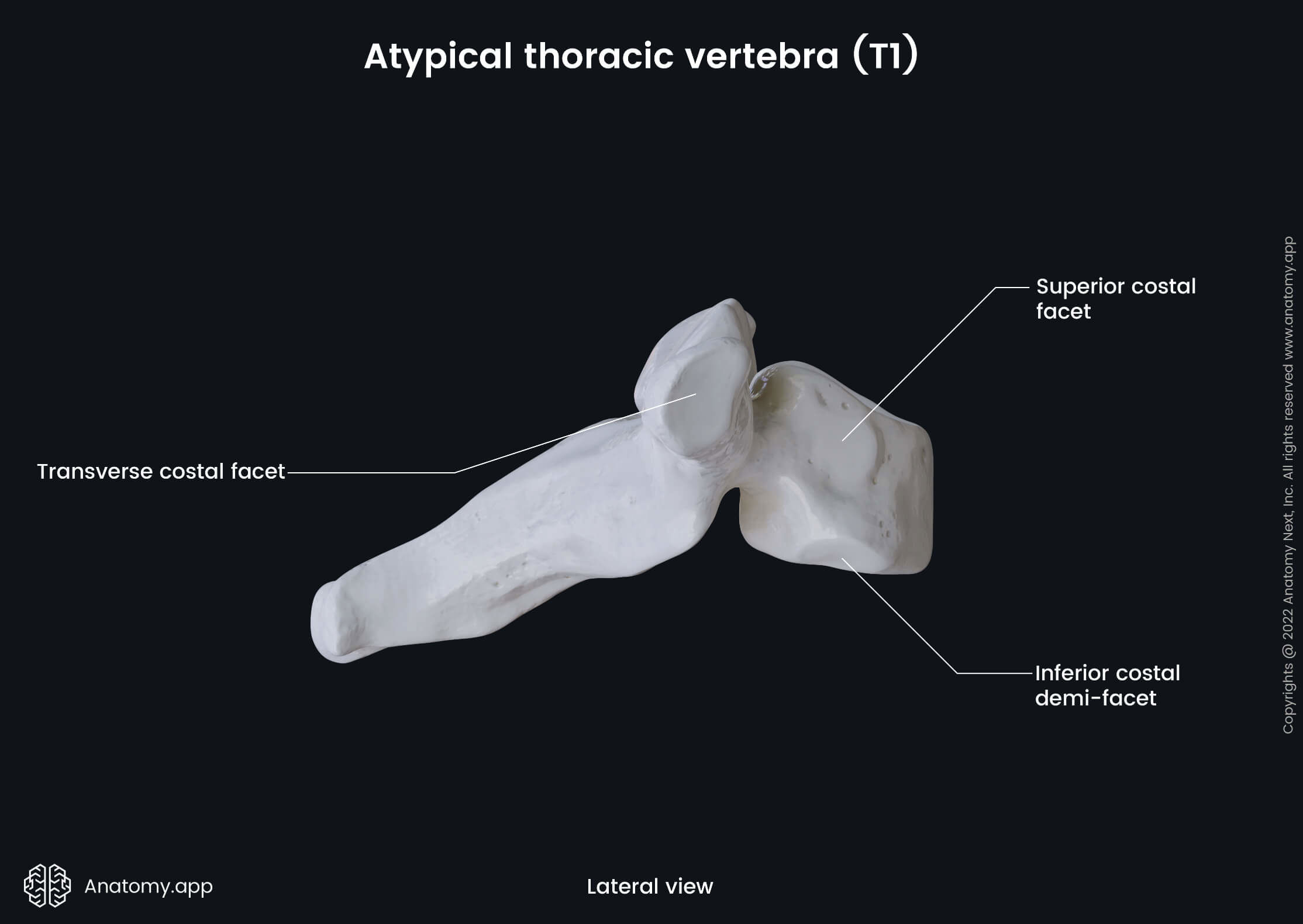
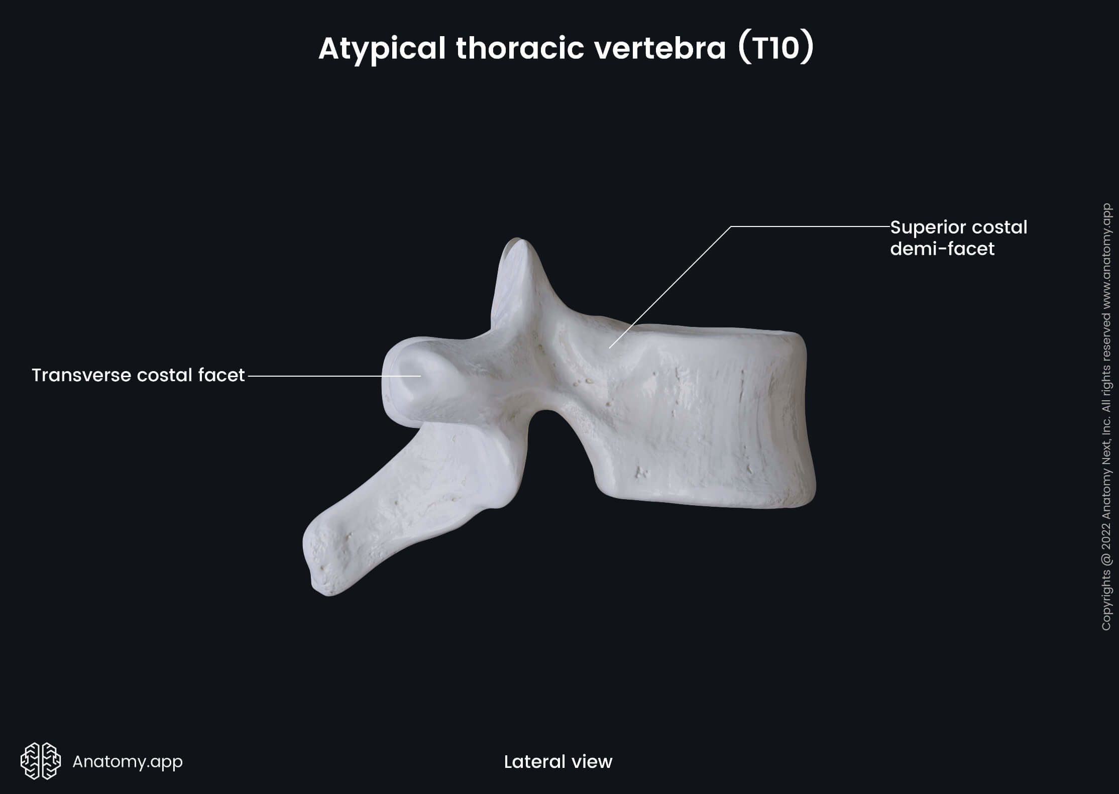
Atypical thoracic vertebrae
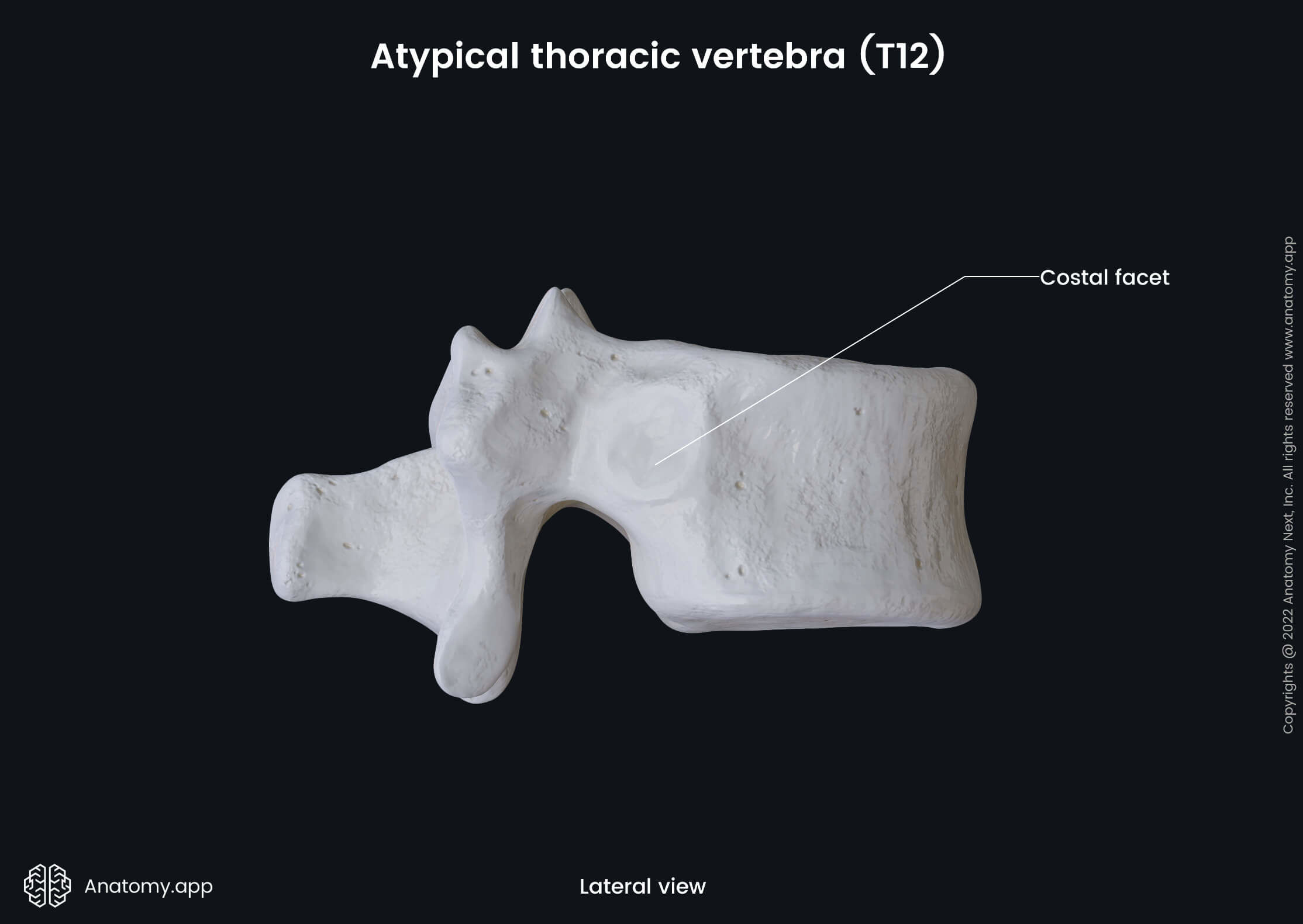
The atypical thoracic vertebrae include the first, tenth, eleventh and twelfth vertebrae. These vertebrae present the same anatomical landmarks as typical vertebrae, but the shape and presence of costal facets differ.
- The first thoracic vertebra (T1) contains a whole superior costal facet and an inferior costal demi-facet. It also has a transverse costal facet.
- The tenth thoracic vertebra (T10) presents a superior costal demi-facet and lacks an inferior costal demi-facet. It contains a transverse costal facet.
- The eleventh thoracic vertebra (T11) has whole costal facets on its vertebral body but lack transverse costal facets.
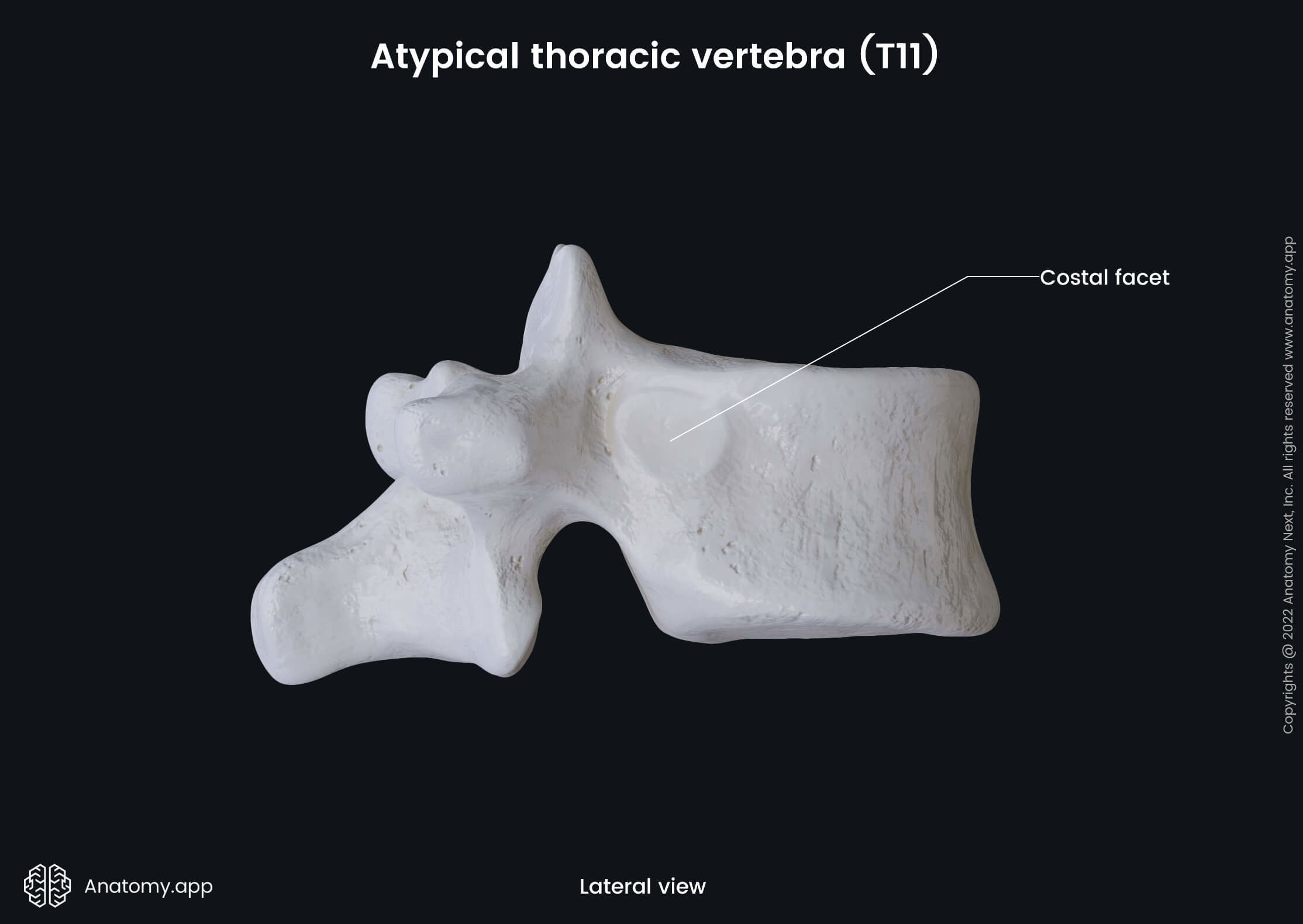
- The twelfth thoracic vertebra (T12) has whole costal facets on its vertebral body but lack transverse costal facets.
Anatomy.app
Contact information
- For questions regarding business inquiries. Please contact:
- info@anatomy.app