- Anatomical terminology
- Skeletal system
- Joints
- Muscles
- Heart
- Blood vessels
- Lymphatic system
- Nervous system
- Respiratory system
- Digestive system
- Urinary system
- Female reproductive system
- Male reproductive system
- Endocrine glands
- Eye
- Ear
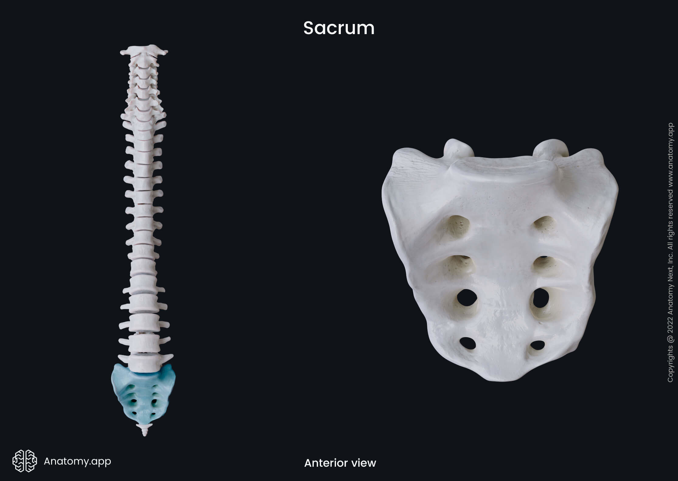
Sacrum
The sacrum (Latin: os sacrum), also known as the sacral bone, is a triangular-shaped bone located at the base of the spine. It is formed by five sacral vertebrae (S1 - S5) that fuse with each other between ages 18 and 30.
The sacrum articulates with four bones - the last (fifth) lumbar vertebra above, the coccyx below, and the ilium on each its lateral side. The sacrum also contributes to the formation of the skeleton of the pelvis.
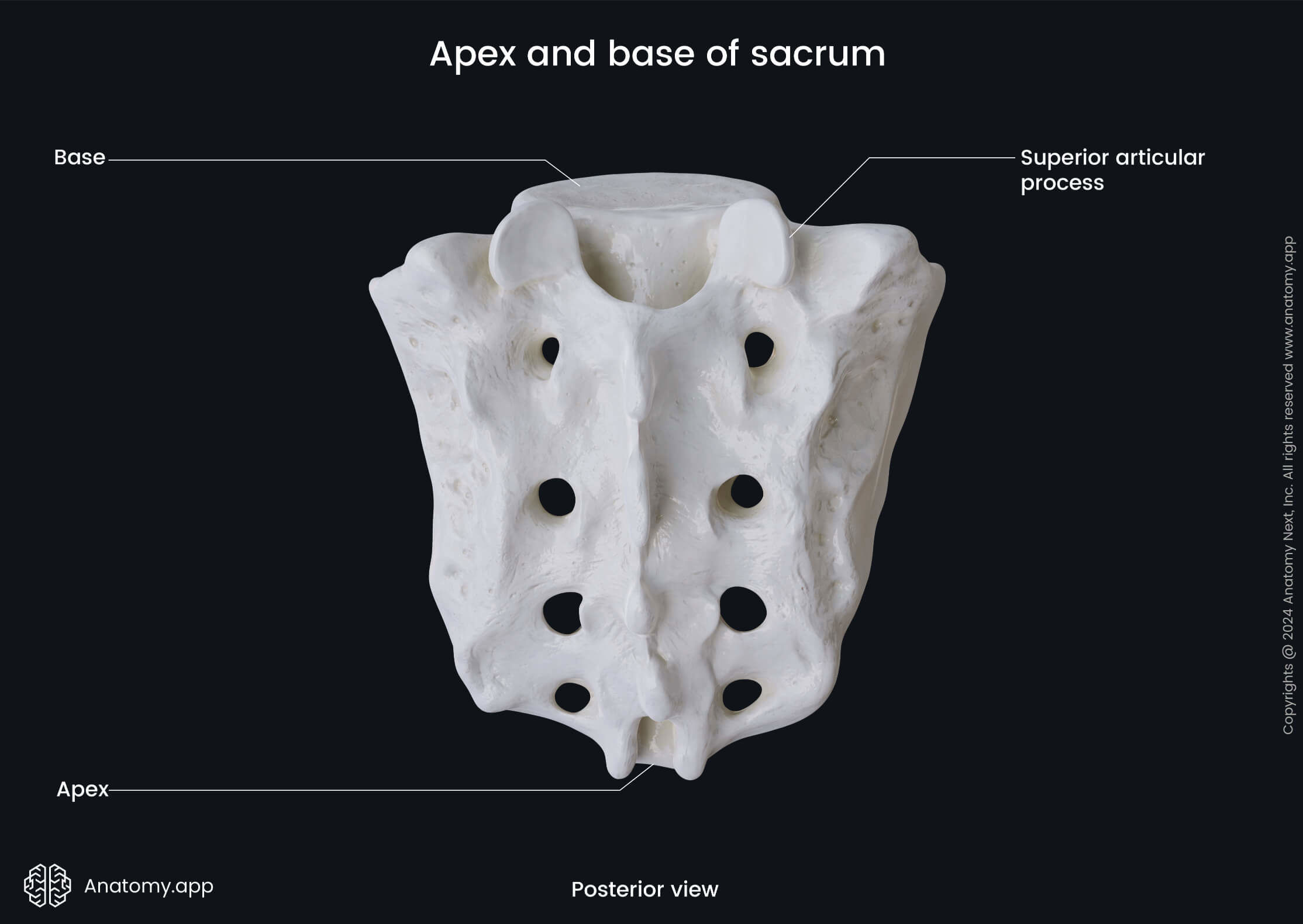
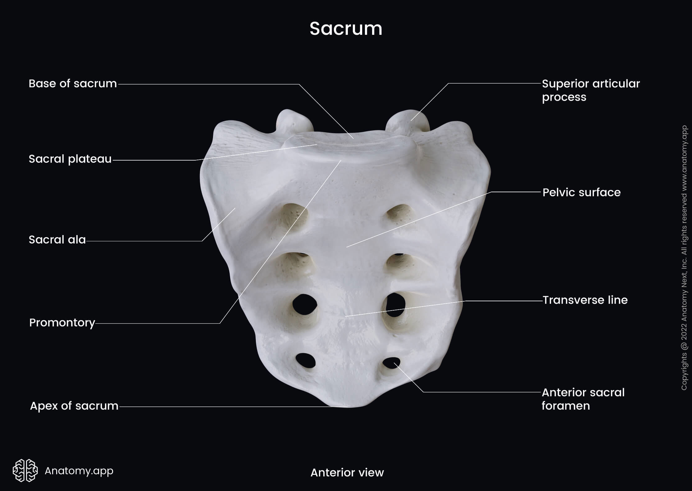
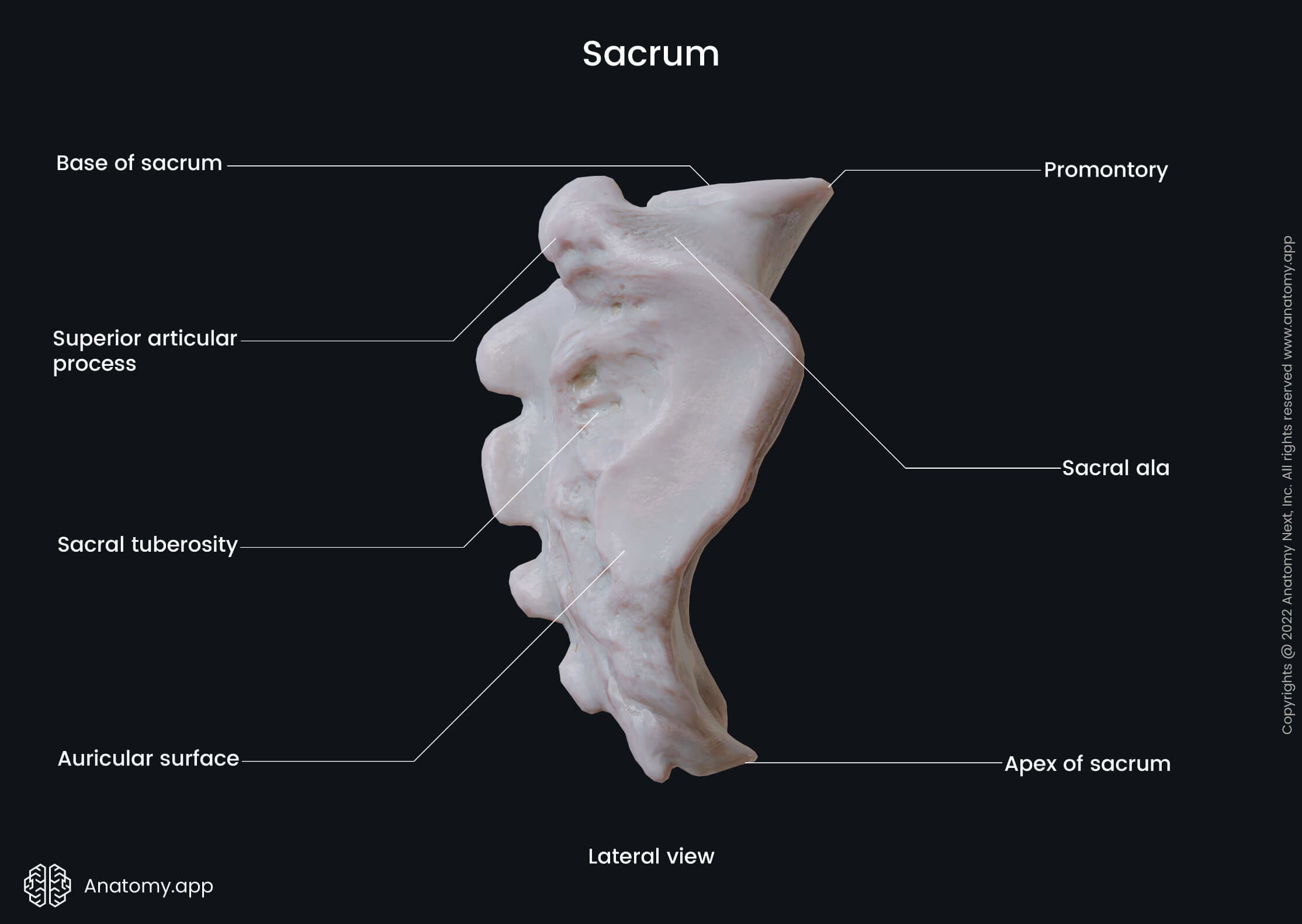
The sacrum has four surfaces - pelvic, dorsal and two lateral surfaces. As this bone is triangular-shaped, it presents a downward-directed apex and an upward-directed base.
Apex and base of sacrum
The apex is the lower tip of the sacrum, and it is connected to the coccyx. The joint between both bones is known as the sacrococcygeal symphysis. In contrast, the base is the broad upper part featuring the promontory and two superior articular processes.
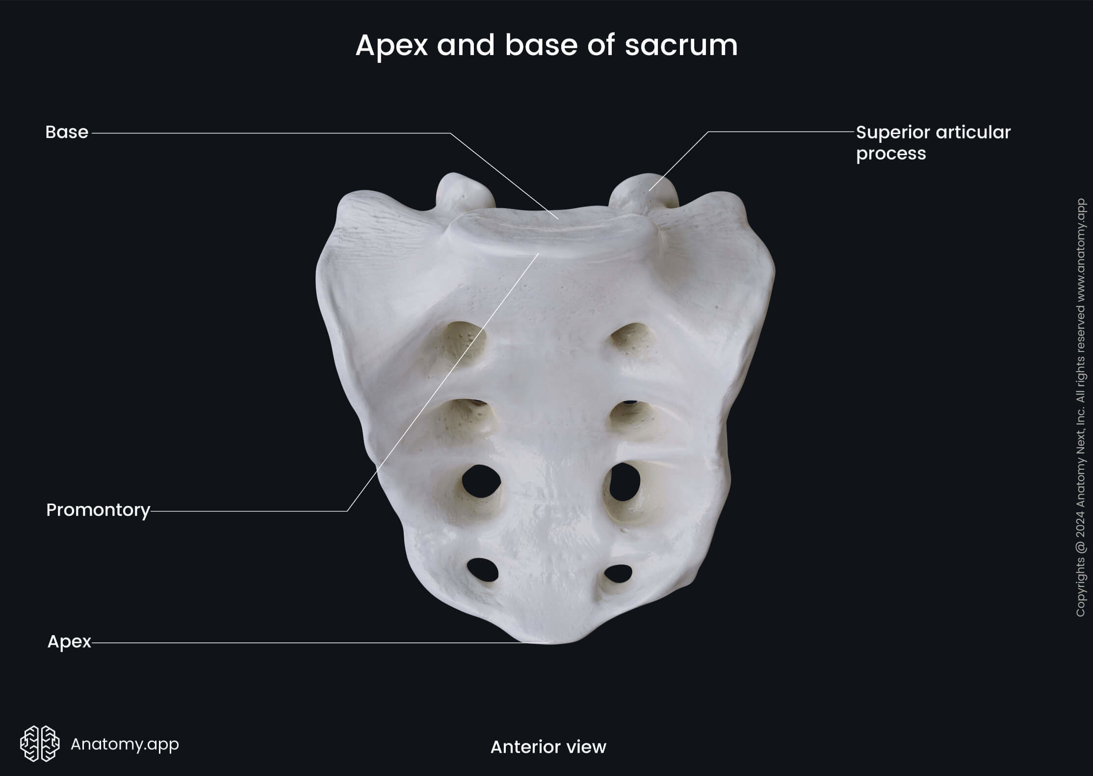
The promontory, also known as the sacral promontory or promontory of sacrum, is a prominent upper anterior margin of the body of the first sacral vertebra. Its anterior aspect projects far into the pelvic cavity.
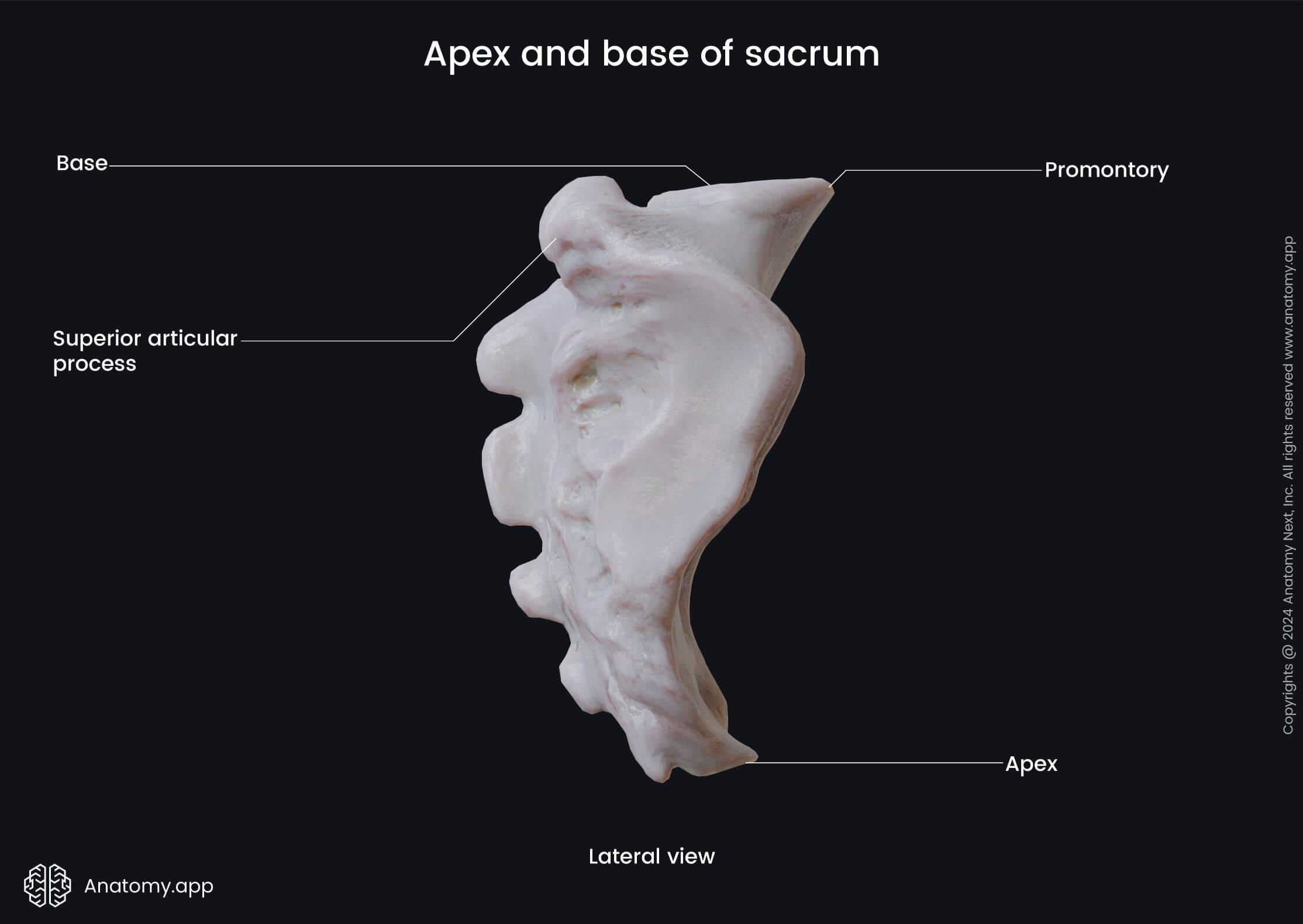
The superior articular processes are two large projections on the posterior aspect of the base. Both processes articulate with the corresponding inferior articular processes of the fifth lumbar vertebra, forming the lumbosacral joint.
Pelvic surface of sacrum
The pelvic surface of the sacrum is the anterior surface that faces the pelvic cavity. It is concave and presents the following landmarks:
- Transverse lines (4)
- Anterior sacral foramina (4 pairs)
The transverse lines are four lines corresponding to the fusion sites between the bodies of all five sacral vertebrae. They are more or less parallel to each other.
The anterior sacral foramina are four pairs of anterior openings in the sacrum. Through the openings pass nerves and blood vessels.
Dorsal surface of sacrum
The dorsal surface is the posterior convex surface of the sacrum. It contains the most significant amount of various anatomical landmarks, and they include the following:
- Median sacral crest
- Posterior sacral foramina (4 pairs)
- Intermediate sacral crests (2)
- Lateral sacral crests (2)
- Sacral canal
- Sacral hiatus
- Sacral cornu (2)
The median sacral crest is a ridge located in the midline of the surface. It is formed by the remnants of the spinous processes of the sacral vertebrae (S1-S4).
The posterior sacral foramina are four pairs of posterior openings in the sacrum. Through these openings pass nerves and blood vessels.
The intermediate sacral crests are two ridges located on either side of the median crest. They are formed by the fusion of articular processes of the sacral vertebrae.
The lateral sacral crests are paired ridges laterally from the posterior sacral foramina on either side of the surface. They are remnants of transverse processes of the sacral vertebrae.
The sacral canal is the inferior end of the vertebral canal running through most of the sacrum. The canal does not contain the spinal cord as the filum terminale starts at the L2 level.
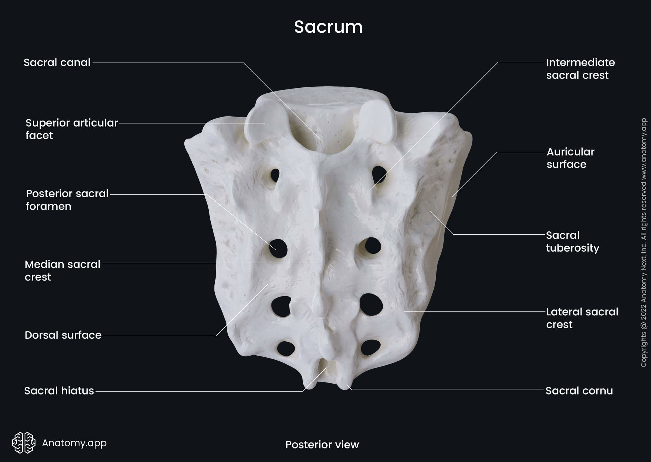
The sacral hiatus is the inferior opening of the sacral canal. It is located in the posterior wall of the sacral canal, usually at the level of the third to fourth sacral vertebrae (S3 - S4).
The sacral cornu (sacral horns) are two processes extending downward on each side of the sacral hiatus. They are remnants of the inferior articular processes of the fifth sacral vertebra.
Lateral surfaces of sacrum
The lateral surfaces, also known as the lateral parts or masses, are two lateral aspects of the sacrum. They are derived from the fusion of rudimentary ribs and transverse processes of the sacral vertebrae. Each lateral surface presents the following landmarks:
- Auricular surface
- Sacral tuberosity
The auricular surface is an ear-shaped articular surface situated on each lateral part of the sacrum. This surface articulates with the same-named surface of the ilium, and both bones form the sacroiliac joint.
The sacral tuberosity is a rough and elevated area on each lateral surface of the sacrum posterior to the auricular surface. It serves as an attachment site for the posterior sacroiliac ligaments.
Anatomy.app
Contact information
- For questions regarding business inquiries. Please contact:
- info@anatomy.app