- Anatomical terminology
- Skeletal system
- Skeleton of trunk
- Skull
- Skeleton of upper limb
- Skeleton of lower limb
- Joints
- Muscles
- Heart
- Blood vessels
- Lymphatic system
- Nervous system
- Respiratory system
- Digestive system
- Urinary system
- Female reproductive system
- Male reproductive system
- Endocrine glands
- Eye
- Ear
Rib cage
The rib cage (also known as the thoracic cage) is a bony framework of the thoracic wall. It encloses the thoracic cavity and is composed of various bones, cartilages, and joints. The shape of the thoracic cage resembles a domed birdcage. Internal organs such as the heart, lungs, spleen, and liver, and major blood vessels rest in the protective frame of the rib cage.
Rib cage anatomy
The rib cage is formed by the sternum, 12 pairs of ribs and their cartilages and 12 thoracic vertebrae and their respective intervertebral discs. The sternum is located anteriorly, the thoracic spine lies posteriorly, and the ribs - laterally. The ribs are found between the thoracic spine and sternum on each side, connecting both structures.
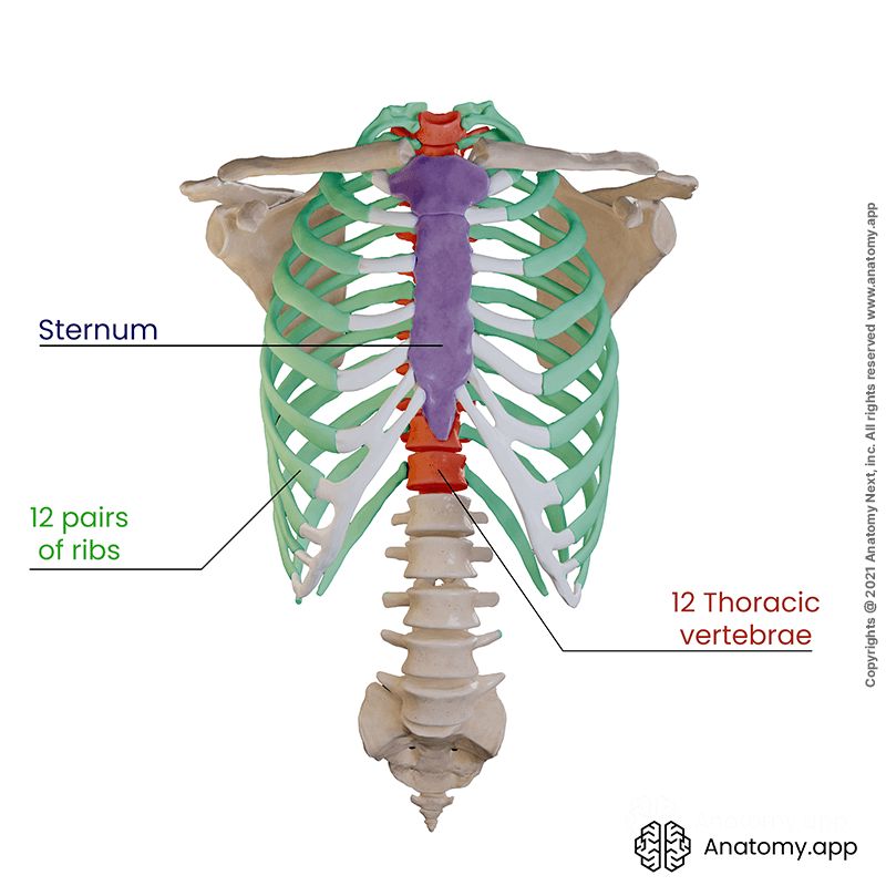
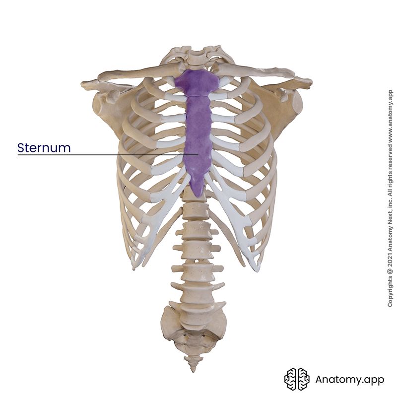
Sternum
The sternum (also known as the breastbone) is a bone located in the anterior midline of the thorax, supporting the rib cage anteriorly. It articulates with the upper seven ribs (1 - 7) and both clavicles. The sternum can be divided into three parts:
- Manubrium at the top - it articulates with clavicles and the first pair of ribs;
- Body (also called corpus) - the main part that articulates to ribs 3 through 7;
- Xiphoid process at the bottom - a small cartilaginous extension.
On the upper aspect of the manubrium in the midline is the jugular notch. It is a noticeable anatomical landmark for the anterior part of the upper thoracic opening (also called the thoracic outlet).
Another important anatomical landmark is found between the manubrium and the body of the sternum. It is the angle of Louis, also called the sternal angle. It is noticeable on palpation, during which a slight angulation marks the following anatomical structures:
- Transition between the superior mediastinum and inferior mediastinum (mediastinal cavity)
- Horizontal thoracic vertebral level T4/ T5
- Tracheal bifurcation
- Articulation with the cartilage of the 2nd rib
Note: the sternum can be useful in bone marrow biopsy in adults. Similar to the ilium, it contains red bone marrow.
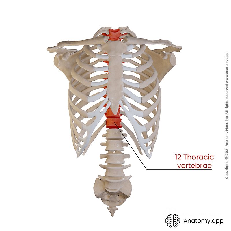
Thoracic spine and intervertebral discs
The thoracic spine consists of 12 thoracic vertebrae with associated intervertebral discs between them. The thoracic part of the spine supports the rib cage posteriorly. All vertebrae together form a column with a bony canal in the middle. The canal is called the spinal canal, and it contains the spinal cord.
When viewed from above, the typical thoracic vertebra has a somewhat heart-shaped vertebral body and a circular vertebral foramen. Each vertebra has a vertebral body, pedicles, transverse processes laterally and a spinous process posteriorly.
A distinguish characteristic of the thoracic vertebrae is their articulation with ribs. The vertebral body of a typical thoracic vertebra has two partial facets (superior and inferior costal demi-facets) on each side. They articulate with the head of its corresponding rib and the rib below. Each transverse process of these vertebrae has a transverse costal facet that articulates with the tubercle of its corresponding rib.
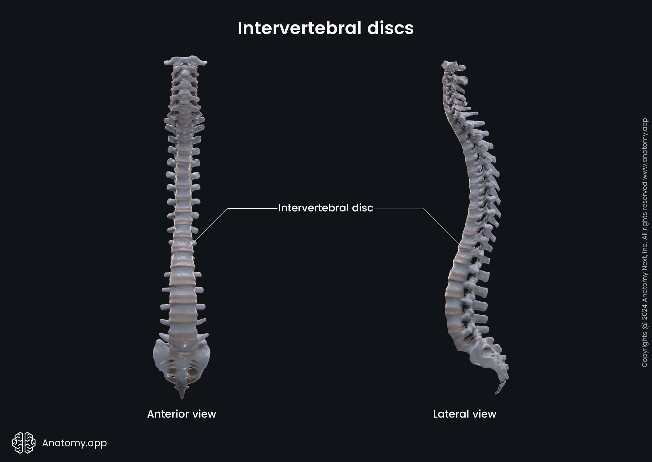
Between the vertebral bodies lie structures known as intervertebral discs. They act as cushions and support the vertebrae, providing movement in the spine and joints. Each disc is composed of two main parts. The outer fibrous layer of the intervertebral disc is called the annulus fibrosus. And the inner weaker core structure - nucleus pulposus.
Ribs
There are a 12 pairs of ribs in the thoracic cage. Ribs are long, flat and curved bones that have a bony part posteriorly and cartilaginous part in the front. Each rib articulates with a vertebral body at two joints:
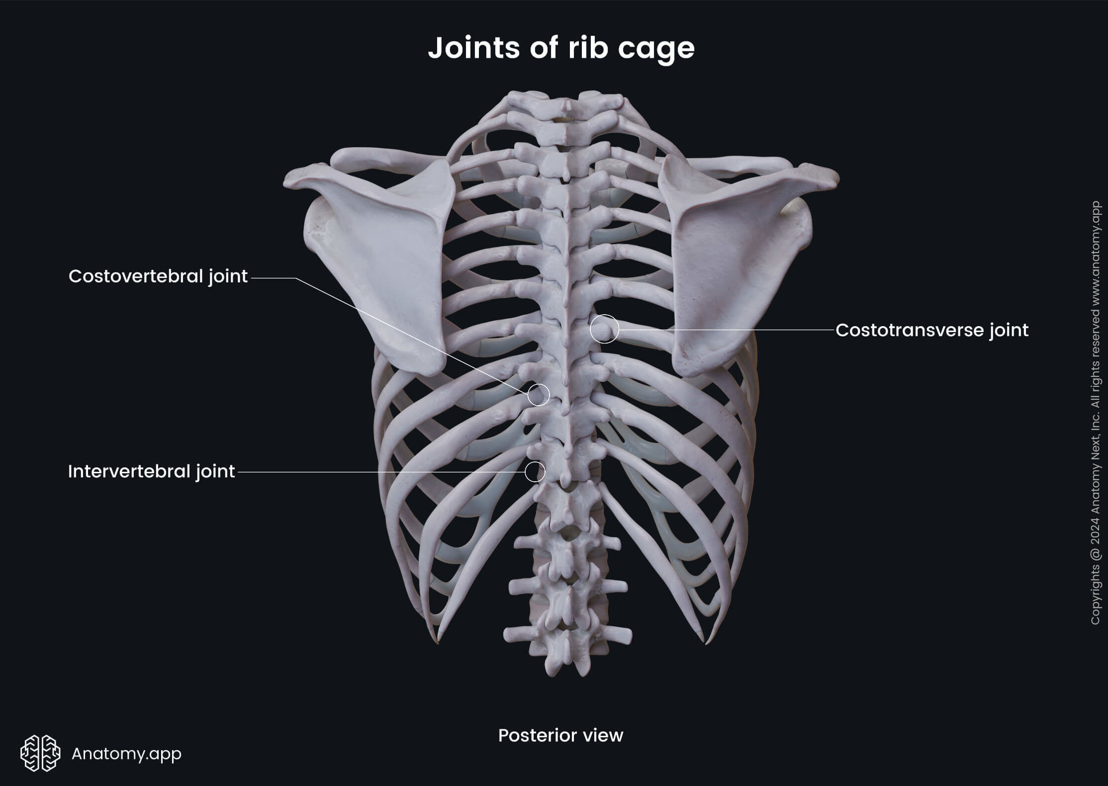
- Costovertebral joint - between the head of the rib and facets of two adjacent vertebral bodies;
- Costotransverse joint - between the costal tubercle and the transverse process of the vertebrae.
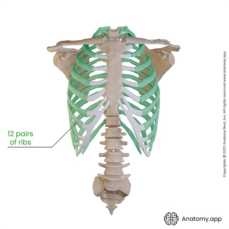
The first seven pairs of ribs (1 - 7) are directly connected with the sternum at the sternocostal joints. They are called the true ribs. Ribs 8 through 10 join the costal cartilages of upper ribs and attach indirectly by interchondral joints, thus termed as false ribs. Ribs 11 and 12 are not connected to the sternum or other ribs and are known as floating or free ribs. Their cartilages tend to end within the abdominal musculature.
Intercostal spaces
The spaces between adjacent ribs and costal cartilages are called the intercostal spaces. These spaces are mainly filled with layers of muscles and their respective membranes called aponeuroses. Overall, there are 11 intercostal spaces found in the thoracic cage. Since it has no ribs below, the 12th space under the 12th rib is called the subcostal space.
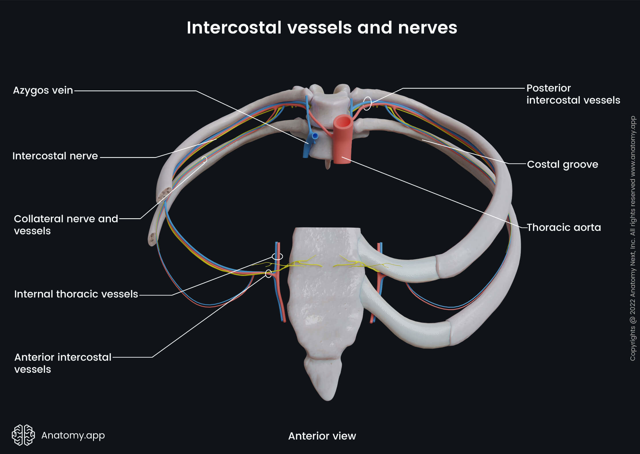
The intercostal spaces also contain intercostal nerves and blood vessels - arteries and veins. This neurovascular bundle lies in between the inner two layers of intercostal muscles. The bundle is located at the inferior margin of the superior rib in a depression called the costal groove.
The neurovascular structures in the costal groove are organized in the following order: the highest is the vein; the artery lies in the middle; the nerve is located inferiorly. Because of its location, the intercostal nerve is usually not protected by the costal groove. The order of the structures in the costal groove can be memorized with the help of the acronym VAN (Vein, Artery, Nerve).
The inner surface of the thoracic wall is lined by the endothoracic fascia. It is also called the parietal fascia of the thorax. This fascia also covers the primary respiratory muscle - the diaphragm.
Joints of rib cage
The joints forming the rib cage are mainly cartilaginous or synovial in type. A cartilaginous joint means that the connected bones are lined by hyaline or fibrous cartilage. These joints are usually fixed (synarthroses) or provide slight mobility (amphiarthroses). Cartilaginous joints can be divided into primary and secondary, depending on the type of hyaline involved. Primary cartilaginous joints (synchondrosis) are between the bones and the hyaline cartilage. Secondary joints (called symphysis) are made up either of hyaline cartilage or fibrocartilage.
Synovial joints are usually diarthroses - they can move in various planes but are restricted in motion to the surrounding muscles and ligaments. The main characteristic of these joints is a synovial cavity, synovial capsule and synovial membrane. The bones articulating in synovial joints are lined by hyaline cartilage. To learn more about the types of joints and their functions, see our topic on the classification of joints.

The joints forming the rib cage are the following:
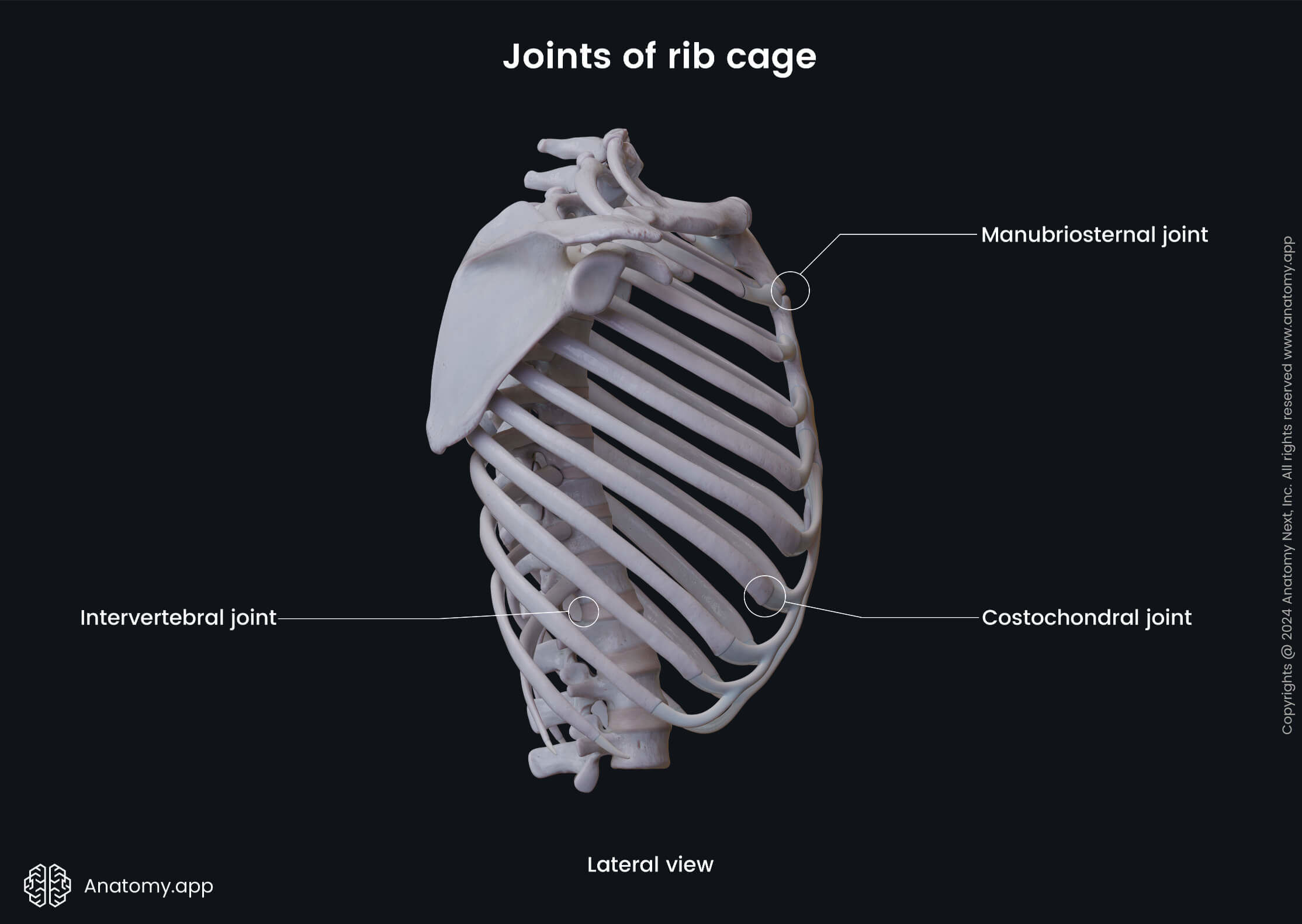
- Intervertebral joints - articulations between adjacent vertebrae. Each intervertebral joint consists of an intervertebral disc and two facet or zygapophyseal joints. The intervertebral joints are symphyses, but the intervertebral discs contain a gel-like substance (nucleus pulposus).
- Sternoclavicular joints - between the manubrium of the sternum and the clavicles. These joints are synovial saddle-type joints.
- Sternochondral joints - between the sternum and costal cartilages of ribs 1 to 7. They are synovial plane-type joints, except for the first sternochondral joint (it is considered to be primary cartilaginous).
- Manubriosternal joint - between the manubrium and body of the sternum. This type of joint is a secondary cartilaginous joint and a kind of symphysis.
- Xiphisternal joint - between the xiphoid process and the body of sternum. This joint is a primary cartilaginous joint or synchondrosis. But with age, the cartilage becomes ossified, and the joint turns into synostosis.
- Costochondral joints - between costal cartilages and ribs. These are primary cartilaginous joints, called synchondroses.
- Costovertebral joints - between the head of the rib and the body of the corresponding vertebra. It is a synovial plane-type joint.
- Costotransverse joints - between the costal tubercle and the transverse process of the vertebrae; also a synovial plane joint.
- Interchondral joints - articulations between two or more costal cartilages. The cartilages are connected by the synovial plane joints and stabilized by attaching interchondral ligaments. The exception is the tenth rib that attaches to the ninth rib by a fibrous type of joint.
Thoracic apertures
There are two thoracic openings or apertures in the rib cage - superior and inferior. They are also referred to as the thoracic inlet and outlet. In some literature, the superior thoracic opening is described as an outlet, while other authors define it as an inlet.
Superior thoracic aperture
The superior thoracic aperture is around 5 x 10 cm wide opening in the upper part of the thoracic cage. Here we will classify it as the thoracic outlet because of its connection to the so-called thoracic outlet syndrome (described below). The borders of the thoracic outlet are as follows:
- Posteriorly - the first thoracic vertebra (T1);
- Laterally - the internal surfaces of the 1st ribs;
- Anteriorly - the sternal jugular notch.
The main structures located in the upper thoracic opening are the trachea and apical parts of both lungs. The lung apex is the upper part of the lung; it also slightly extends into the inferior neck. It rises above the level of the first rib, approximately at the level of the mid-third of the clavicle. The apex projects to the horizontal level of the last cervical vertebra (C7) or the first thoracic vertebra (T1).
There are membranes that cover the pleura and lung apexes. Also, the apex is then covered by various blood vessels, nerves and muscles that pass over the first rib. It is also important to note that the brachial plexus, subclavian artery and subclavian vein are located between the clavicle and the first rib.
Clinical note: the thoracic outlet syndrome occurs when the compression of nerves (brachial plexus) and blood vessels (subclavian artery and vein) in the thoracic outlet results in upper extremity symptoms. Various causes such as trauma, tumors, inflammation, congenital anomalies can impact the spaces of these important anatomic structures. The diagnosis is challenging because of the variability in presentation. The symptoms vary from mild to very severe, including pain, numbness, swelling, and discoloration of skin.
Inferior thoracic aperture
The inferior thoracic opening is much more prominent in dimensions than the upper thoracic aperture. It is bordered by:
- Posteriorly - the last thoracic vertebra (T12);
- Posterolaterally - by the 11th and 12th ribs and their cartilages;
- Anterolaterally - by the 7th to 10th rib ends and their interchondral joints;
- Anteriorly - by the xiphoid process of the sternum.
Rib cage function
The structures of the rib cage altogether form a frame of protection for vital organs and major blood vessels. The main functions of the thoracic cage include:
- Supporting the thorax
- Protecting thoracic and abdominal organs from injury
- Assisting in respiration
- Providing support for the upper limbs
- Providing attachment for muscles involved in upper limb motion
References:
Gray, H., & Carter, H., V. (2021). Gray’s Anatomy (Leatherbound Classics) (Leatherbound Classic Collection) by F.R.S. Henry Gray (2011) Leather Bound (2010th Edition). Barnes & Noble.
Paulsen, F., Waschke, J., Hombach-Klonisch, S., Klonisch, T., & Peeler, J. (2019). Sobotta Clinical Atlas of Human Anatomy, one volume, English (1st ed.). Urban & Fischer.
Pope, T., Bloem, H., Beltran, H., & Morrison, W. (2014). Musculoskeletal Imaging: Expert Radiology Series (2nd ed.). Saunders.
Skirven, T. M., Osterman, A. L., Fedorczyk, J., Amadio, P. C., Felder, S., & Shin, E. K. (2020). Rehabilitation of the Hand and Upper Extremity, E-Book. Elsevier Gezondheidszorg.
Anatomy.app
Contact information
- For questions regarding business inquiries. Please contact:
- info@anatomy.app