- Anatomical terminology
- Skeletal system
- Joints
- Muscles
- Heart
- Blood vessels
- Blood vessels of systemic circulation
- Aorta
- Blood vessels of head and neck
- Arteries of head and neck
- Veins of head and neck
- Blood vessels of upper limb
- Arteries of upper limb
- Veins of upper limb
- Blood vessels of thorax
- Blood vessels of abdomen
- Blood vessels of pelvis and lower limb
- Blood vessels of systemic circulation
- Lymphatic system
- Nervous system
- Respiratory system
- Digestive system
- Urinary system
- Female reproductive system
- Male reproductive system
- Endocrine glands
- Eye
- Ear
Subclavian artery
The subclavian artery (Latin: arteria subclavia) is a paired vessel found within the upper thorax below the clavicle. It supplies arterial blood to the head, brain, neck region, upper thorax, shoulder and upper extremity. The subclavian artery originates within the rib cage and exits through the superior thoracic aperture.
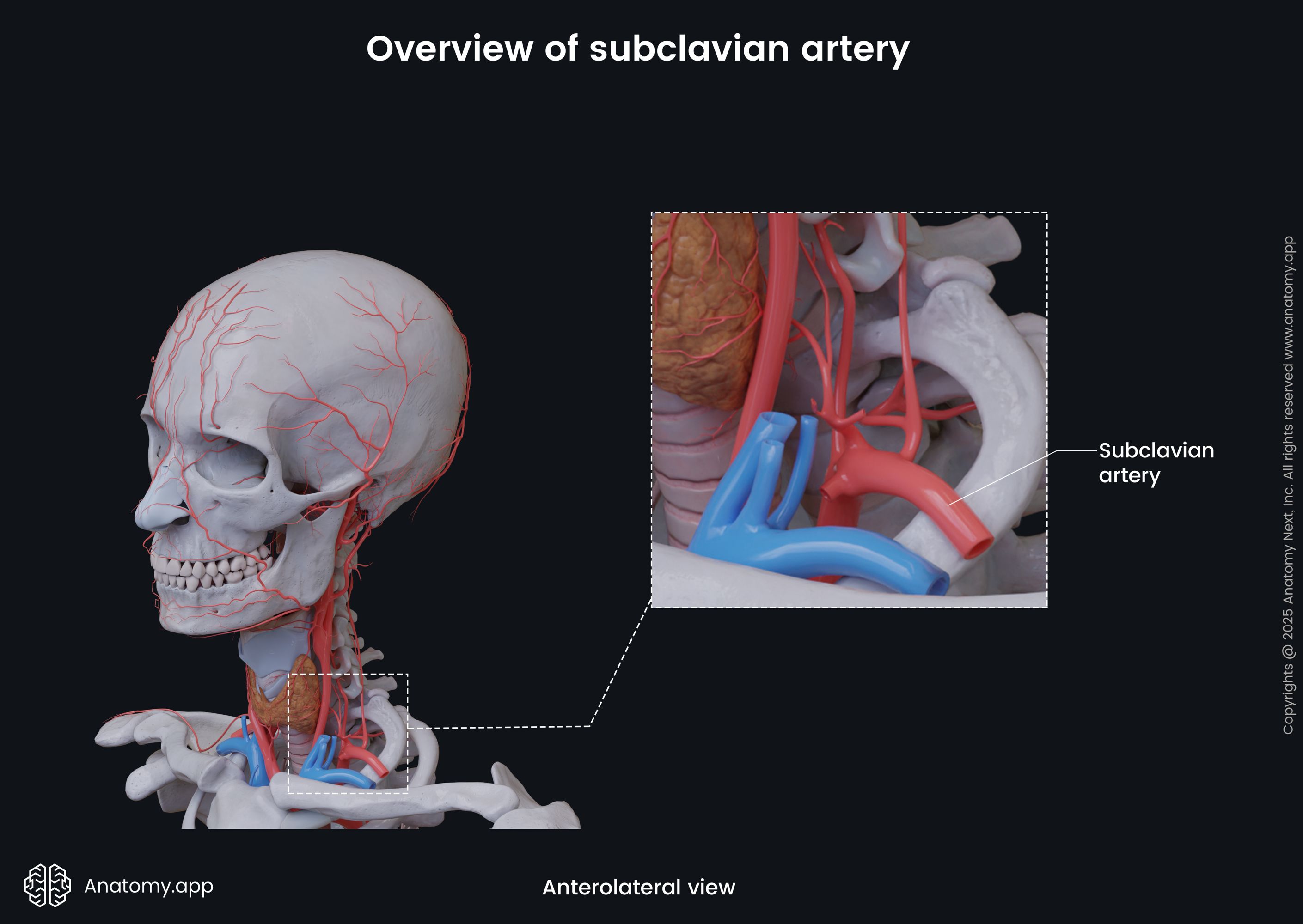
Each person has a right and left subclavian artery. Both arteries have different origins. The right subclavian artery arises from the brachiocephalic trunk, while the left subclavian artery originates from the aortic arch. When reaching the outer border of the first rib, both arteries continue as the axillary arteries.
Right subclavian artery
The right subclavian artery originates from the brachiocephalic trunk posterior to the right sternoclavicular joint approximately at the first or second thoracic vertebra (T1 or T2) level. From its origin, the right subclavian artery arches in the superolateral direction. It exits the superior mediastinum and rib cage through the superior thoracic aperture and enters the root of the neck. At first, it is located posterior to the anterior scalene muscle and the sternal end of the clavicle.

The right subclavian artery then passes between the anterior and middle scalene muscles. It goes across the superior surface of the first rib, right below the clavicle. Here it travels within a neurovascular bundle consisting of the subclavian artery, subclavian vein, and brachial plexus. At the external border of the first rib, the right subclavian artery becomes the right axillary artery.
Left subclavian artery
The left subclavian artery usually arises directly from the aortic arch. It is the smallest of the three vessels arising from the aortic arch, following the brachiocephalic trunk and the left common carotid artery. Although both subclavian arteries have different origins, they have the same courses.
The left subclavian artery also leaves the rib cage through the superior thoracic aperture and enters the neck. Here, it is located posterior to the anterior scalene muscle and the sternal end of the clavicle. As it goes further, it is positioned between the anterior and middle scalene muscles.
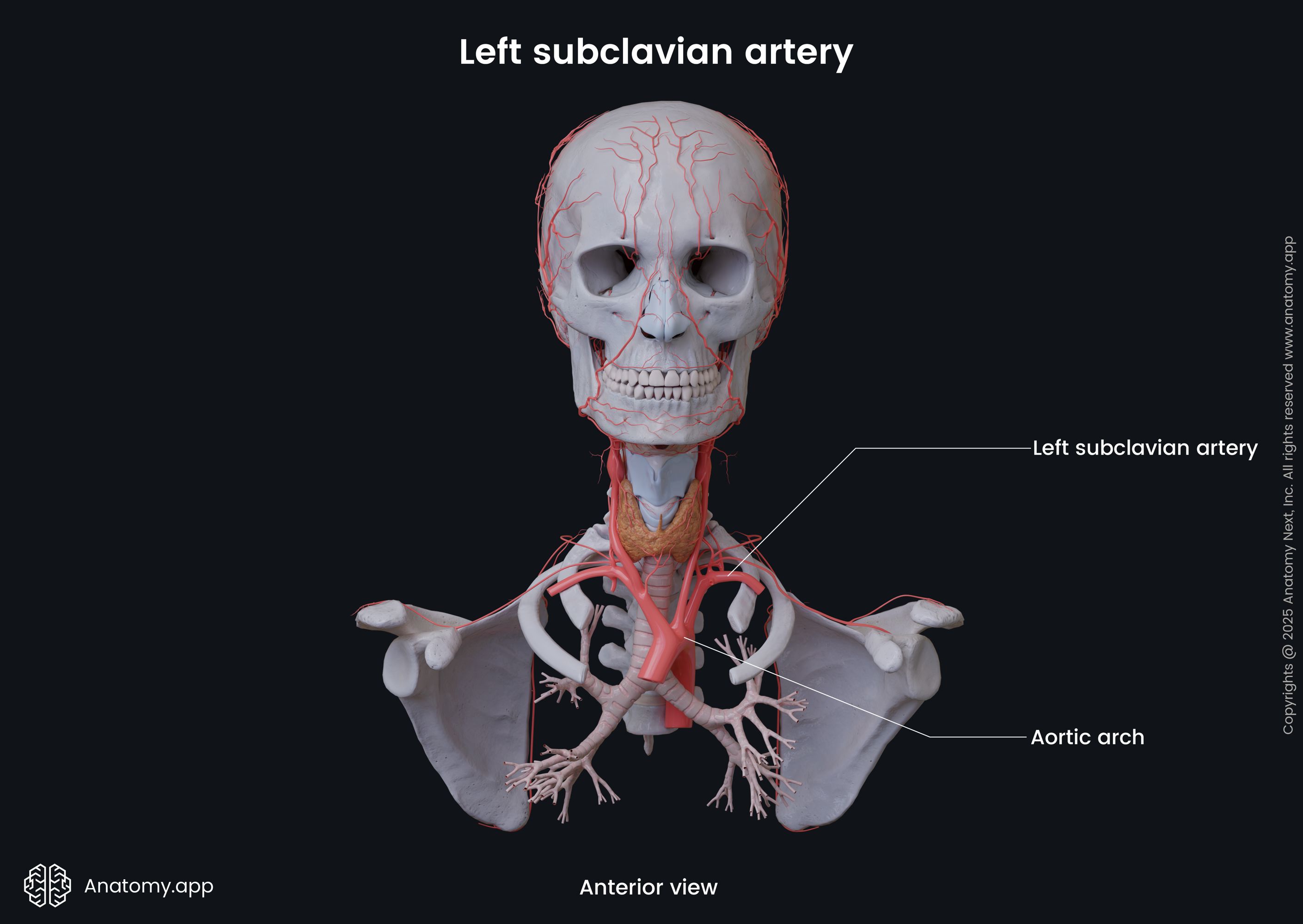
And finally, the left subclavian artery passes between the first rib and clavicle as a part of a neurovascular bundle consisting of the subclavian artery, subclavian vein and brachial plexus. At the external border of the first rib, the left subclavian artery becomes the left axillary artery.
Clinical note: Damage or compression of the structures within the neurovascular bundle can cause a variety of symptoms referred to as thoracic outlet syndrome. This condition usually occurs due to trauma when nerves or blood vessels become compressed by the rib, clavicle or neck muscles at the top of the thoracic outlet. It is relatively rare and usually presents with nervous symptoms, including pain and tingling or weakness in the shoulder and arm. But more severe cases can present with edema of the arm, hand or fingers, bluish or white skin color of the arm and hand, painful tingling in the hand and arm and cold hand. These symptoms are usually present when the vascular structures also become involved.
Subclavian artery parts
The subclavian artery can be subdivided into three parts based on its relation to the anterior scalene muscle. Each subclavian artery has the following parts:
- Pre-scalene part
- Retro-scalene part
- Post-scalene part
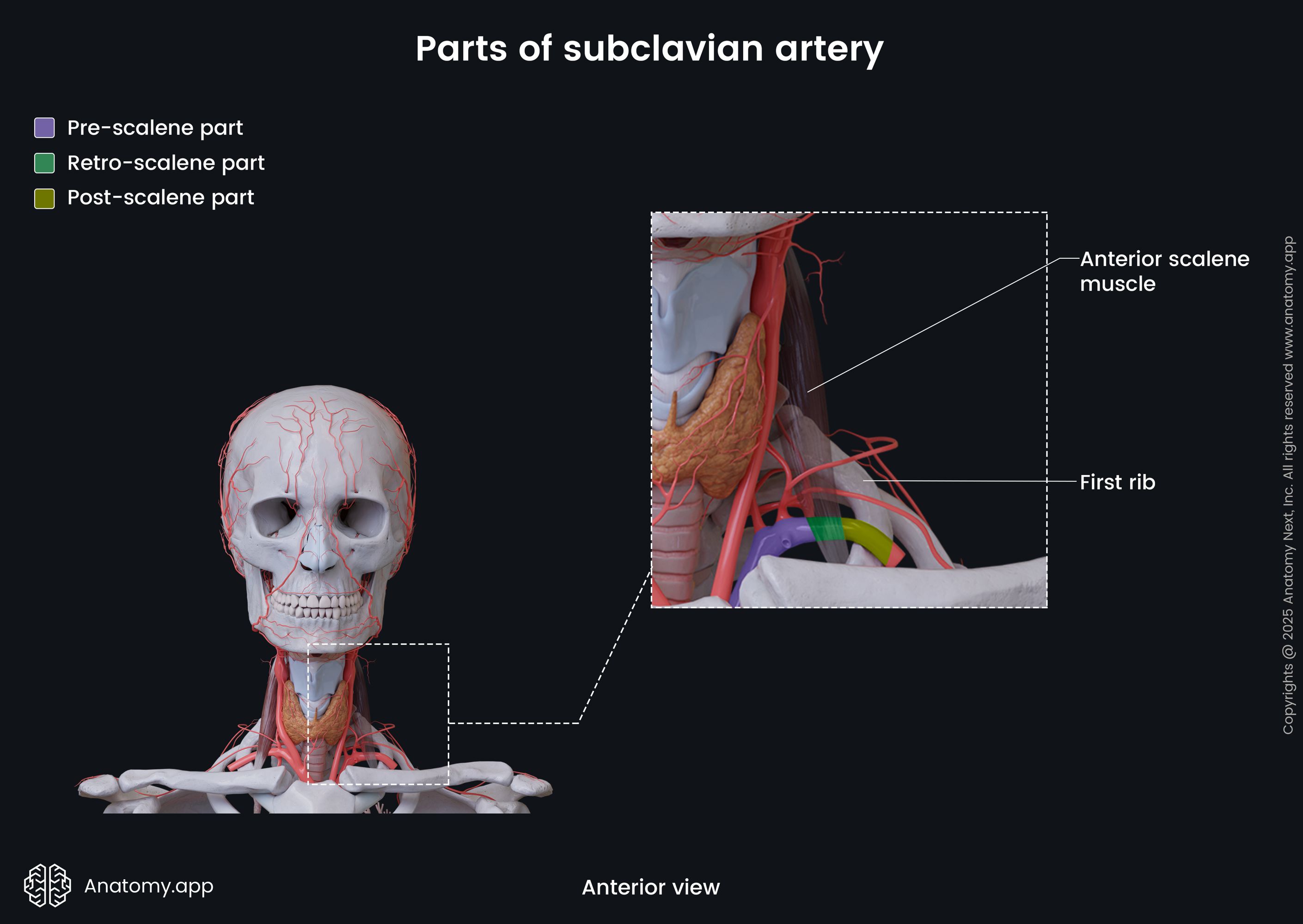
The pre-scalene part is the first portion of the subclavian artery that starts at its origin site and ends at the medial border of the anterior scalene muscle. Overall, branches of this part supply the thyroid gland, upper thorax and breasts.
The retro-scalene part is the second portion that begins at the medial border of the anterior scalene muscle. This segment goes between the anterior and middle scalene muscles, extending to the lateral border of the anterior scalene. The retro-scalene portion supplies arterial blood to the back of the neck and upper part of the rib cage.
The post-scalene part is the third and final segment that starts at the lateral border of the anterior scalene muscle and terminates at the external margin of the first rib, where the artery continues as the axillary artery. This segment supplies the shoulder and upper extremity.
Subclavian artery branches
The subclavian artery gives off several branches along its course. Each segment has different branches described below.
Branches of pre-scalene part
The pre-scalene part of the subclavian artery gives off the following branches:
- Vertebral artery
- Internal thoracic artery
- Thyrocervical trunk
The vertebral artery is the first branch of the subclavian artery. It arises from the superior aspect of the artery and ascends towards the external cranial base. It goes through the transverse foramina of the sixth to first cervical vertebrae (C6 - C1). It unites with the contralateral vertebral artery on the clivus of the occipital bone, forming the basilar artery and contributing to the circle of Willis. Overall, the vertebral arteries supply the deep neck muscles, spinal cord, cerebrum, cerebellum, medulla oblongata and dura mater of the posterior cranial fossa.

The internal thoracic artery, also called the internal mammary artery, is the second branch that arises from the pre-scalene part of the subclavian artery. Overall, it supplies remnants of the thymus, breast tissue, sternum, pericardium, mediastinum and anterior thoracic wall. This artery originates a few inches away from the origin site of the subclavian artery, arising from its inferior aspect.
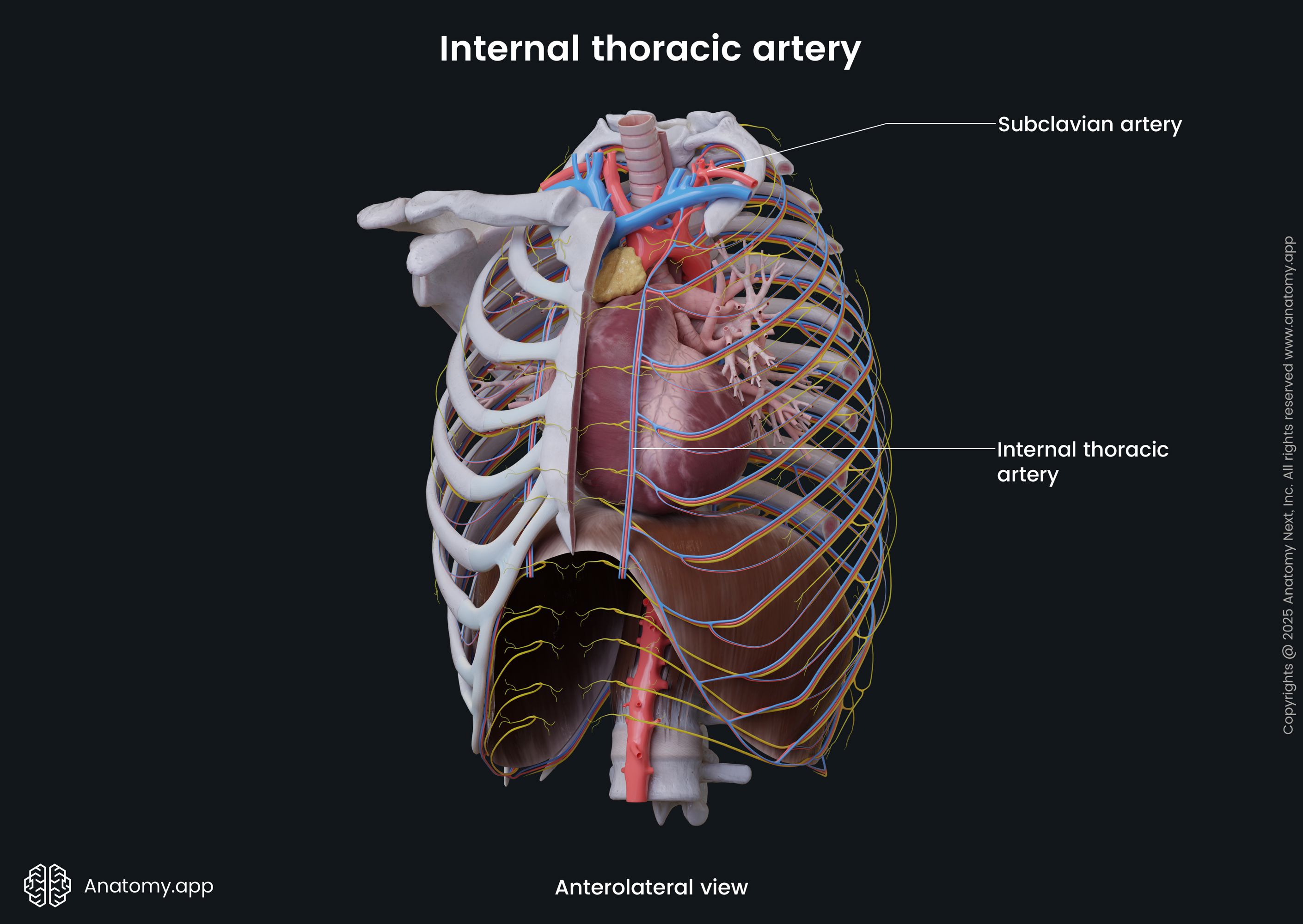
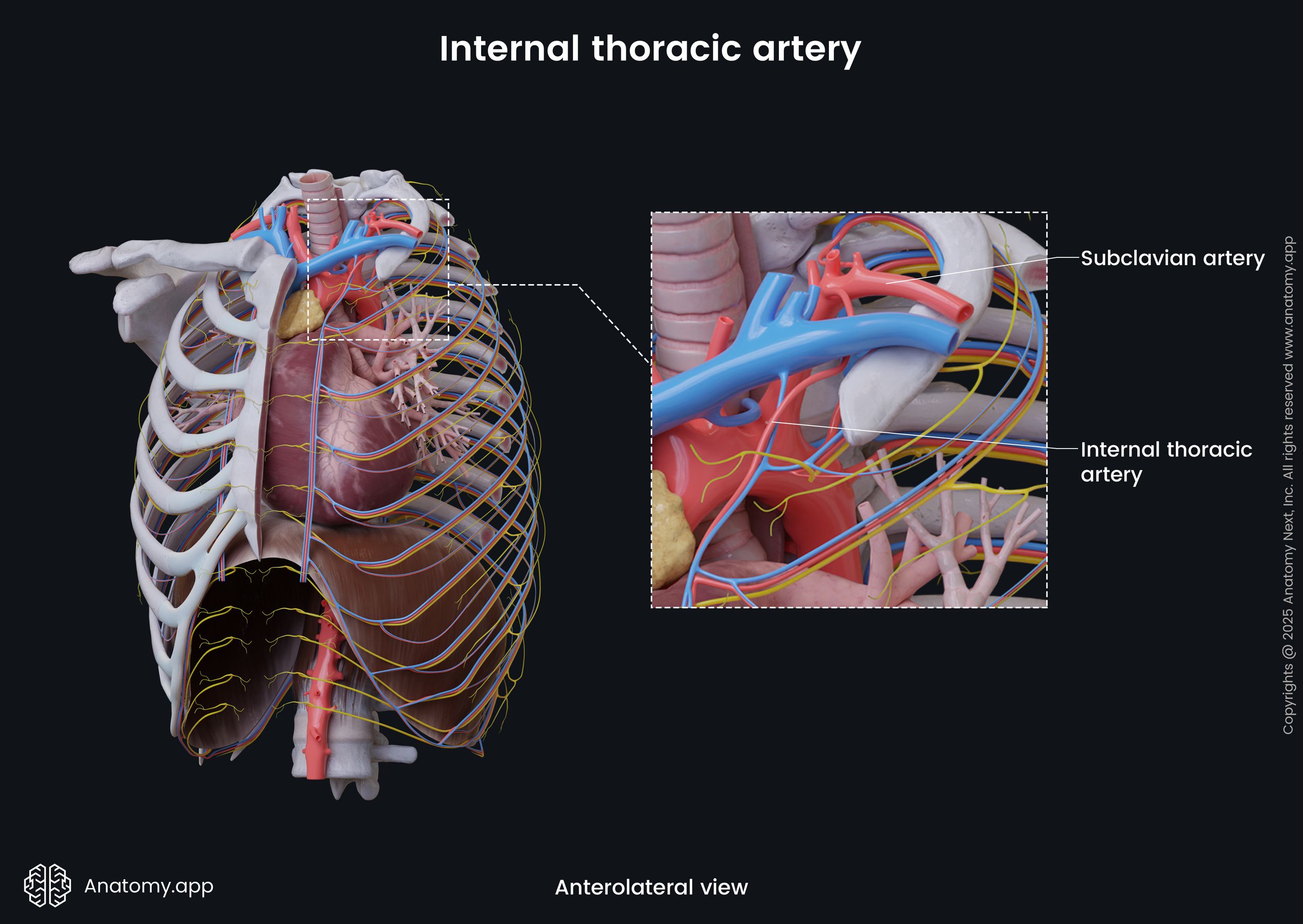
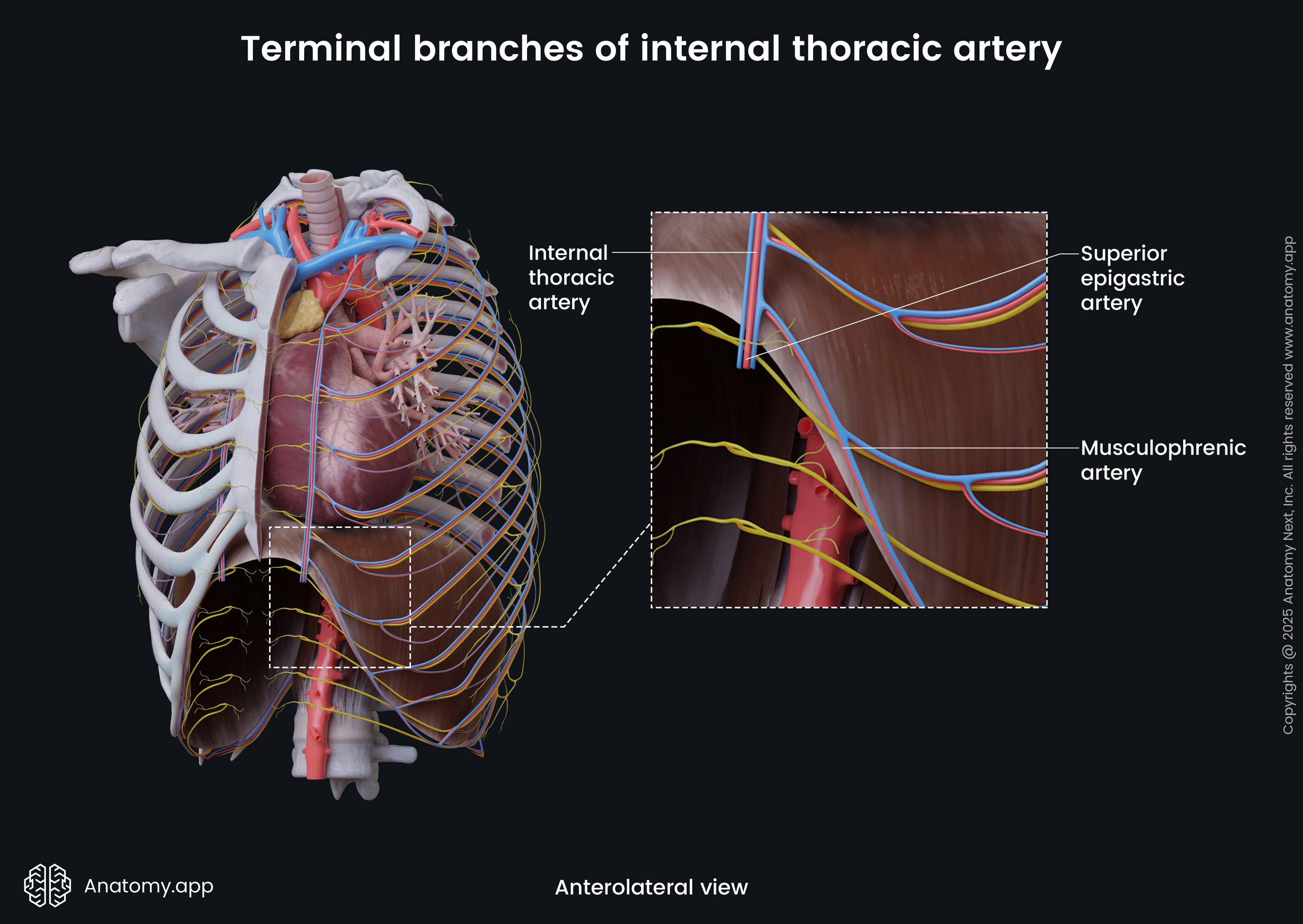
Further, the internal thoracic artery descends along the inner surface of the anterior thoracic wall. It goes within the thoracic cage along the side of the sternum - approximately 0.4 - 0.8 inches (1 - 2 cm) laterally from it. The internal thoracic artery is accompanied by a pair of internal thoracic veins. It ends at the sixth or seventh costal cartilage and their respective intercostal spaces by dividing into the superior epigastric artery and musculophrenic artery.
- The superior epigastric artery descends along the internal surface of the anterior abdominal wall from the lower border of the thoracic cage to the umbilicus. It ends by anastomosing with the inferior epigastric artery above the umbilicus. The superior epigastric artery supplies muscles (rectus abdominis) and skin of the anterior abdominal wall.
- The musculophrenic artery courses downwards and laterally along the inferior border of the thoracic cage behind the costal cartilages. It goes from the sixth or seventh costal cartilage to the ninth costal cartilage. It then passes through the diaphragm, supplying it. The musculophrenic artery ends in the last (eleventh) intercostal space, anastomosing with the inferior phrenic artery, last two posterior intercostal arteries and ascending branches of the deep circumflex iliac arteries. Overall, this artery supplies the pericardium, last three to four intercostal spaces, abdominal muscles and anterior abdominal wall.
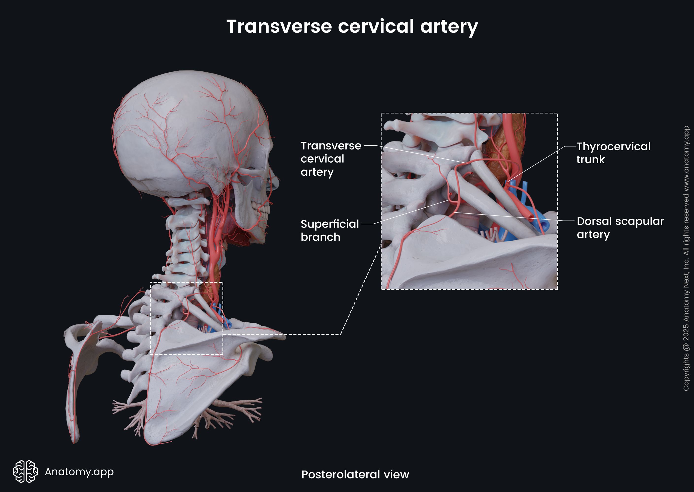
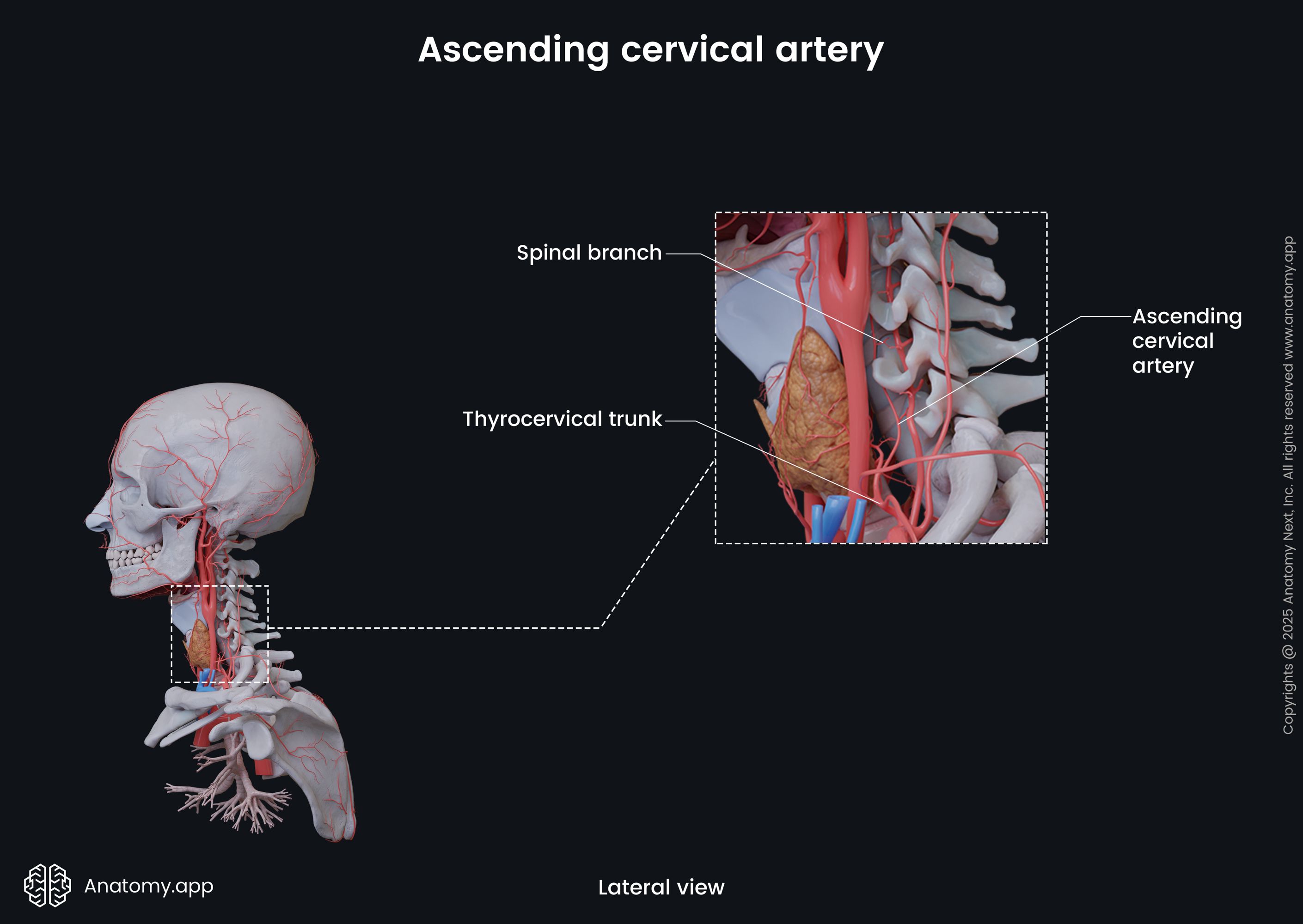
The thyrocervical trunk is the third branch of the pre-scalene portion. It arises from the superior aspect of the subclavian artery distally to the vertebral artery. It is a relatively short and wide artery that is only about 0.6 inches (1.5 cm) long. The thyrocervical trunk almost immediately divides into three or four branches - inferior thyroid, transverse cervical, suprascapular and ascending cervical arteries. Overall, it supplies the pharynx, esophagus, larynx, trachea, thyroid and parathyroid glands, brachial plexus and muscles of the neck.

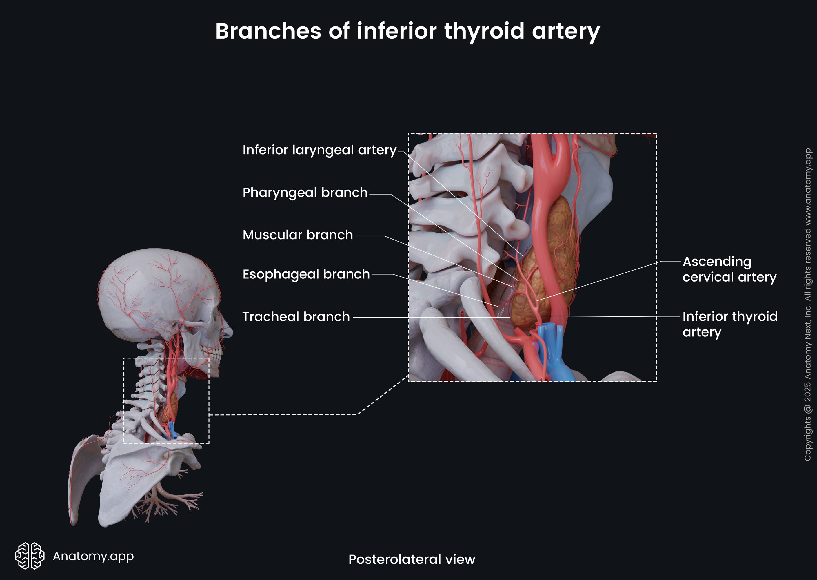
- The inferior thyroid artery is the largest branch of the thyrocervical trunk. At first, it vertically ascends towards the neck and then arches in the anteromedial direction and descends towards the base of the thyroid gland. Overall, it supplies the posterior and inferior aspects of the thyroid, as well as the parathyroid glands. The inferior thyroid artery also gives off small muscular, pharyngeal, tracheal and esophageal branches, as well as the inferior laryngeal artery to the respective organs. The inferior thyroid artery sometimes gives off the ascending cervical artery that can alternatively arise directly from the thyrocervical trunk.
- The transverse cervical artery travels posteriorly towards the scapula, crossing the phrenic nerve, brachial plexus and anterior scalene muscle. It divides into superficial and deep branches - the superficial cervical artery and the dorsal scapular artery, respectively. Both branches supply muscles of the neck and upper back.
- The suprascapular (transverse scapular) artery descends laterally and backward across the anterior scalene muscle and phrenic nerve. It then goes parallel behind the clavicle, crossing the subclavian artery and brachial plexus and reaching the upper aspect of the scapula. Overall, it supplies the sternocleidomastoid, subclavius, supraspinatus and infraspinatus muscles, as well as the acromioclavicular and shoulder joints.

- The ascending cervical artery is a small artery that might arise from the inferior thyroid artery or directly from the thyrocervical trunk. Overall, it supplies muscles of the neck, spinal cord, spinal meninges and vertebral bodies.
Branches of retro-scalene part
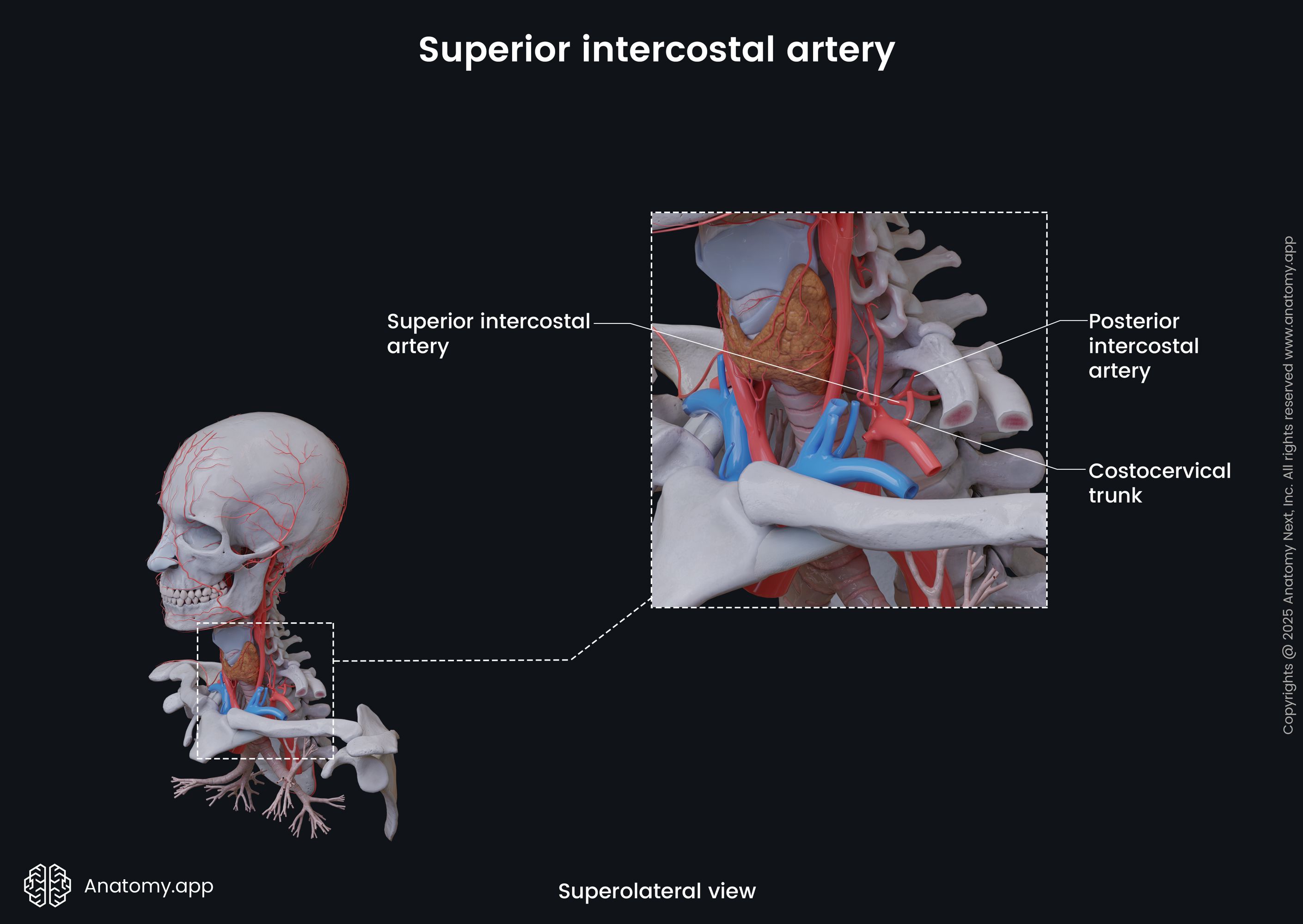
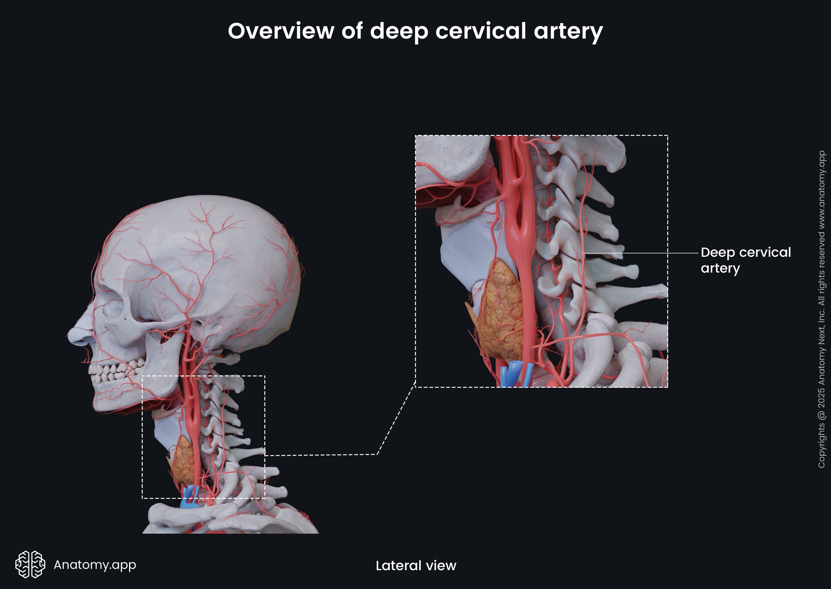
The retro-scalene part of the subclavian arteries gives rise to the costocervical trunk. It arises from the posterior surface of the subclavian artery. The costocervical trunk is a short artery that passes in a posterosuperior direction and divides into two branches - the superior intercostal artery and the deep cervical artery.
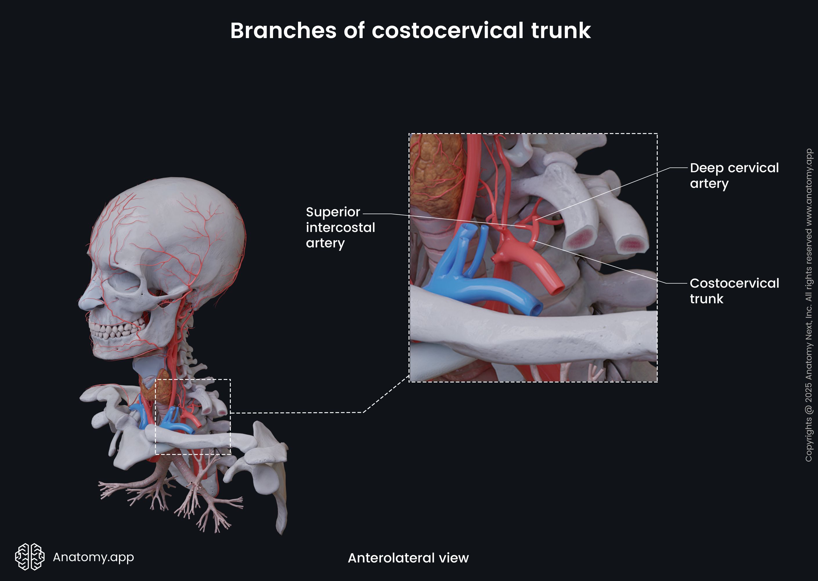
The superior (supreme) intercostal artery goes inferiorly and posteriorly along the posterior and medial aspects of the thorax, passing between the necks of the first and second ribs and the adjacent pleura. Here it divides into two posterior intercostal arteries that supply the upper two to three intercostal spaces. Overall, the superior intercostal artery supplies the wall of the rib cage, parietal pleura and skin and muscles of the back.
The deep cervical artery travels between the facet (zygapophyseal) joints towards the deep posterior muscles of the neck, supplying them.
Branches of post-scalene part
The post-scalene part of the subclavian artery gives off only one artery - the dorsal scapular artery. Please note that vascular anatomy variations are common, and the dorsal scapular artery sometimes branches off the transverse cervical artery instead of the subclavian artery, as mentioned above. This artery accompanies the dorsal scapular nerve and runs posteriorly to supply the levator scapulae and rhomboid muscles.

Related structures
Both subclavian arteries are positioned on the anterior aspect of the pleural cupula, traveling between the anterior and middle scalene muscles. They are surrounded by nerve fibers from the inferior cervical ganglia and subclavian loops. Also, both arteries are positioned behind the sternal ends of the clavicles and sternoclavicular joints.
They are crossed by the vagus (CN X) and phrenic nerves. On the right side, the recurrent laryngeal nerve - a branch of the vagus nerve - loops around the subclavian artery and ascends posterior to it. Anterior goes the subclavian vein, and subclavian arteries are also related to their confluences with the internal jugular veins.
Another important neural structure located next to each subclavian artery is the brachial plexus. The main trunks of the brachial plexus are positioned superior and posterior to the subclavian arteries. Overall, the subclavian arteries are surrounded by various neurovascular structures and muscles.
References:
- Alabduladhem, T.O., Lasrado, S. (2021) Anatomy, Head and Neck, Costocervical Trunk Arteries. StatPearls [Internet]. StatPearls Publishing; Treasure Island (FL): Aug 11, 2021.
- Barral, J.P., Croibier, A. (2011). Visceral Vascular Manipulations. Churchill Livingstone.
- Gray, H., & Carter, H. (2021). Gray’s Anatomy (Leatherbound Classics) (Leatherbound Classic Collection) by F.R.S. Henry Gray (2011) Leather Bound (2010th Edition). Barnes & Noble.
- Louie, P., An, H.S. (2022). Atlas of Spinal Imaging. Elsevier.
- Rahimi, O., Geiger, Z. (2021) Anatomy, Thorax, Subclavian Arteries. StatPearls [Internet]. StatPearls Publishing; Treasure Island (FL): Jul 26, 2021.
- Shahoud, J.S., Kerndt, C.C., Burns, B. (2021) Anatomy, Thorax, Internal Mammary (Internal Thoracic) Arteries. StatPearls [Internet]. StatPearls Publishing; Treasure Island (FL): Jul 26, 2021.
- Wei, F., Mardini, S. (2009). Flaps and Reconstructive Surgery. Saunders.
- Young-wook, K. (2022). Subclavian, vertebral, and upper limb arteries: a difficult surgical field. Vascular Surgery : A Clinical Guide to Decision Making, 01 Jan 2022, pages 45 – 57 DOI: 10.1016/B978-0-12-822113-6.00016-4.
Anatomy.app
Contact information
- For questions regarding business inquiries. Please contact:
- info@anatomy.app