- Anatomical terminology
- Skeletal system
- Joints
- Muscles
- Heart
- Blood vessels
- Blood vessels of systemic circulation
- Aorta
- Blood vessels of head and neck
- Arteries of head and neck
- Veins of head and neck
- Blood vessels of upper limb
- Blood vessels of thorax
- Blood vessels of abdomen
- Blood vessels of pelvis and lower limb
- Blood vessels of systemic circulation
- Lymphatic system
- Nervous system
- Respiratory system
- Digestive system
- Urinary system
- Female reproductive system
- Male reproductive system
- Endocrine glands
- Eye
- Ear
Common carotid artery
The common carotid artery (Latin: arteria carotis communis) is a paired large magistral artery that is mainly positioned in the neck. Therefore, it is responsible for arterial blood supply to the head and neck region. Each common carotid artery supplies one side of the head and neck with arterial blood coming directly from the heart. Both arteries terminate at the level of the superior border of the thyroid cartilage by dividing into two main branches - the external carotid artery and the internal carotid artery.
The right common carotid artery is located only in the neck, and thus, it has only a cervical part. A part of the left common carotid artery is situated in the superior mediastinum; therefore, it is longer and has both - cervical and thoracic portions. In the neck, the common carotid arteries go within the carotid triangle - a part of the anterior neck triangle. The carotid triangle is formed by the posterior belly of the digastric muscle, the superior belly of the omohyoid muscle and the anterior margin of the sternocleidomastoid muscle.
Clinical note: The common carotid arteries can be auscultated (listened to their sounds) or palpated (felt by touch) on either side of the neck in the carotid triangles. A person can palpate the carotid pulse by gently pressing the index and middle fingers at the upper neck between the sternocleidomastoid muscle and trachea approximately at the cricoid cartilage level.
Left and right common carotid arteries: origins and courses
The common carotid arteries begin asymmetrically. The left common carotid artery is longer, which is why both arteries do not have the same origin, and they also have slightly different courses described below.
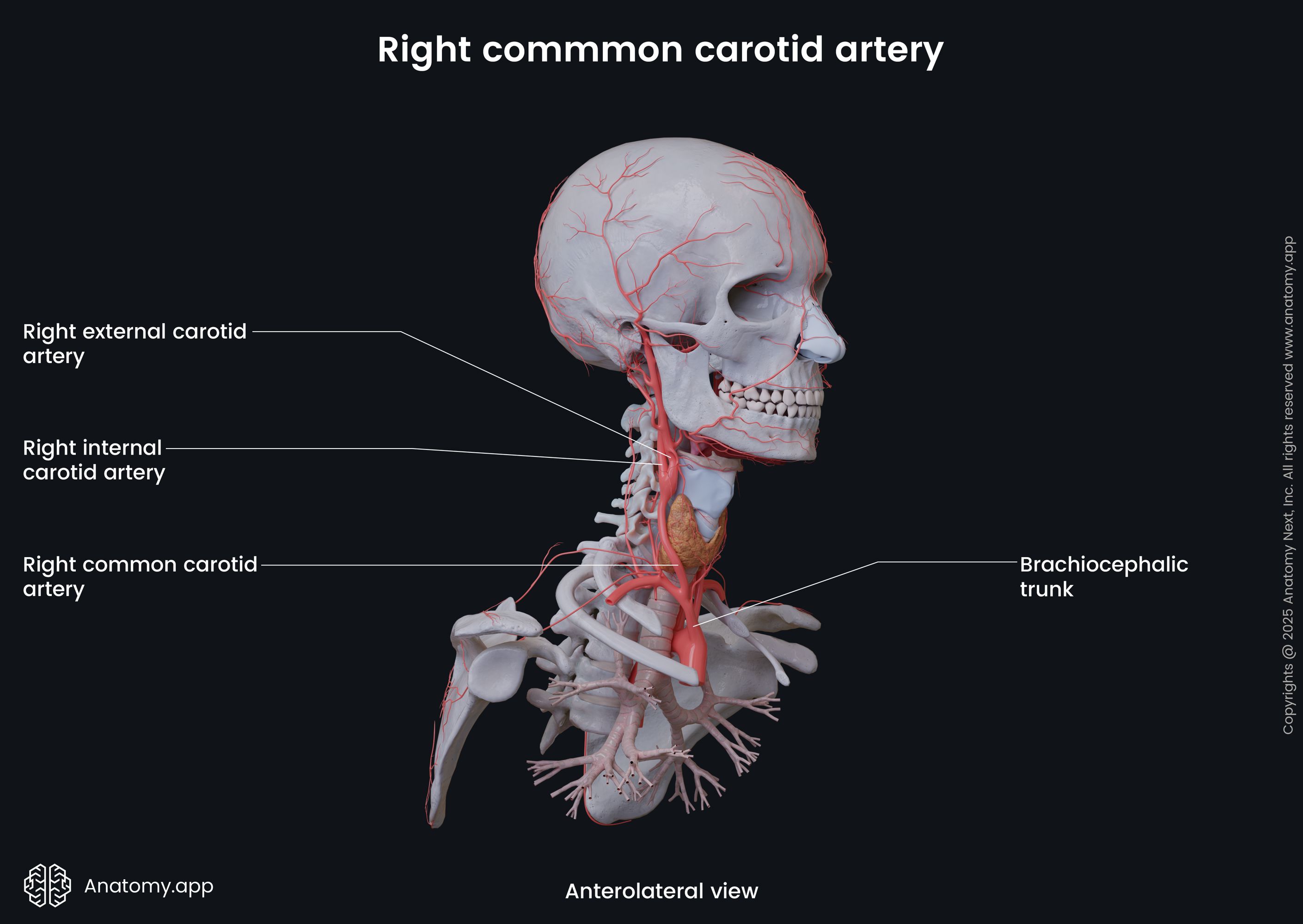
The right common carotid artery is located exclusively in the anterior neck region. It usually originates from the brachiocephalic trunk after it divides into two terminal branches, the other being the right subclavian artery. The bifurcation is situated behind the right sternoclavicular joint.
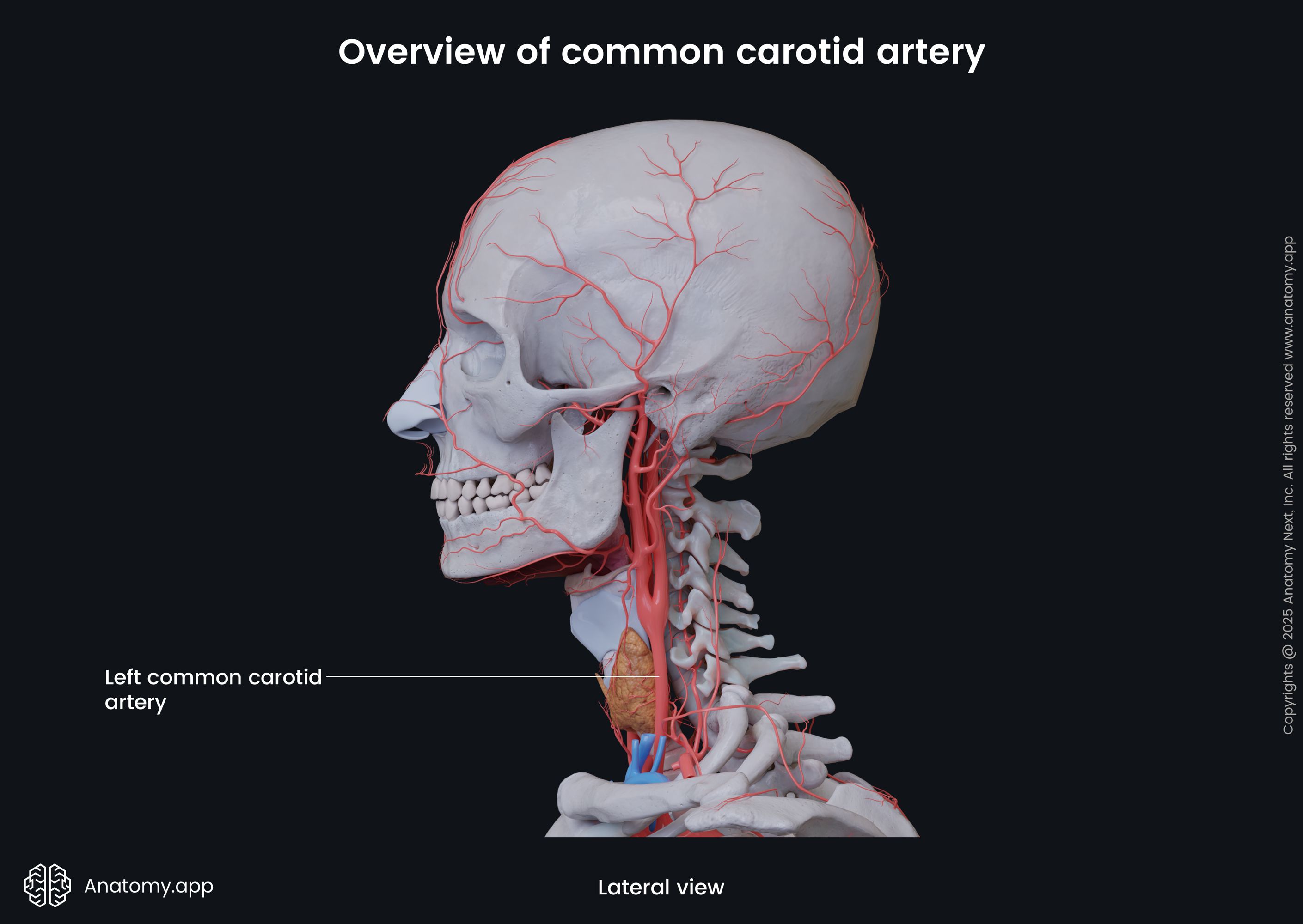
The left common carotid artery arises directly from the aortic arch. It is its second branch located in between the brachiocephalic trunk and the left subclavian artery. Because its course begins within the superior mediastinum, the left common carotid artery is longer than the right one. Also, in contrast to the right common carotid, the left artery has two portions - a thoracic and a cervical part.
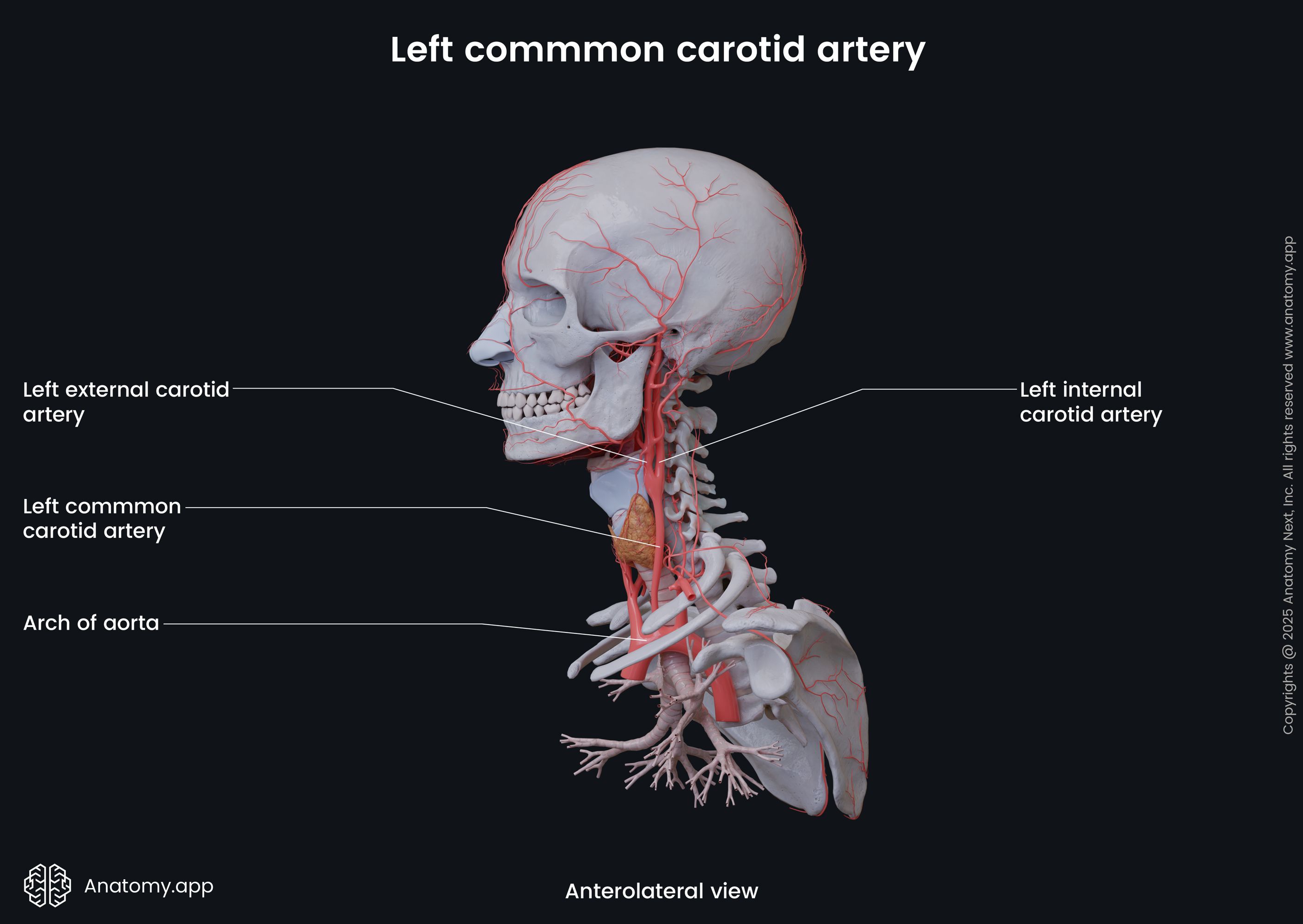
The thoracic part of the left common carotid artery is situated within the superior mediastinum - at first, anterior and then on the left side of the trachea. The artery exits the thorax and superior mediastinum via the superior thoracic aperture. At this point, it is located behind the left sternoclavicular joint.
In the neck, each common carotid artery ascends within the carotid sheath, which is a condensation of the deep cervical fascia. Within the carotid sheath, the common carotid artery goes together with the internal jugular vein (lateral to the artery) and the vagus nerve (CN X) (posterior to the artery).
Overall, the cervical parts of the common carotid arteries have similar courses. Both arteries ascend laterally towards the upper neck and thyroid cartilage. And they both terminate by dividing into two branches - internal and external carotid arteries. The bifurcation usually happens at the level of the upper border of the thyroid cartilage of the larynx, approximately between the third and fourth cervical vertebrae (C3 - C4 junction). In around 90% of the cases, the internal carotid artery lies posterolaterally to the external carotid artery.
Common carotid artery branches
The common carotid arteries are magistral arteries, meaning they usually do not give any branches along their course. As mentioned previously, they have two terminal branches - external and internal carotid arteries.
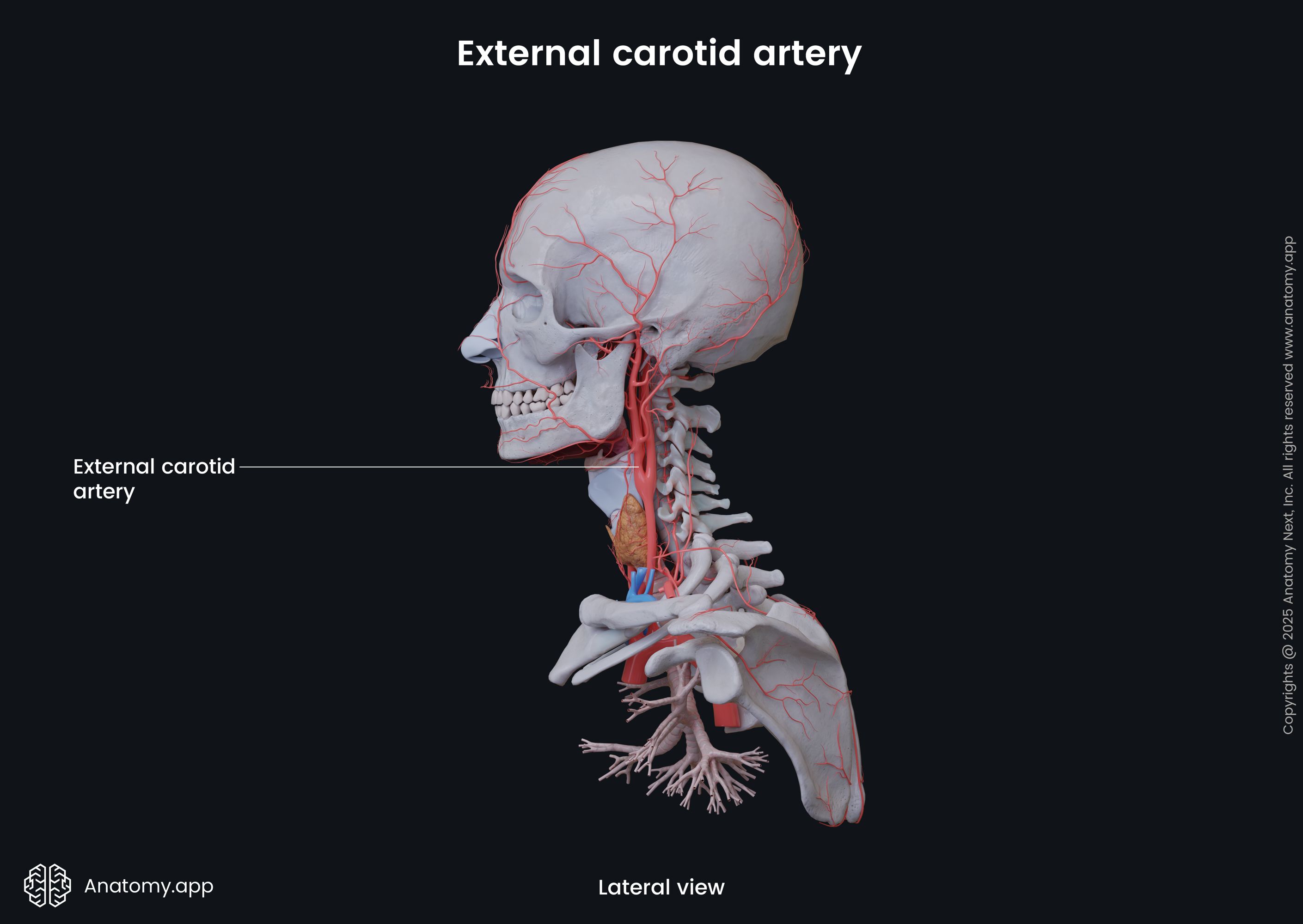
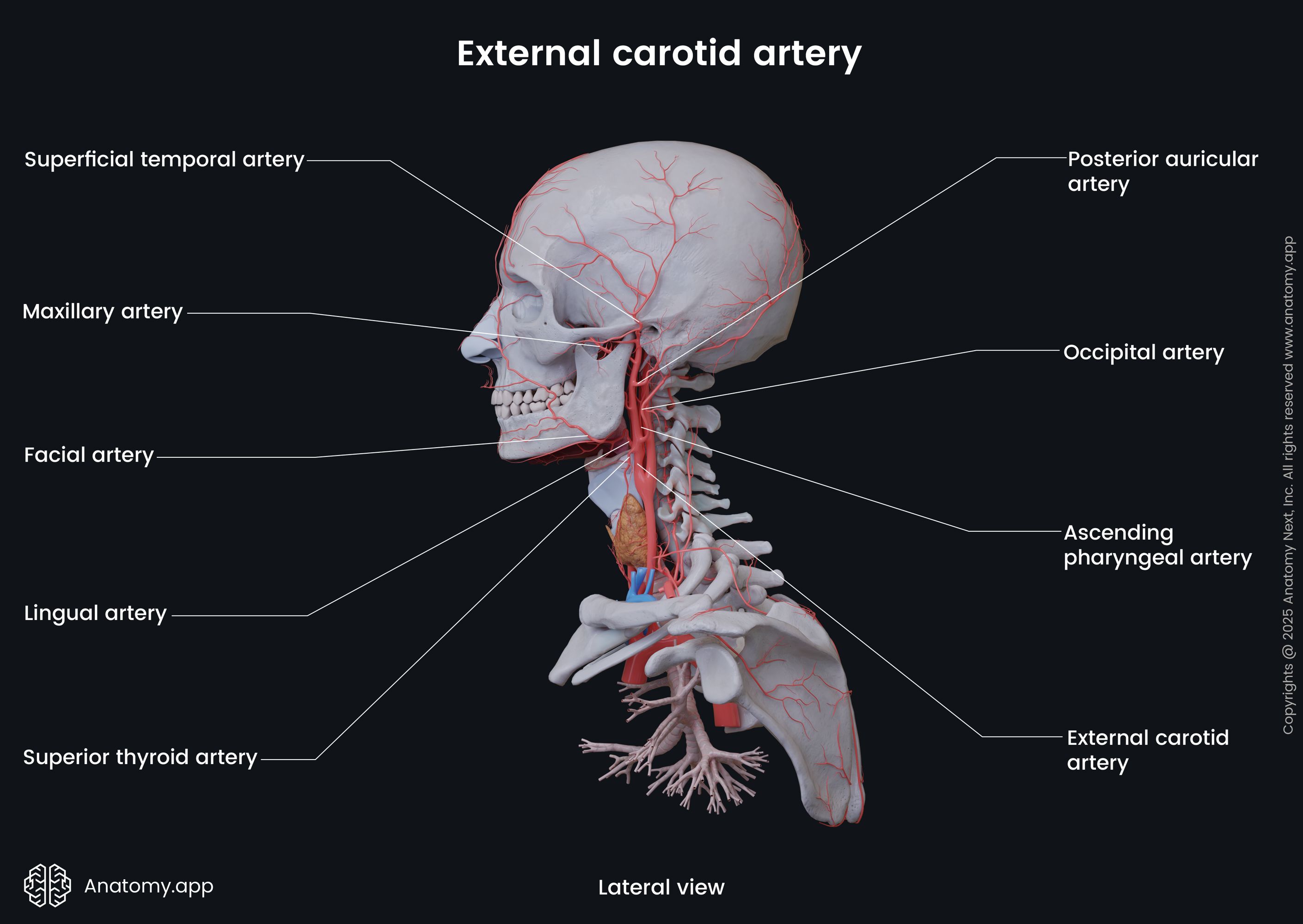
External carotid artery
The external carotid artery supplies the structures of the face, neck and scalp. It has eight branches: maxillary, superficial temporal, superior thyroid, lingual, facial, posterior auricular, occipital and ascending pharyngeal arteries.
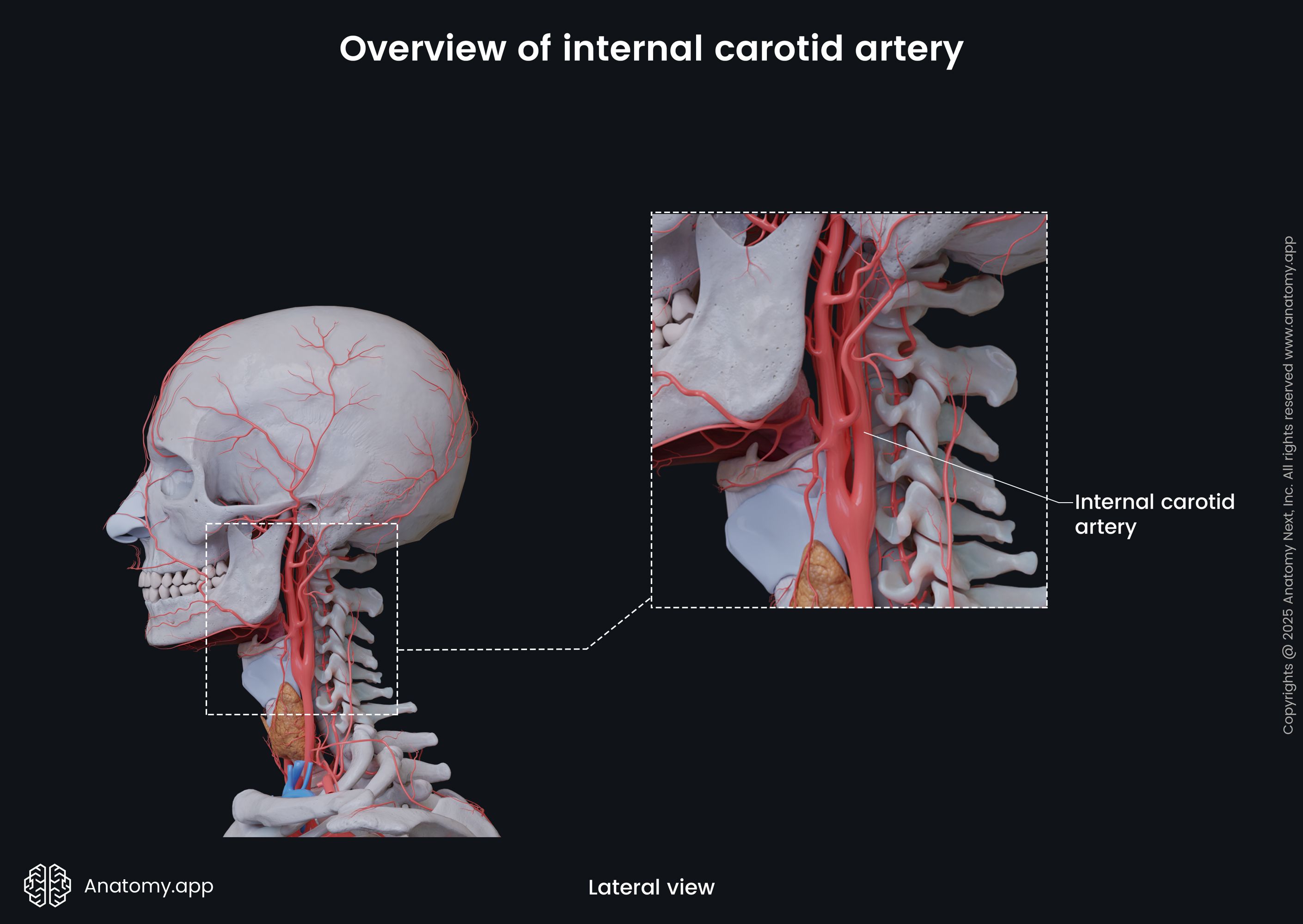
Internal carotid artery
The internal carotid artery usually does not give any branches until it reaches the skull base. Therefore, it does not supply the neck region. The internal carotid artery enters the cranial cavity via the carotid canal located in the petrous part of the temporal bone. Within the skull, it gives off various branches that supply the cerebral hemispheres, eyes, nose, nasal cavity, paranasal sinuses and forehead.
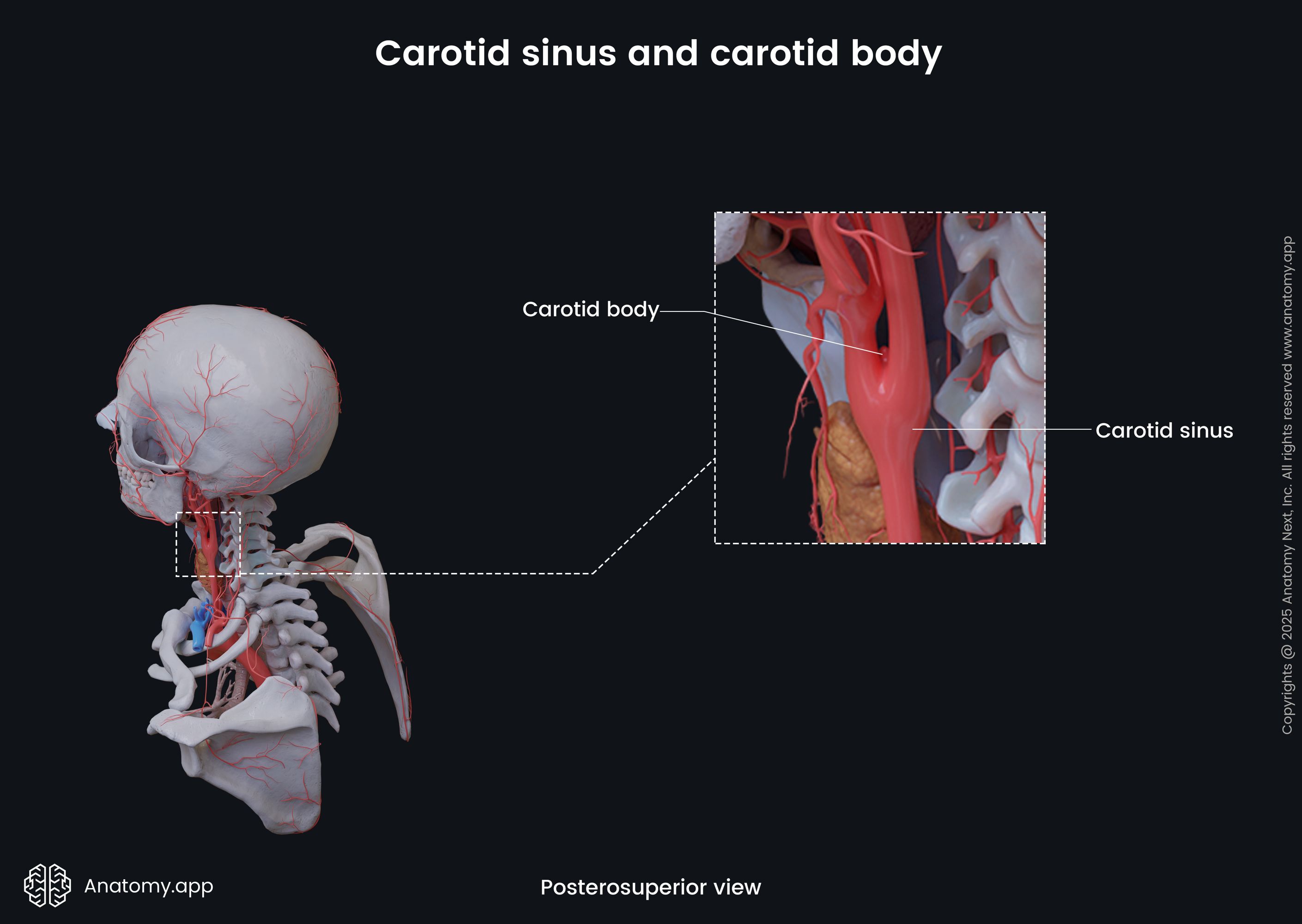
Carotid sinus and carotid body
Near the bifurcation, the common carotid artery is slightly dilated, and this area contains two specialized structures known as the carotid sinus and the carotid body. These structures are responsible for receiving information about the pressure (carotid sinus) and chemical composition (carotid body) of the arterial blood that flows through the common carotid artery. The carotid sinus and carotid body are both innervated mainly by the carotid branches of the glossopharyngeal nerve (CN IX).
Carotid sinus
The carotid sinus, or carotid bulb, is a neurovascular structure. It appears as a dilation of the proximal part of the internal carotid artery, occasionally involving the common carotid artery and its bifurcation. The carotid sinus functions as a baroreceptor and is sensitive to arterial blood pressure changes.
The carotid sinus is involved in the regulation of the heart rate and blood pressure. Detected changes are transmitted to the midbrain of the central nervous system via nerve impulses. The central nervous system interprets these impulses and sends signals back via the vagus nerve (CN X) that change the heart rate. This is known as the carotid sinus baroreflex.
Carotid body
The carotid body is an oval sensory structure located behind the carotid bifurcation or between both terminal branches of the common carotid artery. It is in close relation with the carotid sinus. The carotid body sometimes may appear as a group of separate nodules. Overall, it is composed of multiple lobules, which are separated by septa. Each lobule contains sensory cells (glomus cells). A fibrous capsule encloses the carotid body.
The carotid body functions as an arterial chemoreceptor, and it mainly monitors the oxygen, carbon dioxide and pH levels in the arterial blood. Low oxygen levels, high carbon dioxide, and low pH stimulate glomus cells. They send nerve signals to the respiratory centers in the brainstem, which increase the rate and volume of ventilation as a response. These changes also increase blood pressure and cardiac rate.
Common carotid artery relations
Both common carotid arteries are located next to various structures along their courses. Between both arteries (medial to each artery) in the lower neck lies the trachea, while both arteries are separated by the larynx and pharynx in the upper neck. Also, anteromedially to both arteries in the upper neck lies the thyroid gland.
As mentioned previously, within the carotid sheath, the common carotid artery is accompanied by the internal jugular vein on its lateral side and the vagus nerve (CN X) posteriorly. The esophagus and recurrent laryngeal nerve are situated medially to the carotid sheath. Anterior to the sheath is the ansa cervicalis, while posterior to it is the sympathetic trunk.
The anterior and lateral sides of the common carotid artery are crossed by the intermediate tendon or sometimes even the superior belly of the omohyoid muscle. It usually happens at the cricoid cartilage level.
- Below the omohyoid muscle, the common carotid artery is covered by the skin, superficial cervical fascia, platysma, deep cervical fascia and some of the neck muscles, such as the sternocleidomastoid, sternothyroid and sternohyoid.
- Above the omohyoid muscle, the common carotid artery is covered only by the medial border of the sternocleidomastoid, deep cervical fascia, platysma, superficial cervical fascia and skin.
Various neurovascular structures may cross the common carotid artery along its course, including the following structures:
- Sternocleidomastoid branch of the superior thyroid artery
- Superior root of the ansa cervicalis
- Superior thyroid vein
- Middle thyroid vein
- Anterior jugular vein
Posterior to the common carotid artery lies the sympathetic trunk and ascending cervical branch of the inferior thyroid artery. Also, deep to the common carotid artery can be found the transverse processes of the fourth to sixth cervical vertebrae (C4 to C6) together with the muscles attached to them - longus colli, longus capitis and anterior scalene muscles.
References:
- Gray, H., & Carter, H. (2021). Gray’s Anatomy (Leatherbound Classics) (Leatherbound Classic Collection) by F.R.S. Henry Gray (2011) Leather Bound (2010th Edition). Barnes & Noble.
- Kaufman, J. A., & Lee, M. J. (2014). Vascular and Interventional Radiology: The requisites. Saunders.
- Mauro, M. A., Murphy, K., Thomson, K. R., Venbrux, A. C., & Morgan, R. A. (2021). Image-guided interventions. Elsevier.
- Moore, K.L., Dalley, A.F., Agur, A.M. (2018). Clinically Oriented Anatomy, 8th Edition, Lippincott Williams & Wilkins.
Anatomy.app
Contact information
- For questions regarding business inquiries. Please contact:
- info@anatomy.app