- Anatomical terminology
- Skeletal system
- Joints
- Muscles
- Heart
- Blood vessels
- Blood vessels of systemic circulation
- Aorta
- Blood vessels of head and neck
- Arteries of head and neck
- Veins of head and neck
- Blood vessels of upper limb
- Blood vessels of thorax
- Blood vessels of abdomen
- Blood vessels of pelvis and lower limb
- Blood vessels of systemic circulation
- Lymphatic system
- Nervous system
- Respiratory system
- Digestive system
- Urinary system
- Female reproductive system
- Male reproductive system
- Endocrine glands
- Eye
- Ear
Vertebral artery
The vertebral artery (Latin: arteria vertebralis) is a paired artery found on each side of the neck. It provides arterial blood supply to the spinal cord, lower portion of the brainstem, posterior aspects of the cerebellum and cerebral hemispheres, as well as it supplies the meninges. Both vertebral arteries are not identical - the left is usually larger than the right.

The vertebral artery is the first branch of the subclavian artery. It arises from its initial (pre-scalene) segment in approximately 94 - 95 % of the cases. In about 5 - 6 % of individuals, the vertebral artery may arise directly from the aortic arch on the left side or the brachiocephalic trunk on the right side.
The vertebral artery ascends posteromedially towards the skull, passing through the transverse foramina of the cervical vertebrae, except the seventh cervical vertebra (C7). When reaching the first cervical vertebra (C1, atlas), the vertebral artery curves around its lateral mass and enters the cranial cavity through the foramen magnum.
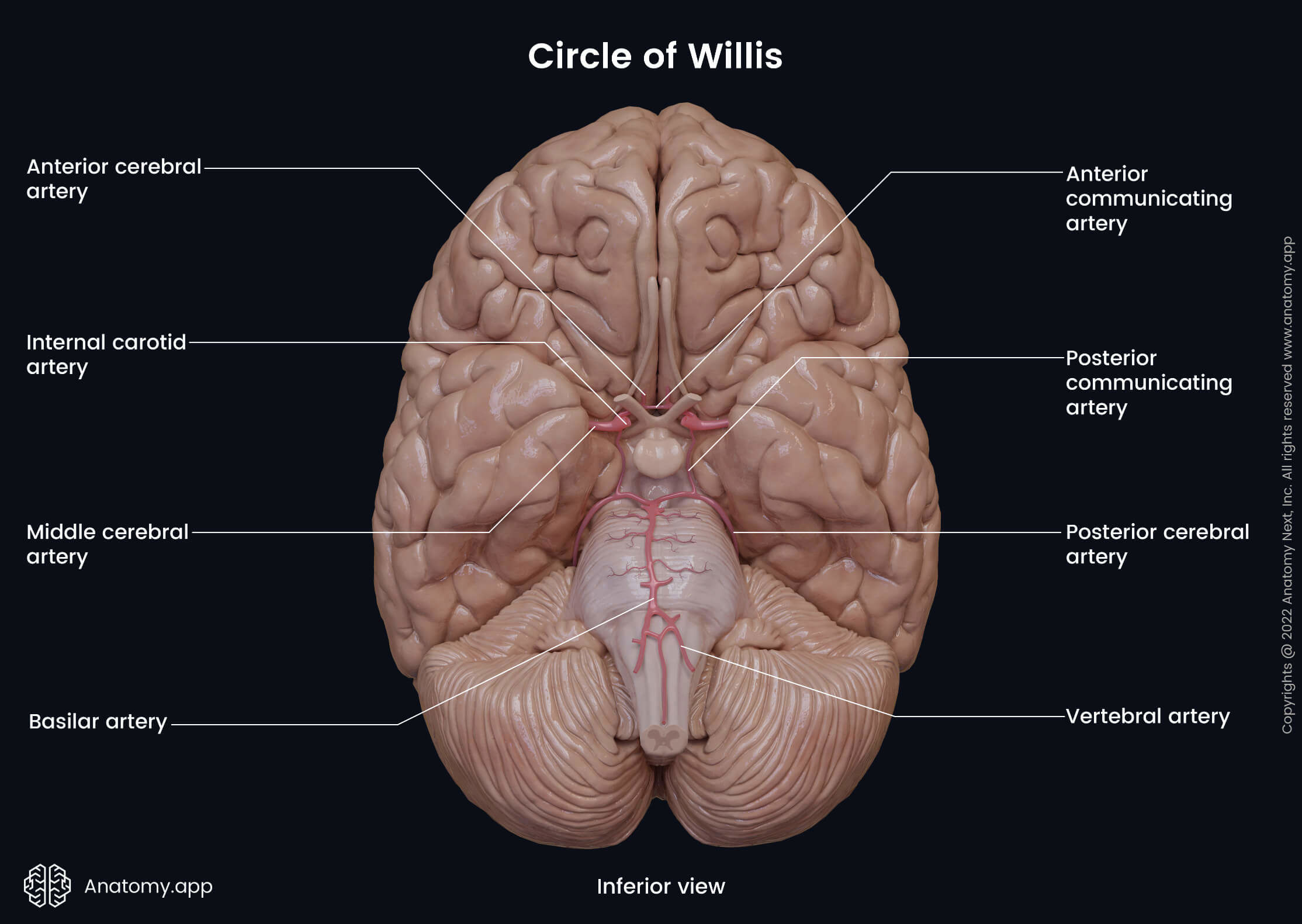
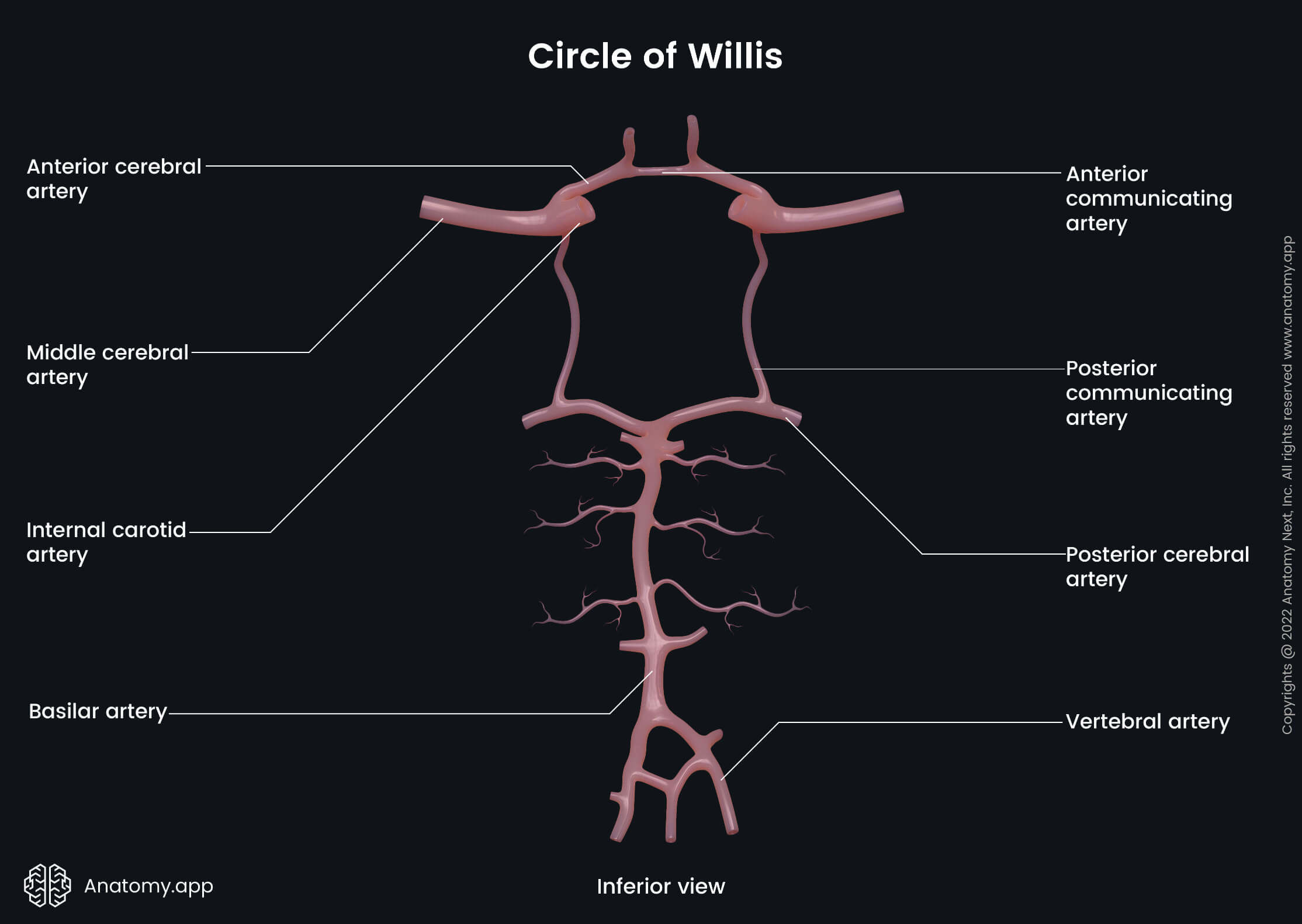
Both vertebral arteries approach each other on the clivus of the occipital bone, uniting into a single artery at its midline called the basilar artery. The basilar artery arises approximately at the level of the pontomedullary junction.
The basilar artery and vertebral arteries form the vertebrobasilar system, which is the posterior circulation system of the brain. The vertebrobasilar system comprises the posterior aspect of the circle of Willis.


Vertebral artery course and segments
The vertebral artery usually is subdivided into four segments based on its course:
- V1 - first or preforaminal segment;
- V2 - second or foraminal segment;
- V3 - third, extradural or extraspinal segment;
- V4 - fourth, intradural or intracranial segment.
The first segment (V1) starts at the origin of the vertebral artery when it branches off the subclavian artery. It ascends between the longus colli and anterior scalene muscles towards the transverse process of the seventh cervical vertebra (C7). The first segment does not go via the transverse foramen, but it crosses the anterior aspect of the transverse process. From there, the first segment further reaches the transverse foramen of the sixth cervical vertebra (C6). At this level, the first segment transitions into the second segment of the vertebral artery.
The second segment (V2) ascends through the transverse foramina of the sixth through third cervical vertebrae (C6 - C3). When the second segment reaches the transverse process and transverse foramen of the second cervical vertebra (C2, axis), it transitions into the third segment.
The third segment (V3) starts within the transverse foramen of the second cervical vertebra (C2; axis). After leaving the transverse foramen of the axis, this segment makes a turn laterally and posteriorly before entering the transverse foramen of the atlas (C1). It then curves around the lateral mass and courses posteromedially. It travels in a groove called the groove for vertebral artery that is found on the superior surface of the posterior arch of the atlas (C1). This segment then pierces the posterior atlantooccipital membrane to enter the vertebral canal, where it transitions into the final segment.
The fourth segment (V4) is the final portion starting at the site where the vertebral artery pierces the dura mater and arachnoid mater. It reaches the cranial cavity via the foramen magnum. Eventually, it joins the contralateral vertebral artery on the clivus to form a single artery called the basilar artery. The fourth portion of the vertebral artery courses along the anterior surface of the medulla oblongata and ends at its junction with the pons (pontomedullary junction).
Vertebral artery relations
The first segment (V1) passes behind the common carotid artery and the vertebral vein, between the longus colli and anterior scalene muscles. The thoracic duct lies anterior to the left vertebral artery, while the right lymphatic duct is positioned anterior to the right vertebral artery. Both arteries are located anteromedially to the inferior thyroid artery. The transverse process of the seventh cervical vertebra (C7), ventral rami of the seventh and eighth cervical spinal nerves (C7 - C8) and the inferior cervical ganglion are posterior to the first segment.
The second segment (V2) travels via the transverse foramina. It is usually accompanied by sympathetic nerves and a venous plexus, which later forms the vertebral vein. Behind the V2 segment lie the ventral rami of the cervical spinal nerves C2 to C6.
The third segment (V3) goes within the suboccipital triangle, where it is surrounded by the rectus capitis posterior major, obliquus capitis superior and obliquus capitis inferior muscles. It curves around the lateral mass of the atlas; therefore, it is lateral, posterior and medial to it. The vertebral artery is crossed by the dorsal ramus of the first cervical spinal nerve (C1).
The fourth segment (V4) of the vertebral artery lies anterior to the roots of the hypoglossal nerve (CN XII) and ventral surface of the medulla oblongata.
Vertebral artery branches
The vertebral artery gives off several branches along its course. All branches can be classified into two main groups depending on their origin site. These groups are the cervical and cranial branches.
Cervical branches
The cervical part of the vertebral artery gives off numerous branches as it courses up the neck. They are called cervical branches, and all cervical branches can be classified into two groups - spinal and muscular branches.
- The spinal branches supply the spinal cord via the intervertebral foramina. Moreover, these branches also supply the bodies of the cervical vertebrae.
- The muscular branches supply the deep muscles of the neck. These branches can form anastomoses with the occipital branches of the external carotid artery.
Cranial branches
Within the skull, the vertebral artery usually gives off several branches that are called cranial branches. They include the following:
- Anterior spinal artery
- Posterior spinal artery
- Posterior inferior cerebellar artery
- Meningeal arteries
- Medullary arteries
The anterior spinal artery is a small artery that originates near the basilar artery. It arises from the confluence of two smaller arteries given by both vertebral arteries. These arteries originate near the site where both vertebral arteries unite. From their origin, the anterior spinal branches descend and merge on the clivus, forming a single anterior spinal artery at the level of the foramen magnum. It reaches the vertebral canal via the foramen magnum and descends along the anterior surface of the spinal cord. It passes within the anterior median fissure found on the midline of the anterior surface of the spinal cord. The anterior spinal artery is the primary source that supplies arterial blood to the spinal cord.
The posterior spinal artery is a paired artery that usually arises near the medulla oblongata and descends along the right or left posterolateral sulcus of the spinal cord. These arteries supply the posterior one-third of the spinal cord. The posterior spinal arteries can also arise from the posterior inferior cerebellar arteries.
The posterior inferior cerebellar artery is the largest branch of the vertebral artery. It is a paired artery that originates near the lower aspect of the olive of the medulla oblongata and passes upwards along the side of the medulla near the roots of the glossopharyngeal (CN IX), vagus (CN X) and accessory (CN XI) nerves. The artery then reaches the inferior part of the pons and moves along the fourth ventricle. And finally, it reaches the cerebellum. The posterior inferior cerebellar artery supplies the lower part of the medulla oblongata and cerebellum.
The meningeal branches of the vertebral artery originate near the foramen magnum and supply the dura mater of the posterior cranial fossa and falx cerebelli. The medullary arteries supply the medulla oblongata.
References:
- Gray, H., & Carter, H. (2021). Gray’s Anatomy (Leatherbound Classics) (Leatherbound Classic Collection) by F.R.S. Henry Gray (2011) Leather Bound (2010th Edition). Barnes & Noble.
- Kaufman, J. A., & Lee, M. J. (2014). Vascular and Interventional Radiology: The requisites. Saunders.
- Shen, F. H., Samartzis, D., & Fessler, R. G. (2015). Textbook of the cervical spine. Elsevier/Saunders.
- Winn, H. R., & Youmans, J. R. (2022). Youmans and Winn neurological surgery. Elsevier.