- Anatomical terminology
- Skeletal system
- Skeleton of trunk
- Skull
-
Skeleton of upper limb
- Bones of shoulder girdle
- Humerus
- Bones of forearm
- Bones of hand
- Skeleton of lower limb
- Joints
- Muscles
- Heart
- Blood vessels
- Lymphatic system
- Nervous system
- Respiratory system
- Digestive system
- Urinary system
- Female reproductive system
- Male reproductive system
- Endocrine glands
- Eye
- Ear
Humerus
The humerus (Latin: humerus) is a long bone that forms the skeleton of the free upper arm. It extends between the shoulder and the elbow, and it is also the longest and largest bone of the upper limb.
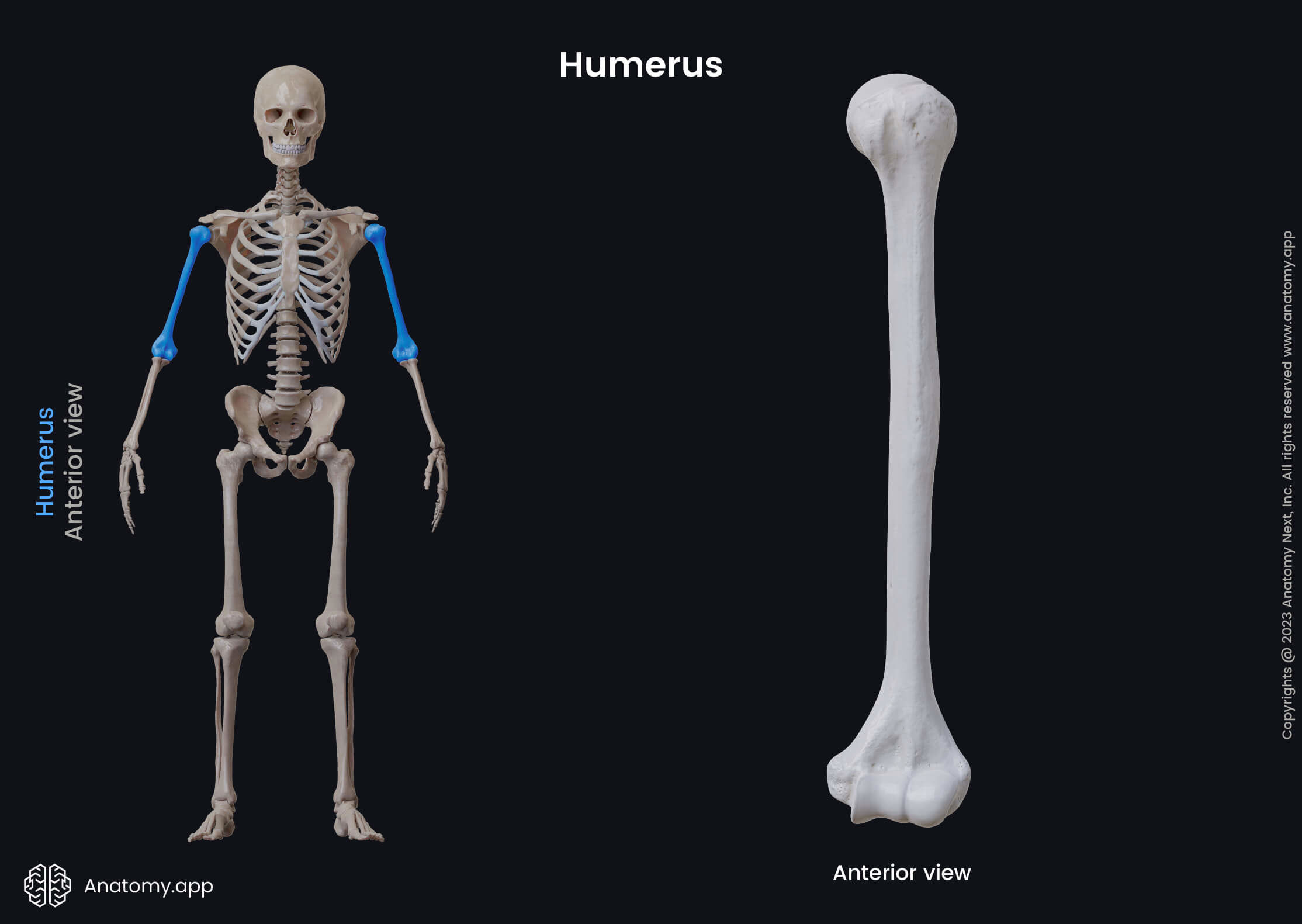
The humerus is connected with the scapula at its proximal end, and with both forearm bones (radius and ulna) at its distal end. Both articulations are known as the shoulder or the glenohumeral and elbow joints.
The proximal end of the humerus articulates with the glenoid cavity of the scapula. At the distal end, the humerus articulates with the head of the radius and the trochlear notch of the ulna, forming the elbow joint.
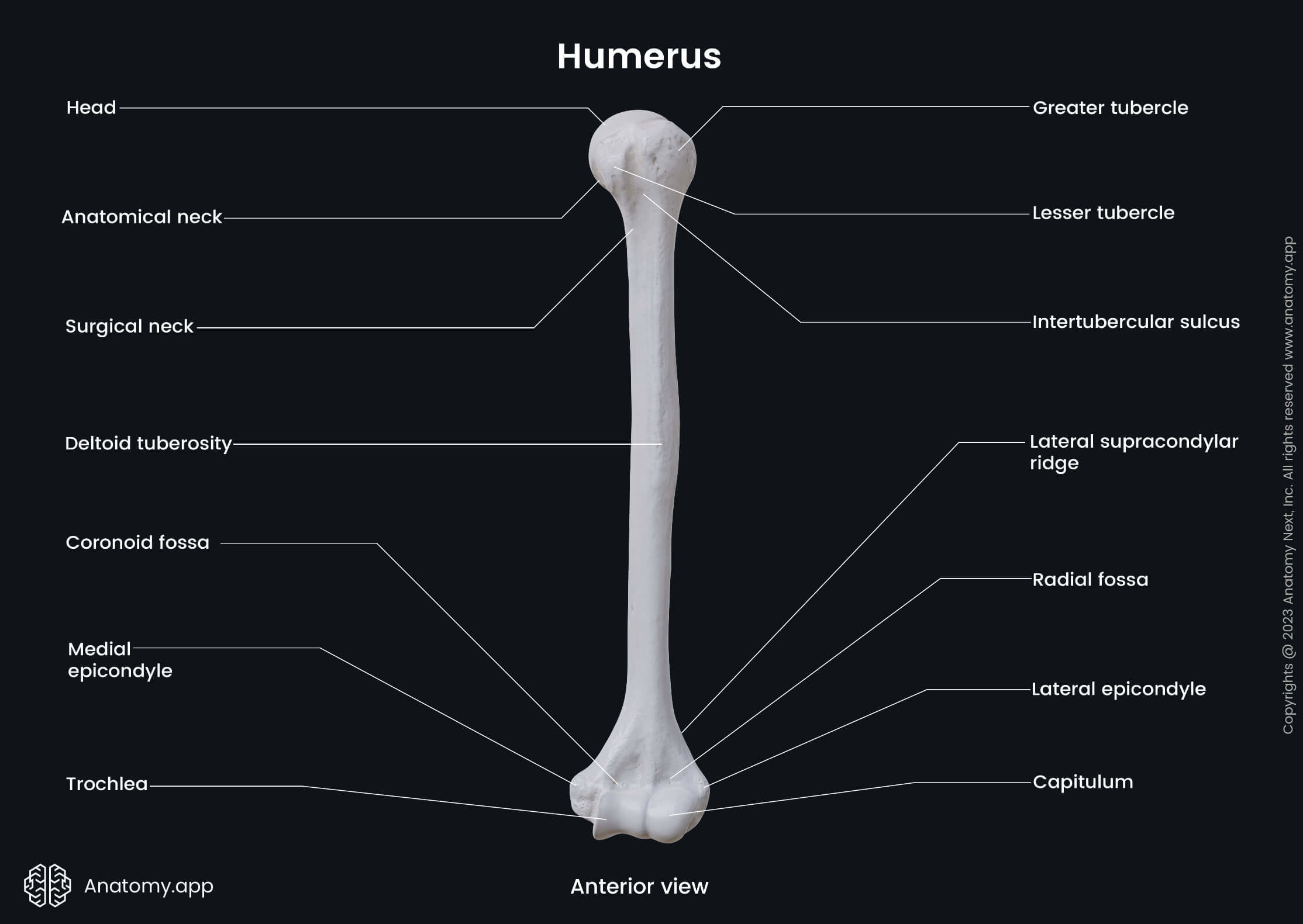
Like other long bones, the humerus is made of three main parts - a proximal end or epiphysis, a shaft or diaphysis, and a distal end or epiphysis. All of these parts contain important anatomical landmarks.
Proximal end of humerus
The proximal end or epiphysis of the humerus is the part of the humerus that is located closer to the shoulder girdle. It consists of the head, anatomical and surgical necks, and the greater and lesser tubercles.
The head of the humerus has a hemispherical shape. It contains a smooth articular surface, which is covered by hyaline cartilage. The head faces in medial, superior, and posterior directions in the anatomical position. It articulates with the glenoid cavity of the scapula.
The anatomical neck of the humerus is a circular and oblique narrowing below the articular surface of the head. It encircles the head of the humerus. The joint capsule of the shoulder joint is attached to the sides of the anatomical neck.
The greater tubercle is the most lateral part of the proximal end. The upper posterior aspect of this tubercle presents with three impressions, all of which serve as attachment sites for muscles. Muscles that attach at these impressions are three of the rotator cuff muscles:
The lateral aspect of the greater tubercle is covered by the deltoid muscle. Multiple vascular foramina (openings) also mark the lateral aspect of the greater tubercle. From the greater tubercle downward extends the crest of greater tubercle.
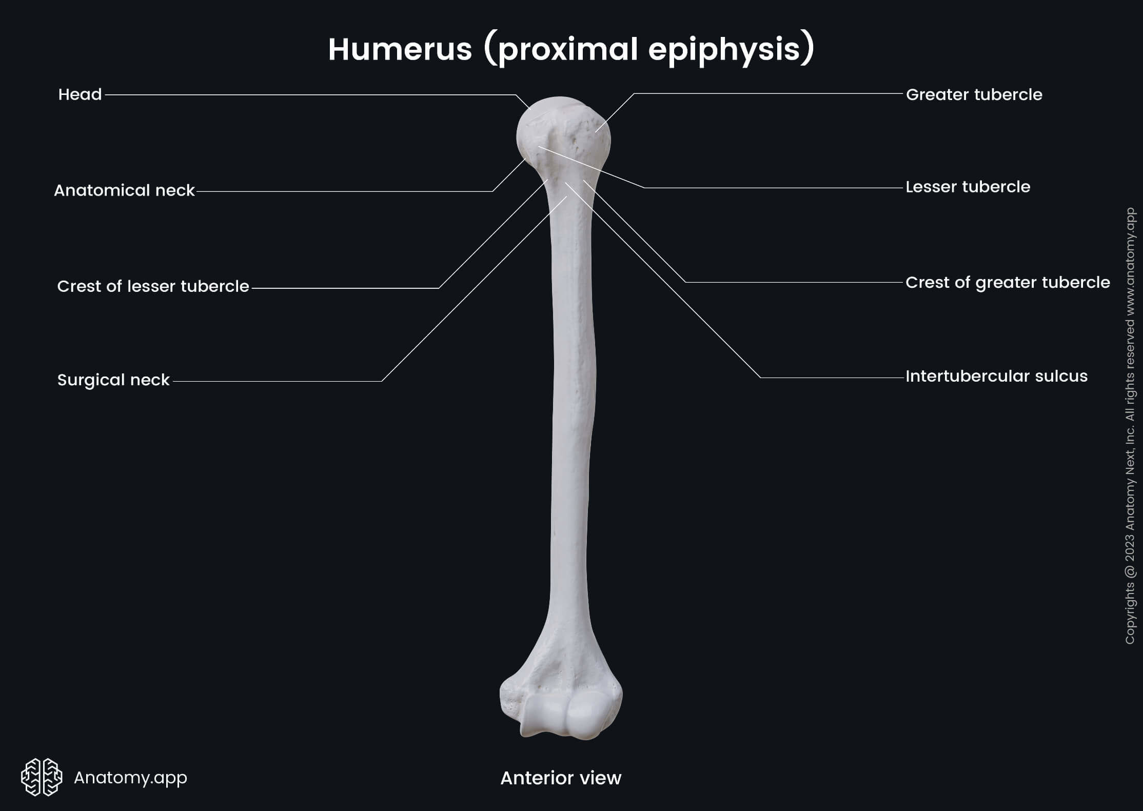
The lesser tubercle is found anterior to the anatomical neck, and it has a smooth, palpable muscular impression. The lateral aspect of this tubercle forms the medial margin of the intertubercular sulcus - a deep groove separating both tubercles.
The intertubercular sulcus is also known as the bicipital groove. The long tendon of the biceps brachii and an ascending branch of the anterior circumflex humeral artery lie within the intertubercular sulcus. It is formed by two lips named the lateral and medial lips.
The lateral lip is also known as the crest of greater tubercle, and it gives attachment to the tendon of the pectoralis major muscle. The medial lip is known as the crest of lesser tubercle, and it serves as an attachment site for the teres major muscle.
Also, the tendon of the latissimus dorsi muscle attaches to the posterior aspect of the crest of lesser tubercle. The lesser tubercle of the humerus also gives attachment to the subscapularis muscle and the transverse humeral ligament.
Below both tubercles, the proximal end of the humerus presents with a slight narrowing, which is known as the surgical neck. It is a common fracture site. The axillary nerve and the posterior circumflex humeral artery lie close to the bone at its surgical neck part.
Shaft of humerus
The shaft or diaphysis is the middle part of the humerus, and it gives attachment to several muscles. Cross-section studies reveal that the proximal half of the shaft is circular, while its distal half is more triangular and flattened. The shaft of the humerus presents with three borders and three surfaces.
Borders of shaft
The shaft of the humerus contains three borders:
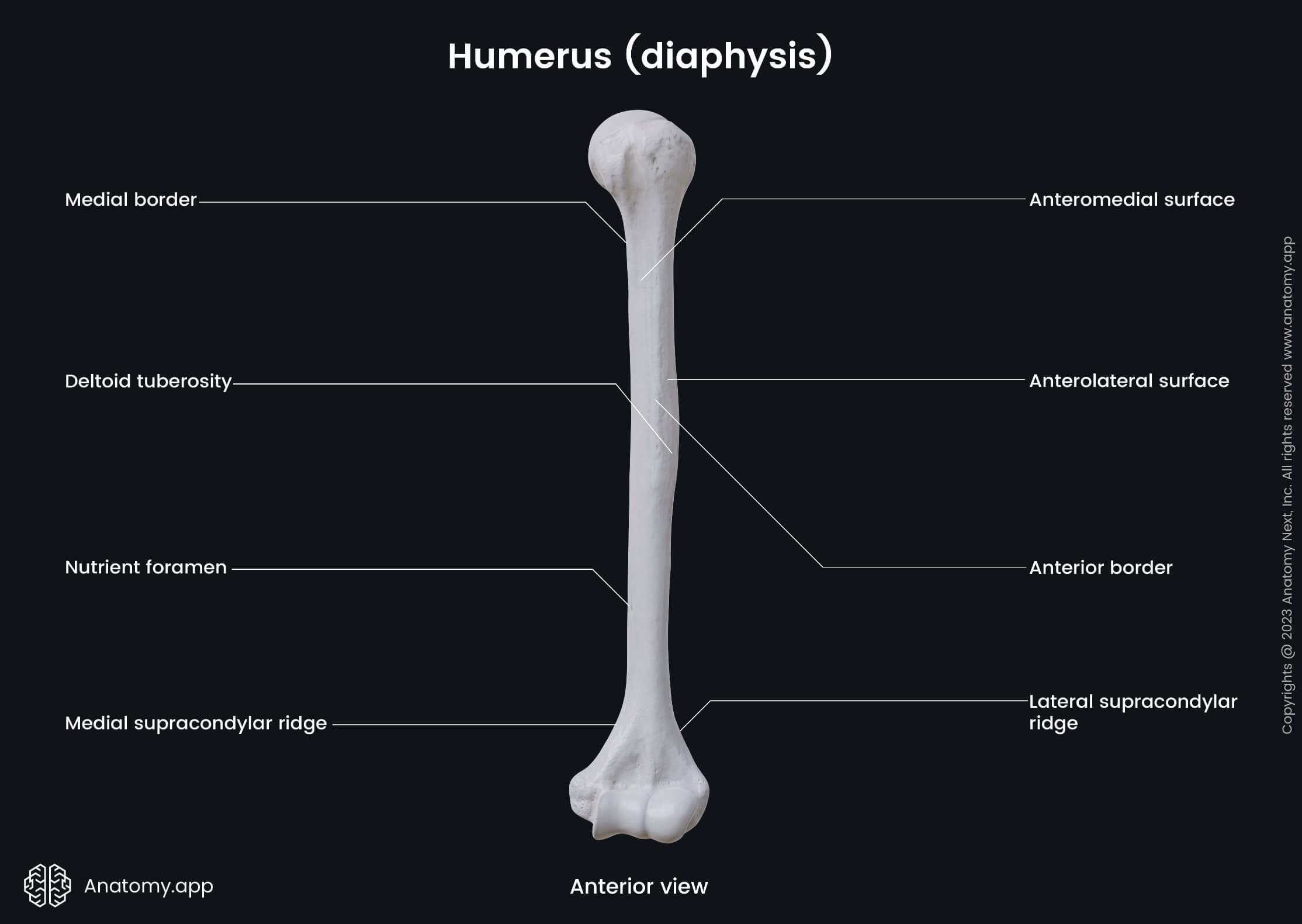
The anterior border starts from the greater tubercle of the humerus and extends downward from the tubercle almost to the end of the humerus. The upper part of the border is continuous with the lateral lip of the intertubercular sulcus.
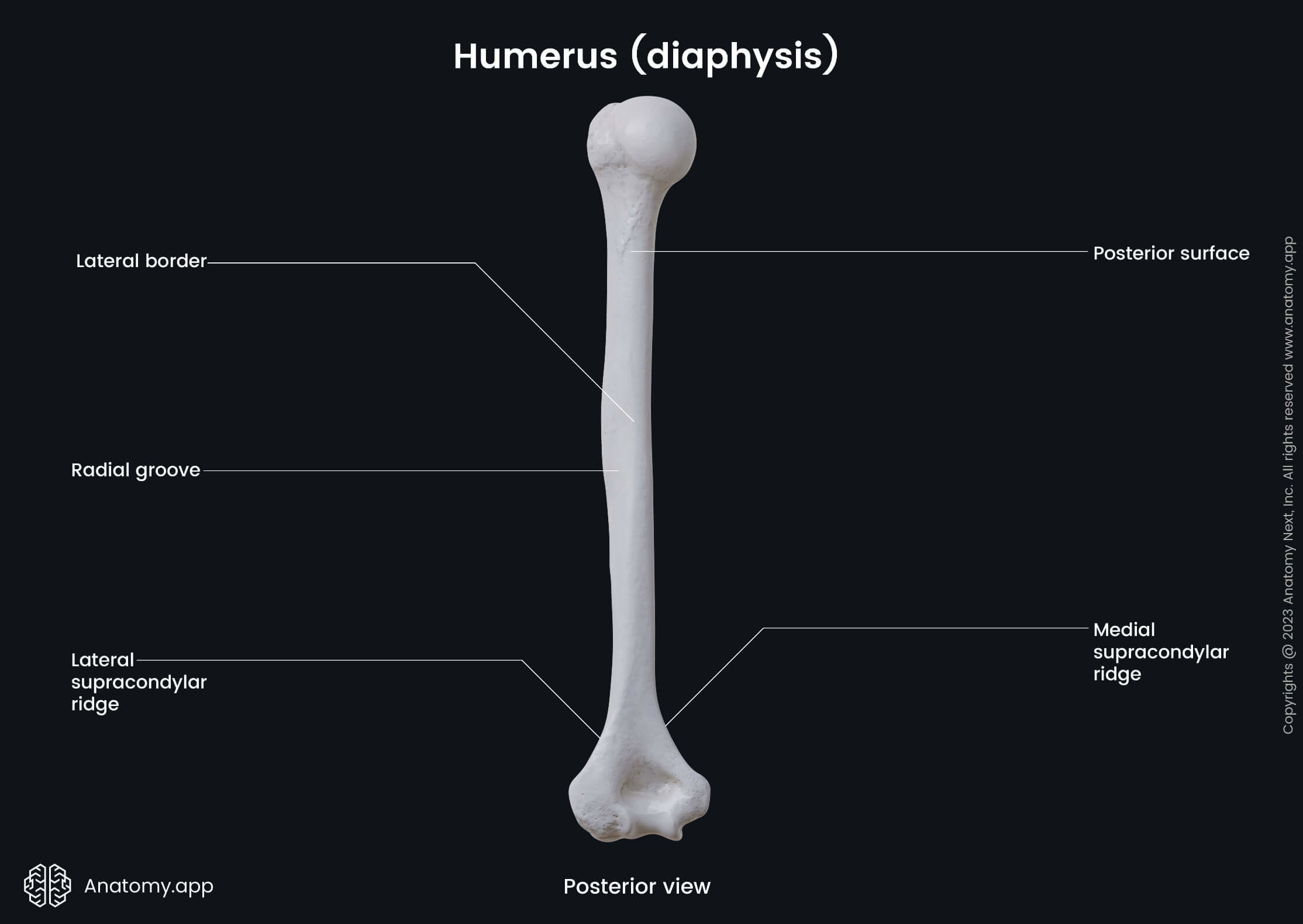
The lateral border extends downward from the posterior part of the greater tubercle. Distally this border gets thicker to form the lateral supracondylar ridge. In the middle of the lateral border is located a rough area called the deltoid tuberosity.
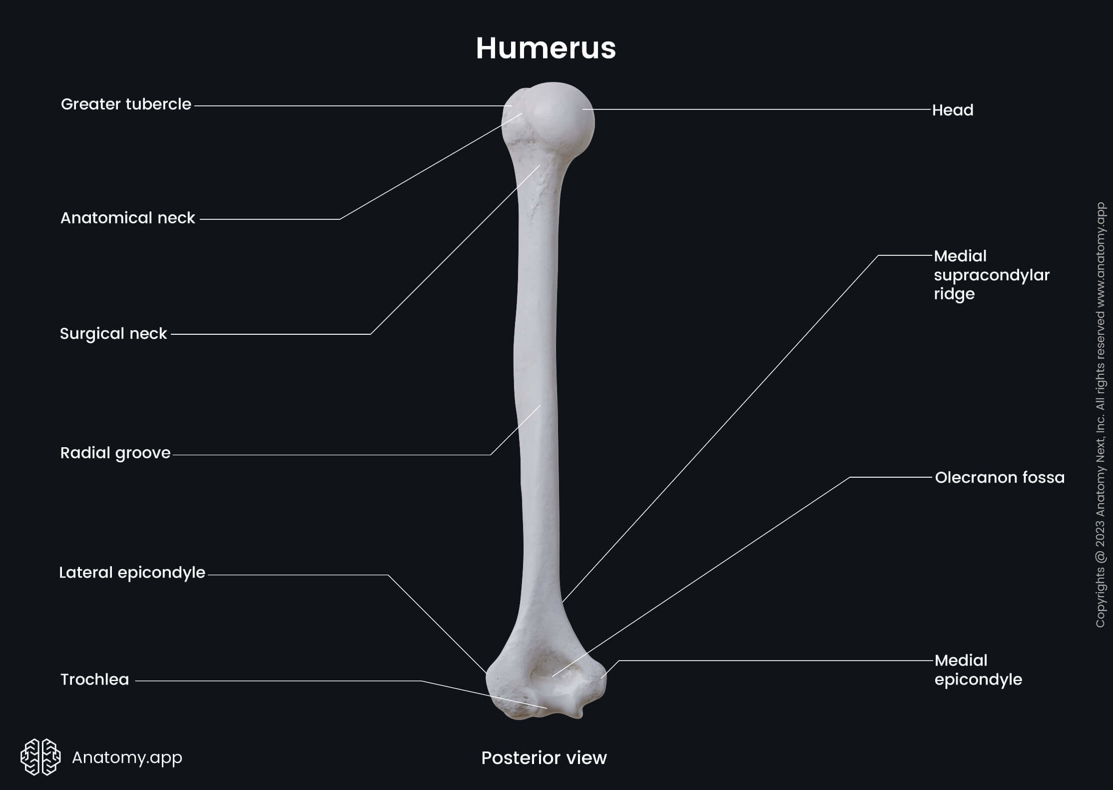
Like the lateral border, the medial border becomes thicker distally and forms the medial supracondylar ridge. This border is interrupted in its medial third by a shallow groove called the radial groove. The radial nerve and the deep brachial artery lie in the groove.
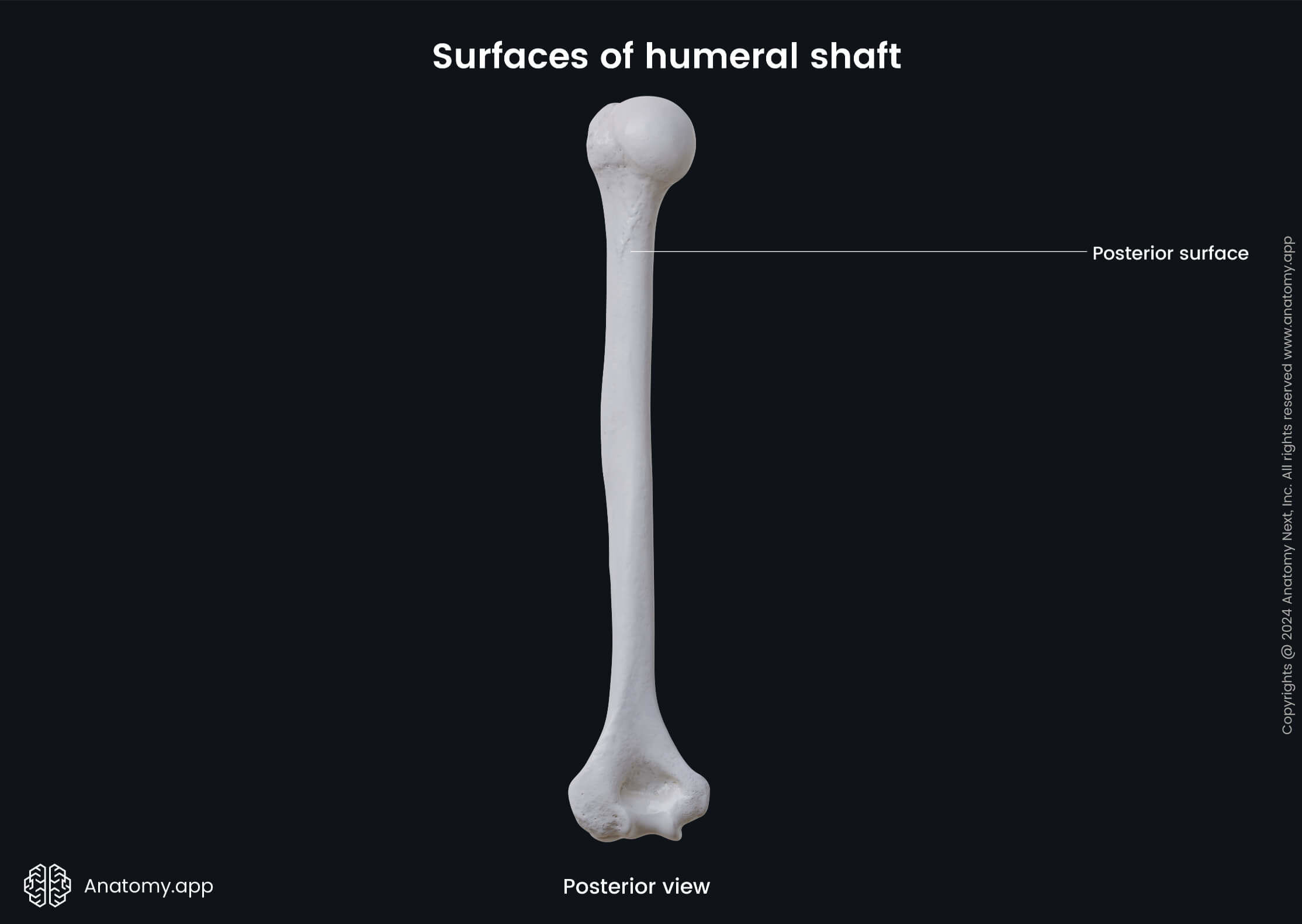
Surfaces of humeral shaft
The shaft of the humerus presents with three surfaces:
The anterolateral surface is the area between the anterior and lateral borders. Its proximal aspect is smooth and mostly covered by the deltoid muscle that inserts into the deltoid tuberosity. From the distal portion of the anterolateral surface and the proximal two-thirds of the lateral supracondylar ridge originates the lateral portion of the brachialis muscle.
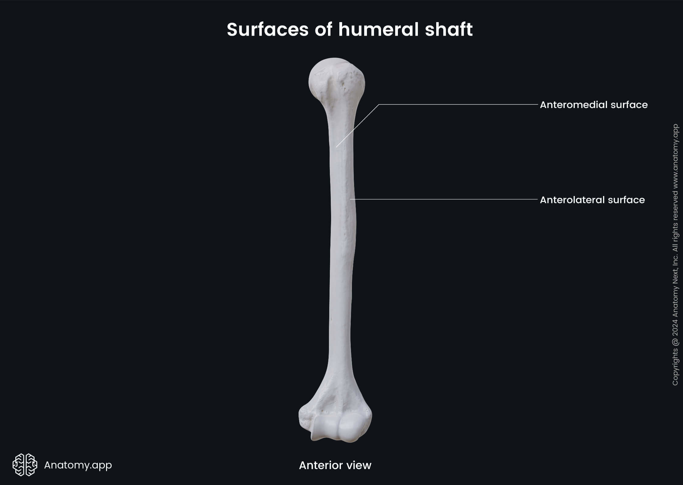
The anteromedial surface is the area between the anterior and medial borders. The coracobrachialis muscle attaches approximately in the middle part of this surface, while the distal half of the anteromedial surface is mainly covered by the medial portion of the brachialis.
The posterior surface is located between the medial and lateral borders of the shaft. Most of this surface is covered by the medial head of the triceps brachii muscle, while a ridge that is located on its proximal third also serves as an attachment site for the lateral head of the triceps brachii muscle.
Distal end of humerus
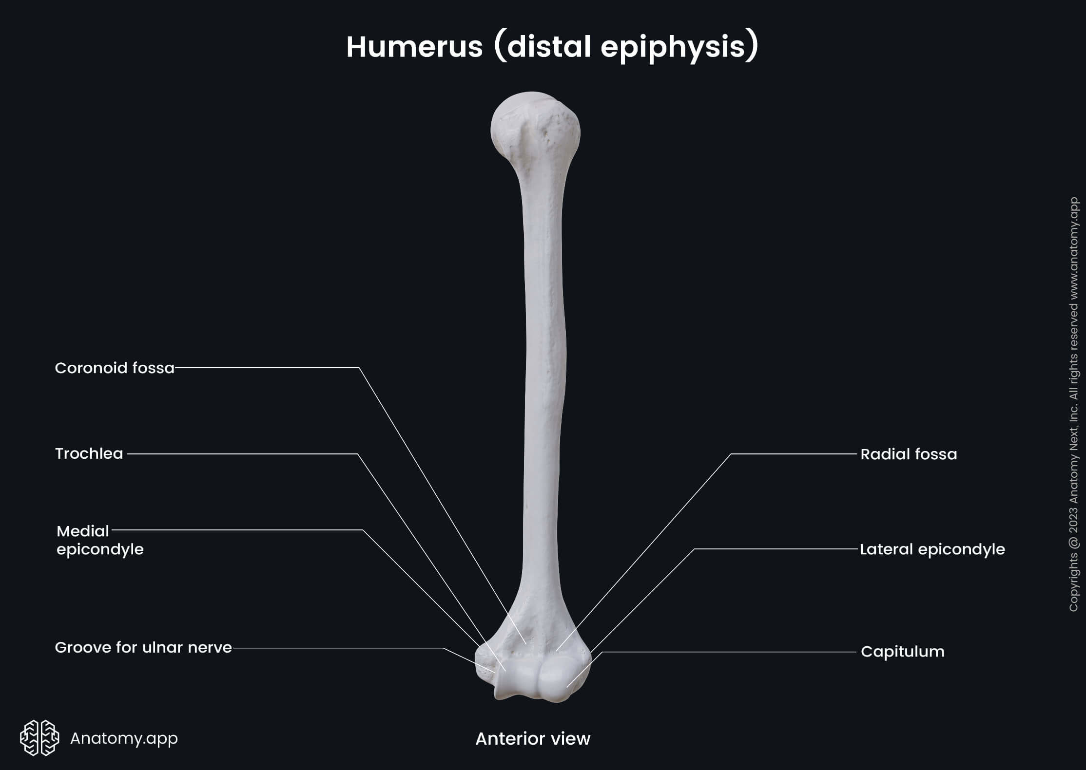
The distal end or epiphysis presents with articular and non-articular parts. The articular part consists of a modified condyle, which is formed by a medially located trochlea and laterally positioned capitulum. Both structures are separated by a groove.
The non-articular part primary consists of two large tuberculated eminences on either side of the humerus known as the epicondyles. This part also includes structures located above the condyle of humerus - the olecranon, coronoid and radial fossae.
Articular part of distal end
As mentioned above, the articular part of distal end includes the condyle of humerus with trochlea and capitulum. Both structures articulate with the bones of the forearm - ulna and radius - and together form the elbow joint.
The trochlea is a projection that is shaped like a pulley. It is located on the medial side of the condyle, extending onto the posterior aspect of the humerus. The trochlea articulates with the ulna at the trochlear notch.
Lateral to the trochlea is located the capitulum - a rounded and convex projection on the distal end of the humerus. It articulates with the head of the radius, and this articulation participates in the formation of the elbow joint.
Non-articular part of distal end
The non-articular part of the distal end includes two epicondyles - medial and lateral epicondyles - and three fossae - olecranon, coronoid and radial fossae. Although all are non-articular structures, they are indirectly involved in the formation of the elbow joint.
The medial epicondyle is a blunt projection forming the end of the medial border of the humerus. It is located superomedially to the medial aspect of the condyle. The posterior aspect of the medial epicondyle contains a groove for ulnar nerve.
The anterior surface of the medial epicondyle provides origin sites for several superficial muscles of the anterior compartment of the forearm. These superficial muscles of the anterior compartment of the forearm are as follows:
The lateral epicondyle is smaller than the medial one, and it is located at the end of the lateral border. The lateral epicondyle contains an impression to which are attached seven superficial muscles of the lateral and posterior compartments of the upper limb:
- Brachioradialis
- Extensor carpi radialis longus
- Extensor carpi radialis brevis
- Extensor digitorum
- Extensor digiti minimi
- Extensor carpi ulnaris
- Anconeus
The olecranon fossa is a deep depression on the posterior surface of the distal end, located superior to the condyle. During the elbow extension, the tip of the olecranon of the ulna lodges into this fossa.
The coronoid fossa is a smaller depression that is located superior to the trochlea on the anterior side of the humerus. Upon elbow flexion, the coronoid process of the ulna lodges into this fossa.
Lateral to the coronoid fossa and superior to the capitulum of the humerus is another hallow area called the radial fossa. The margin of the head of the radius lodges into this fossa upon full elbow flexion.
Anatomy.app
Contact information
- For questions regarding business inquiries. Please contact:
- info@anatomy.app